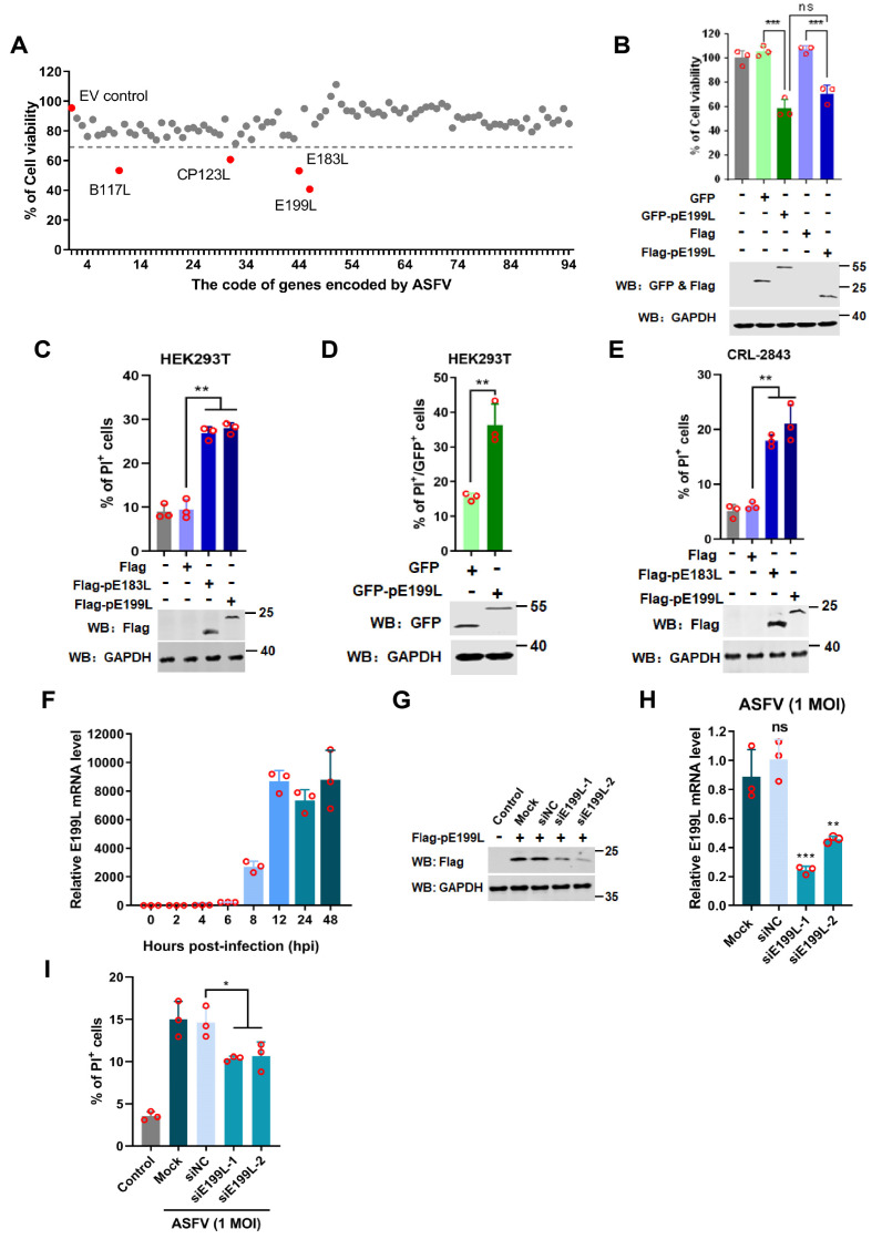
Figure 2.
ASFV pE199L induces cell death. (A) Screening of the ASFV genes involved in cell death in vitro. HEK293T cells were transfected with a plasmid expressing 1 of the 94 ASFV-encoded proteins for 36 h and then examined for cell viability. The genes corresponding to relative cell viability (normalized to the empty vector control) below the dotted line were considered. (B) Detection of the cell viability induced by pE199L. HEK293T cells were transfected with pFlag-E199L or pGFP-E199L for 36 h, followed by ATP activity examination. The HEK293T cells transfected with pCAGGS-Flag (pFlag) or pGFP-C1 (pGFP) were used as control. (C,D) Detection of cell death induced by pE199L in HEK293T cells. HEK293T cells were transfected with pFlag-E199L, pFlag-E183L as a positive control (C), or pGFP-E199L (D) for 36 h. The cells were stained with PI, and the percentage of the PI-labeled cells in the total cells (C) or in the GFP-expressed cells or the GFP-pE199L-expressed cells (D) were analyzed by flow cytometry. (E) Detection of cell death induced by pE199L in CRL-2843 cells. CRL-2843 cells were transfected with pFlag-E199L or pFlag-E183L as a positive control for 36 h and then stained with PI. In total, 10,000 cells were analyzed to determine the percentage of PI-labeled cells by flow cytometry. (F) Detection of the mRNA level of E199L in ASFV-infected PAMs. PAMs were infected with ASFV HLJ/18 at 1 MOI and the mRNA of E199L was analyzed at 0, 2, 4, 6, 8, 12, 24, and 48 hpi by qRT-PCR. (G,H) Testing of siE199L knockdown efficiency. HEK293T cells were transfected with non-targeting siRNA (siNC) or siRNA targeting ASFV E199L gene (siE199L-1, siE199L-2) for 12 h followed by transfection with the pFlag-E199L for 24 h and then Flag-pE199L expression testing by Western blotting (G). PAMs were transfected with siE199L or siNC for 12 h and then infected with HLJ/18 at 1 MOI for 24 h. The PAMs were subjected to detection of the knockdown effect of ASFV E199L by qRT-PCR (H). (I) Analysis of the effect of pE199L on cell death during ASFV infection. The 10,000 cells in (H) were stained with PI and analyzed by flow cytometry. Control: untreated cells; Mock: ASFV-infected cells with non-transfected siRNA. In the Figure 2, + means transfection of indicated plasmids while − means non-transfection of indicated plasmids. The significance of the differences between the groups was determined by the Student’s unpaired t-test with 2-tails (parametric test) or the ordinary ANOVA test with Dunnett (* p < 0.05, ** p < 0.01, and *** p < 0.001).

