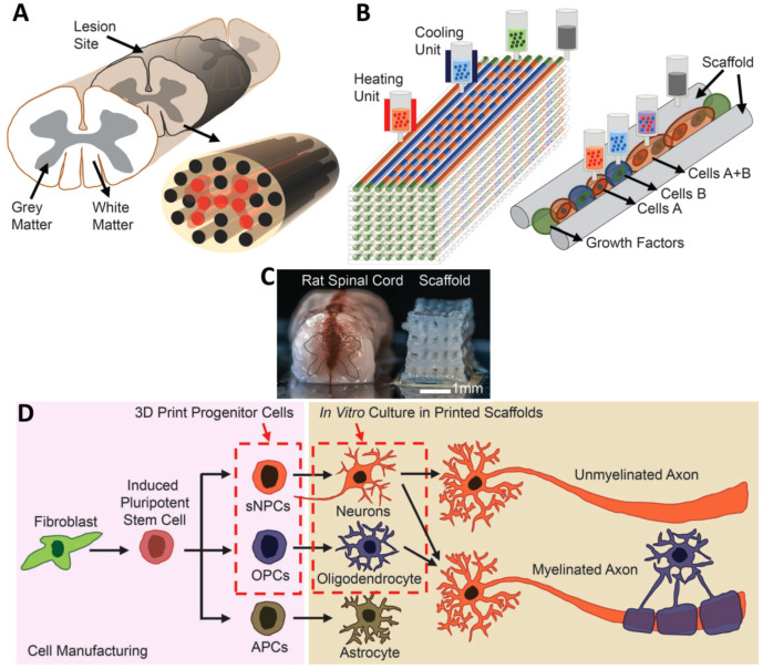
Figure 5.
(A) Schematic of the spinal cord and 3D bioprinted scaffold. (B) Schematic of the extrusion bioprinting process. (C) Comparison of a real spinal cord and the fabricated scaffold. (D) Schematic of differentiation of iPSCs into three different types of neuronal cells. Reprinted from [75] with permission from Wiley Online Library.

