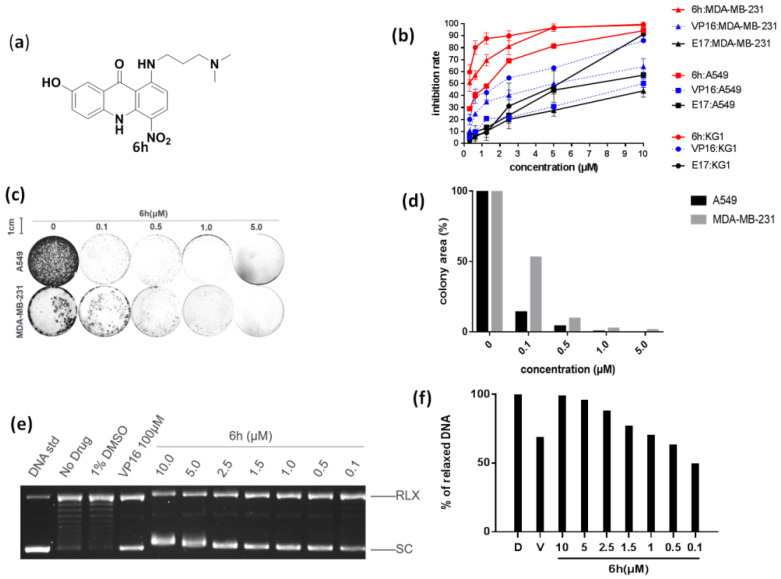
Figure 1.
6h exhibited strong anticancer efficacy by inhibiting topo IIα. (a) Chemical structure of 6h. (b) 6h dose-dependently inhibited the proliferation of different cancer cells. Data are presented as mean ± SD (n = 3). (c,d) 6h inhibited colony formation of two cancer cells. Images display the colony formation in 6h-treated cells. 6h dose-dependently inhibited colony formation in A549 and MDA-MB-231 cells. (e,f) 6h inhibited topo IIα-mediated DNA relaxation. VP16 was set as a positive control. Supercoiled pBR322 DNA (SC) and relaxed DNA (RLX) are shown. D: DMSO, V: VP16 at 100 μM.

