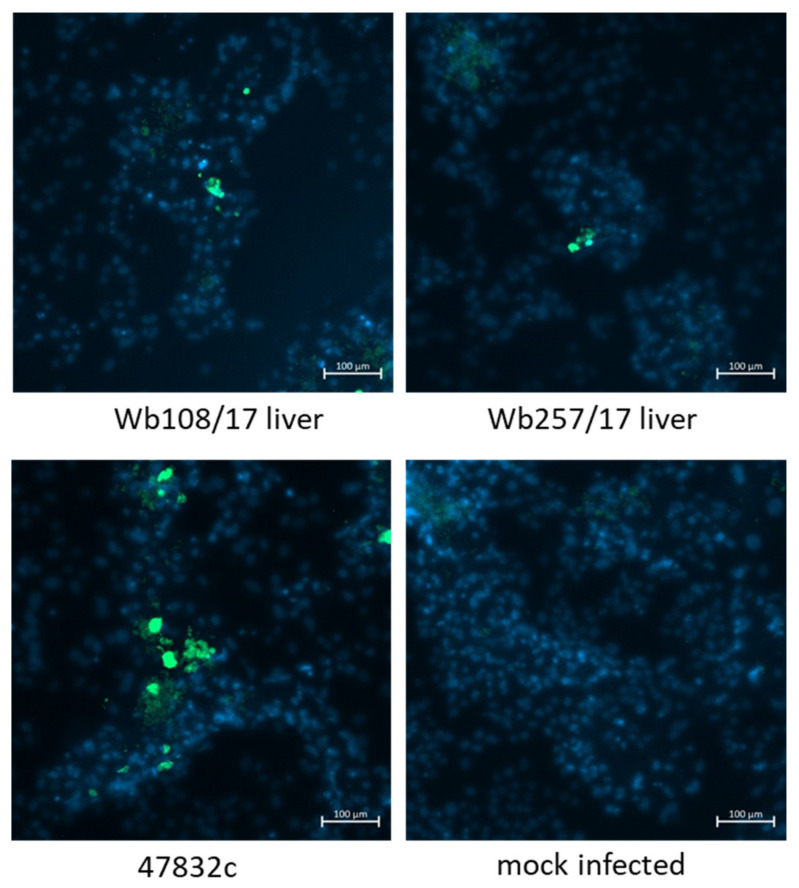
Figure 5.
Immunofluorescence analysis of PLC/PRF/5 cells 2 weeks after infection with cell culture supernatants from 49 d p.i. of the 2nd passage. Staining was done using an anti-HEV capsid protein-specific antiserum (green staining). Cell nuclei were stained with DAPI (blue staining). Scale bar: 100 µm.

