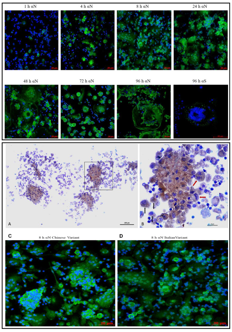
Figure 6.
Time-course of MDM infected with the SARS-CoV-2 strains/variant. The upper panel shows the Italian strain time-course. Cells are stained with the anti-N and anti-Spike protein (green fluorescence) and with DAPI (blue fluorescence), at 40× magnification in confocal microscopy. The lower panel shows the anti-N immunostaining (brown stain) of infected MDM (A,B) and the comparison of “blackberry-shaped” formations in the Chinese-derived (C) and PV10734 strains (D) 8 h p.i. with confocal microscopy. The area squared in (A) is enlarged in panel (B) with multinucleated MDM (red arrows) and individual cells (black arrows).

