Abstract
Bicarbonate (HCO3−) transporter family including the anion exchanger (AE) group is involved in multiple physiological processes through regulating acid-base homeostasis. HCO3− transporters have been extensively studied in mammals, but fungal homologues of AE are poorly understood. Here, we characterized the AE group member (MoAE4) in Magnaporthe oryzae. MoAE4 exhibits more sequence and structure homologies with the reported AE4 and BOR1 proteins. In addition to the common sublocalization on cytomembrane, MoAE4 also localizes on tonoplast. Yeast complementation verified that MoAE4 rescues boron sensitivity and endows NaHCO3 tolerance in the BOR1 deleted yeast. MoAE4 gene is bicarbonate induced in M. oryzae; and loss of MoAE4 (ΔMoAE4) resulted in mycelial growth inhibited by NaHCO3. Lucigenin fluorescence quenching assay confirmed that ΔMoAE4 accumulated less HCO3− in vacuole and more HCO3− in cytosol, revealing a real role of MoAE4 in bicarbonate transport. ΔMoAE4 was defective in conidiation, appressorium formation, and pathogenicity. More H2O2 was detected to be accumulated in ΔMoAE4 mycelia and infected rice cells. Summarily, our data delineate a cytomembrane and tonoplast located HCO3− transporter, which is required for development and pathogenicity in M. oryzae, and revealing a potential drug target for blast disease control.
Keywords: anion exchange protein 4 (AE4), HCO3− transporter, tonoplast, pathogenicity, Magnaporthe oryzae
1. Introduction
The bicarbonate anion (HCO3−)-transporter family, also known as the SLC4 (solute carrier 4) transporter family, functions to transport HCO3− across the plasma membrane and in the maintenance of intracellular pH value. HCO3− transporter proteins have been extensively studied in mammalians and invertebrates. In mammals there are 14 genes which encode proteins with bicarbonate transport activity [1]. According to the physiological activity, bicarbonate transporters can be classed into three major groups: Cl−/HCO3− exchangers (AEs), Na+/HCO3− cotransporters (NBCs), and Na+ dependent Cl−/HCO3− exchangers (NDCBEs). The Cl−/HCO3− exchangers AE1-3 are about 53–56% identical to one another at the amino-acid level. The electrogenic Na+/HCO3− cotransporters NBCe1 and NBCe2 are about 28–34% identical to the AEs. The electroneutral Na+/HCO3− transporters NBCn1, NDCBE, and NBCn2 are about 30–34% identical to the AEs, and about 39–50% identical to the electrogenic NBCs. Therefore, the deduced amino acid sequences of HCO3− transporter proteins show a high degree of similarity to anion exchangers [2].
HCO3− transporters share many common features in membrane topology, glycosylation, and inhibition by stilbene disulfonate inhibitors; but they are different in some ways such as the nature of transport activity and the subsidiary ions carried [1,2]. All function by an electroneutral mechanism, exchanging Cl− for HCO3− across the plasma membrane, driven by the respective gradients of the transport substrates [1].
The three identified anion exchangers (AE1-3) mediate the electroneutral exchange of one monovalent anion for another across the plasma membrane. The well characterized anion exchanger 1 (AE1), the first bicarbonate transporter cloned and sequenced Cl−/HCO3− exchanger, is the erythrocyte band 3 glycoprotein that contains a membrane domain responsible for transport function. The crystal structure of the AE1 anion exchanger domain reveals a transport mechanism with which to understand the many mutations in the protein that lead to diseases [3,4].
The AE4 (SLC4A9) was originally reported to facilitate Cl−/HCO3− exchange [5,6]. Based on the strong phylogenetic clustering of AE4 with reported Na+/HCO3− co-transporters, AE4 was regarded as a Na+/HCO3− co-transporter, not a Cl−/HCO3− exchanger [2]. Native AE4 activity in mouse salivary gland acinar cells supports Na+-dependent Cl−/HCO3− exchange that is comparable with that obtained upon heterologous expression of mouse AE4 and human AE4 in CHO-K1 cells. Particularly, AE4 mediates Cl−/HCO3− exchange activity in the presence of K+ as well as Cs+, Li+, and Rb+ [7,8,9].
The AE4 gene is conserved in a variety of species. Homologous sequences and crystal structures of HCO3− transporters have been identified not only in mammals, but also in fungi and plants [10]. The budding yeast Saccharomyces cerevisiae genome harbors the YNL275w gene (ScAE4), which showed some sequence identity to band 3 [11]. The YNL275w is transcribed at an extremely low level and not induced in response to nitrogen starvation. In addition, the YNL275w disruption mutant did not show any phenotype alteration under normal growth conditions. However, YNL275w homologue BOR1 is involved in tolerance to boric acid and the maintenance of the protoplasmic boron concentration [12,13], and BOR1 regulates a saturable uphill boron efflux, with characteristics consistent with a bicarbonate-independent exchange of extracellular H+ for intracellular H3BO3 [14]. Similarly, Arabidopsis YNL275w (BOR1) also supports plant boron tolerance [15]. Recently, several SLC family members such as SbtA (AN4904), SbtB (AN0218), and SB (AN2730) were characterized in Aspergillus nidulans [16]. SbtB functions as a BOR1 homologue; but SB appears to be a HCO3− transporter.
The rice blast fungus M. oryzae is the causal agent of blast disease worldwide. Host infection is initiated by developed conidia, which occurs outside plant cells and involves conidium germination, tube elongation, appressorium maturation, and differentiation [17,18]. After penetration, successful development of invasive hyphae determines the severity of blast [19,20]. However, during invasive hyphae growth and development in the host plant, M. oryzae undergoes various harsher obstacles involving not only plant-derived passive and active resistance such as the accumulation of reactive oxygen species (ROS), antimicrobial compounds, and pathogenesis-related proteins for instance [21,22,23], but also in planta nitrogen starvation, high- HCO3−, and low-oxygen stresses [24,25,26]. To colonize the host successfully, M. oryzae must ensure a basic standard to survive these adverse environments. In this research, we biologically analyzed the M. oryzae AE4 homologue gene through creating deletion mutant and complementary strains. Biologic and molecular data reveal that MoAE4 is a cytomembrane and tonoplast localized HCO3− transporter. Importantly, MoAE4 required for pathogenicity provides a new target for blast disease control.
2. Materials and Methods
2.1. Sequence Alignment Assays
The MoAE4 (MGG_15203) gene and amino acid sequences were acquired from the NCBI database (https://www.ncbi.nlm.nih.gov/, accessed on 25 March 2021). The protein tertiary and transmembrane structures were predicted using I-TASSER (https://zhanglab.ccmb.med.umich.edu/I-TASSER/, accessed on 25 March 2021), TMHMM Server v. 2.0 (http://www.cbs.dtu.dk/services/TMHMM/, accessed on 16 May 2021), and Softberry (http://www.softberry.com/berry.phtml, accessed on 16 May 2021). In addition, the amino acid sequence was aligned using the DNAMAN program, and the phylogenic tree was drawn using MEGA7.0.9 software.
2.2. Fungal Strains and Culture Conditions
M. oryzae strain JJ88 was used as the wild type. It was isolated and purified from Oryza sativa cultivar Jijing88, a variety that is widely planted in Jilin Province, China. All the fungal strains were cultured on complete media (CM) agar plates and maintained on paper filters at −20 °C (CM [10 g/L glucose, 2 g/L peptone, 1 g/L yeast extract, 1 g/L casamino acids, 0.1% (V/V) trace elements, 0.1% (V/V) vitamin supplement, 0.5 g/L MgSO4, 6 g/L NaNO3, 0.5 g/L KCl, and 1.5 g/L KH2PO4, pH 6.5]). For conidiation, the strains were inoculated on oatmeal–tomato agar medium (OMA) at 24 °C for 7 days in the dark [27]. The strains were grown continually for 3 days while illuminated under fluorescent lights after the aerial hyphae of the strains had been removed by washes with sterile distilled water.
S. cerevisiae BY4741 and the ScBor1 deletion mutant strains (Invitrogen, Beijing, China) were used for functional complementation test. The yeast S. cerevisiae transformation was performed by the lithium acetate procedure. For yeast gene expression, YPB-ADHpt promoter and terminator regions of ADH1 gene in YPB1 was used [28]. All yeast strains were cultured according to Li et al. [29]. ΔScBor1 of S. cerevisiae was transferred in MoAE4 and MoACT, respectively. The mutants of ΔScBor1, ΔScBor1/MoAE4, ΔScBor1/MoACT, and wild type were inoculated on to the Solid YPD medium Plates with 100 mM H3BO3 and 50 mM NaHCO3, respectively.
2.3. Assays for the Subcellular Localization of MoAE4
The localization of MoAE4 in the wild type strain was observed by tagging it with the Bgl Ⅱ-SpeI sites of green fluorescent protein (GFP) of vector pCAMBIA1303 at its C-terminus. We generated transgenic strains expressing GFP-tagged MoAE4 fusion gene in the wild type of M. oryzae (pCAMBIA1303-MoAE4:: GFP). Fluorescent microscopic observation was carried out by using hyphae (6d) and conidia (6d). To visualize the cytoplasmic membrane and vacuolar membrane, vegetative hyphae and conidia were treated with 2 μg/mL FM4-64 (AAT Bioquest, Sunnyvale, CA, USA) solution for 30–60 min before observed [30] under laser scanning confocal microscope (Olympus fluoview FV3000, Olympus, Tokyo, Japan).
2.4. Targeted Gene Deletion and Complementation
To generate the MoAE4 replacement construct pXEH20, the upstream (1155 bp) and downstream (1195 bp) fragments of MoAE4 were amplified using primers MoAE4-L-S/MoAE4-L-A and MoAE4-R-S/MoAE4-R-A, respectively. The resulting PCR products were cloned into the SpeI-KpnI and XbaI-HindIII sites of vector pXEH2.0. The knockout vector was introduced into Agrobacterium tumefaciens strain AGL-1 and then transformed into the wild type M. oryzae using the A. tumefaciens-mediated transformation (ATMT) method as previously described [31]. Transformants were selected and cultured in 200 μg/mL hygromycin. The transformants were identified using PCR with primers HYG-S/HYG-A, MoAE4-LHYG-S/MoAE4-LHYG-A, and MoAE4-G-S/MoAE4-G-A.
The entire MoAE4 sequence was amplified using a PCR technique with MoAE4-C-S/MoAE4-C-A and inserted into the hygromycin resistant vector pCAMBIA1303 for complementation into the mutant strain. The reconstructed pCAMBIA1303-MoAE4 was transformed into the ΔMoAE4 mutant strain and designated ΔMoAE4/MoAE4. The complemented strain was also confirmed by PCR with HYG-S/HYG-A and MoAE4-G-S/MoAE4-G-A.
To further verify the gene deletion and complementation, the expression of the wild type, ΔMoAE4 mutant, and ΔMoAE4/MoAE4 strains was amplified using qRT-PCR with qRT-MoAE4-S/qRT-MoAE4-A and Actin-S/Actin-A, and the strains were identified. The primers for gene deletion and complementation are listed in Table S2.
2.5. Quantitative Real-Time PCR (qRT-PCR)
The total RNA was isolated from mycelia that had been harvested from 5-day-old CM media using the TRIzol reagent (Invitrogen, Carlsbad, CA, USA). First strand cDNA was synthesized using an oligo (dT) primer from total RNA, which had been treated with DNase I. Subsequently, qRT-PCR was performed using an ABI7500 System (Applied Biosystems, Foster City, CA, USA) and SYBR Premix Ex Taq (TaKaRa, Dalian, China). The relative mRNA levels were calculated using the 2−ΔΔCq (Cq = Cqgene − Cqactin) method. The M. oryzae actin gene (MGG_03982.6) was utilized as a reference gene for normalization. Each sample was tested in three replicates in each experiment. The primer sequences used for qRT-PCR are shown in Table S2.
2.6. Assays for Conidial Production, Growth, and Development
The strains (wild type, ΔMoAE4, and ΔMoAE4/MoAE4) were cultured on PDA media to understand the effect on MoAE4 conidial production, and the conidia were cultured on OMA media as previously described. A volume of 200 μL of a 1 × 10⁵/mL conidial solution was placed on OMA medium. After 3 days of cultivation at 28 °C, sterile water was added to remove the hyphae, and a piece of the culture medium was cut with a blade and placed on a glass slide. It was then placed in a moisturizing box and incubated at 28 °C. The piece was observed under a Nikon Eclipse 80i microscope at 6, 12, 24, and 48 h after it had been cut. The strains were then stained with lactophenol cotton blue to observe the conidiophore stalks and hyphae under a light microscope [32]. Additionally, the conidia were collected with 2 mL of sterile water after 3 days of culture on OMA media and counted with a hemocytometer. Each strain was repeated three times, and the experiment was conducted in triplicate.
Conidia of the wild type, ΔMoAE4, and ΔMoAE4/MoAE4 were cultured on OMA media and collected to observe the germination of conidia and formation of appressoria. The conidial suspension was adjusted to 1 × 10⁵/mL and added drop wise to a hydrophobic cover slips under a microscope at 1, 2, 3, 4, 5, and 6 h. Each strain was repeated three times, and the experiment was conducted in triplicate.
2.7. Rice Sheath Penetration and Plant Infection Assays
To determine the pathogenicity of MoAE4, the wild type, ΔMoAE4, and ΔMoAE4/MoAE4 strains were inoculated on OMA media to collect the conidia as previously described. The fourth leaf stage of rice seedlings (Oryza sativa cv. Lijiangxintuanheigu) was assayed for infection following the spraying of 2 mL of a conidial suspension (5 × 10⁴ conidia/mL in 0.2% gelatin). The inoculated plants were placed in the dark in a dew chamber for 24 h at 28 °C and then transferred to a growth chamber with a photoperiod of 16 h for 7 days.
Conidial suspensions (100 μL, 5 × 10⁴ conidia/mL) were injected into seedling leaf sheaths using a 1 mL syringe, and the inoculated plants were placed in a moist chamber as described previously. The formation of lesions and necrosis around the inoculation sites was examined when the injection-wounded leaves unfolded at different time points after the injection. The mean infectious hyphal (IH) growth rates and movement to the adjacent cells were determined from 100 germinated conidia per treatment at 12, 24, and 48 h post inoculation (hpi) and repeated in triplicate as previously described. The leaf sheaths were trimmed at the time points indicated and observed using a Nikon Eclipse 80i microscope. This experiment was performed with three independent replicates, and the representative results from one of these experiments are presented.
2.8. Assays for NaHCO3 Treatment
To illustrate the effect of different concentrations of NaHCO3 on the expression of MoAE4 gene, wild type strains of M. oryzae were cultivated on PDA with 0, 12.5, 25, 37.5, 50, 62.5, and 75 mM NaHCO3 at 28 °C for 7 days.
To investigate the effects of sodium bicarbonate stress on the wild type, ΔMoAE4, and ΔMoAE4/MoAE4 strains, each strain was cultured on PDA with NaHCO3 at final concentrations of 0, 12.5, 25, 37.5, 50, 62.5, and 75 mM at 28 °C for 7 days, and the diameters of fungal strains were photographed using a digital camera (EOS 800D, Canon, Inc., Tokyo, Japan) and measured after inoculation. Each assay was repeated three times independently for each strain, and the experiment was performed in triplicate. Further, the wild type of mycelium treated under different concentrations of NaHCO3 was collected for expression patterns of AE4.
2.9. Assays for HCO3− Transport and Intracellular pH Measurements
To determine the relationship between MoAE4 and HCO3− transport, confocal microscope was performed with lucigenin (bis-N-methylacridinium nitrate) (MCE, Shanghai, China), a compound that is used as a chemiluminescent probe to indicate the presence of superoxide anion radicals in cells in alkaline conditions 28 [33,34]. The microscopy enabled the detection of direction of HCO3− transport in the conidia and hyphae. First, conidia and hyphae from the strains (wild type, ΔMoAE4, and ΔMoAE4/MoAE4) were treated at 28 °C with 0.4 M mannitol in a solution of 50 mM NaHCO3 for 2 h, then washed and added 0.4 M mannitol to continue recovery at 28 °C for 2 h. A solution of only 0.4 M mannitol served as the control. The strains were incubated with 10 mM lucigenin and observed with a 470 nm fluorescence microscope.
The intracellular pH was measured using the dual-excitation ratio method with the pH sensitive dye 20,70-bis-(2-carboxyethyl)-5- (and-6)-carboxyfluorescein acetoxymethyl ester (BCECF-AM) (Mock, Sigma, Shanghai, China) to detect the wild type, ΔMoAE4, and ΔMoAE4/MoAE4 strains treated under 50 mM NaHCO3 as previously described. The pH sensitive dye was excited at 460 and 488 nm using a digital fluorescence microscopy system, and the fluorescence emitted at 520 nm was detected. The ratios of background-corrected emission intensities (I488/I460) were transformed into intracellular pH [35,36].
2.10. H2O2 Treatment and Endogenous H2O2 Measurements
To investigate the effects of exogenous oxidative stress on the wild type, ΔMoAE4, and ΔMoAE4/MoAE4 strains, each strain was cultured on CM agar that contained 2.5 mM or 5 mM H2O2 for 7 days at 28 °C.
The H2O2 content was determined as previously described for plants [37]. Hydrogen peroxide (H2O2) was extracted by homogenizing 3 g of mycelia from the wild type, ΔMoAE4, and ΔMoAE4/MoAE4 strains in 6 mL of cold acetone. The homogenate was then centrifuged at 3500× g for 5 min at room temperature, and the resulting supernatant was designated as the sample extract. Next, 0.1 mL of titanium reagent (5% [w/v] titanic sulfate in concentrated H2SO4) was added to 1 mL of the sample extract, followed by the addition of 0.2 mL of strong aqueous ammonia to precipitate the peroxide-titanium complex. The precipitated sample was centrifuged at 3000× g for 10 min at room temperature; the supernatant was discarded, and the precipitate was then solubilized in 5 mL of 2 M H2SO4. The absorbance of the samples was determined at 415 nm against a blank of 2 M H2SO4. The H2O2 concentration in the samples was determined by comparing the absorbance against a standard curve of a 0–5 mM titanium-H2O2 complex that was prepared according to Cui et al. [38].
The production of H2O2 was monitored by staining with 3,3′-diaminobenzidine (DAB) as an assay [39]. The hyphae of the wild type, ΔMoAE4, and ΔMoAE4/MoAE4 strains were cultured in CM media for 5 days and then incubated in the dark in a 1 mg/mL solution of DAB at room temperature for 8 h. The samples were washed with sterile water and observed under a Nikon light microscope. This experiment was performed in triplicate and repeated three times for each strain. Similarly, leaf sheath cells of rice infected by wild type, mutant, and complementation strains were stained DAB at 36 hpi.
The conidia of the wild type, ΔMoAE4, and ΔMoAE4/MoAE4 strains were extracted with DMSO [40].
2.11. Statistical Analysis
All the experiments were performed at least three times. The mean ± SD of the strain diameter, germination rate, and relative expression were determined using SPSS Statistics 22 (IBM, Inc., Armonk, NY, USA). Error bars represent the standard deviation. * indicates a statistically significant difference at p < 0.05. ** indicates a highly significant difference at p < 0.01. *** indicates a highly significant difference at p < 0.001. **** indicates a highly significant difference at p < 0.0001.
3. Results
3.1. The Bicarbonate Transporter AE4 Homologue in M. oryzae
Homologous sequences of AE4 proteins have been reported in a variety of species. Based on the conserved amino acid sequences of several reported AE4 proteins, a single homolog of AE4 (MGG_15203) was searched in the M. oryzae genome (http://fungalgenomics.ca/wiki/Fungal_Genomes, accessed on 25 March 2021). The M. oryzae AE4, termed as MoAE4, with a length of 2169 bp open reading frame, encodes a protein of 701 amino acids. The protein sequence alignments delineated the 10 transmembrane-spanning domains in the MoAE4 gene (Figure S1A,B).
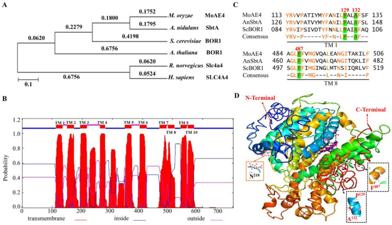
The phylogenic tree indicated that MoAE4 was closely related to the fungal group (Figure 1A), sharing 75.6% identity with A. nidulans SbtA gene, and 57.5% identity with S. cerevisiae YNL275w gene. The transmembrane-spanning domains and tertiary (3D) structures of MoAE4 were predicted with the web-based TMHMM Server v.2.0 (http://www.cbs.dtu.dk/services/TMHMM/, accessed on 16 May 2021) and I-TASSER (http://zhanglab.ccmb.med.umich.edu/I-TASSER/, accessed on 25 March 2021). Both the N-terminal and C-terminal ends are membrane inside (Figure 1B and Figure S1B), which are responsible for transport activity and transmembrane domain anchoring [41]. The two regions TM1 and TM8, together with the N-terminal and C-terminal ends, form the so-called gate domain and core domain. The three conserved ligand sites (P129, A132, and F487) and the active site N218 harbors in the C-terminal end (core domain) (Figure 1C,D and Figure S1C).
Figure 1.
Structure of the MoAE4 and phylogenetic analysis. (A) Phylogenetic tree. It was constructed with reported anion exchange protein 4 homologs from M. oryzae, A. nidulans, S. cerevisiae, A. thaliana, R. norvegicus, H. sapiens, indicating that MoAE4 has a relatively close relationship with the fungal group. (B) TMHMM posterior probabilities for WEBSEQUENCE (on-line analysis). A schematic diagram of the MoAE4 protein transmembrane domain of M. oryzae is shown indicating the 10 transmembrane domains and marked the amino acid inside and outside the membrane. (C) Sequence alignment. The transmembrane domain 1 and 8 of MoAE4 was compared with closely related to the fungal group A. nidulans SbtA gene and S. cerevisiae BOR1 gene. (D) Tertiary structure (3-D). The structure was predicted using the web-based server I-TASSER that shows the ligand binding site residues of transmembrane domain P129, A132, and F487. The active site residue is N218.
According to the 3D structures (Figure 1D and Figure S1C), the conformations with 10 TM helices of MoAE4 were characterized by two inverted repeats that are intertwined to form both core and gate domains, which appears to be similar with that of AE1 or BOR1 [3,10]. The homologous protein sequences and the typical domain patterns reveal MoAE4 is a member of the HCO3− transporter AE4 group.
3.2. MoAE4 Localizes on Cytomembrane and Tonoplast and Functions in Yeast
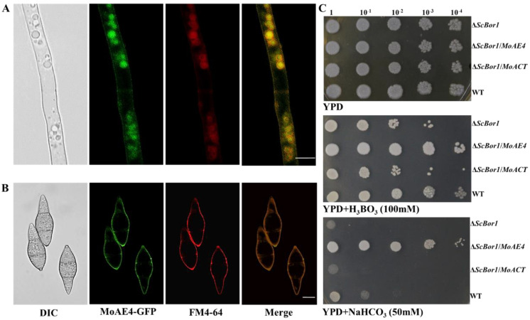
Most HCO3− transporters function on cytoplasm membrane [16]. To test where MoAE4 occurred, we generated transgenic strains expressing GFP-tagged MoAE4 fusion gene in the wild type of M. oryzae (Figure S2A). Fluorescent microscopic observation was carried out by using hyphae (6d) and conidia. The lipophilic dye (FM™ 4-64 Dye, AAT Bioquest, USA) was used for observing the cytoplasmic membrane and vacuolar membrane. A strong green fluorescence signal of the MoAE4-GFP protein co-localized with FM4-64 red fluorescence was detected on the cytoplasmic membrane in young hyphae and conidia (Figure 2A,B); interestingly, a strong co-localized yellow fluorescence signal was also detected on the vacuole membrane in hyphae (Figure 2A,B). By comparison, in the wild type or untransformed strains, the background green fluorescence was too weak to be detected. This result suggests that MoAE4 was targeted to cytomembrane and tonoplast.
Figure 2.
MoAE4 subcellular localization and functions in yeast. (A) Subcellular localization in hyphae (6d). Green fluorescence signals of the MoAE4–GFP protein of 6–day–old hyphae was examined by confocal microscopy and co–localized with FM4–64 on cytomembrane and tonoplast. Scale bar = 10 µm. (B) Subcellular localization in conidia. A strong green fluorescence signal of the MoAE4–GFP protein co–localized with FM4–64 red fluorescence was detected on the cytoplasmic membrane in conidia. Scale bar = 10 µm. (C) Functional complementation of MoAE4 for ScBor1 in S. cerevisiae. In total, 10 μL droplets containing the indicated concentration of yeast cells were inoculated on to the Solid YPD medium Plates (100 mM H3BO3 and 50 mM NaHCO3 added, respectively). The MoAE4 gene could functionally reverse the defect of the ΔScBor1 mutant in boric acid and NaHCO3 tolerance. Representative plates were photographed 3 days post-inoculation.
Yeast bicarbonate transporters are boric acid tolerant [12,13,14]. Based on the ScBor1/AE4 deletion mutant (ΔScBor1), the complementary yeast strains were created by using MoAE4. As a result, the MoAE4 gene could functionally reverse the defect of the ΔScBor1 mutant in boric acid tolerance (Figure 2C), suggesting MoAE4 functions as yeast Bor1. Different from the yeast BOR1, MoAE4 also endowed the NaHCO3 tolerance in the mutant strains (Figure 2C), implying MoAE4 may be a HCO3− transporter.
3.3. MoAE4 Transports Cytosolic HCO3− to Vacuole and Cell outside
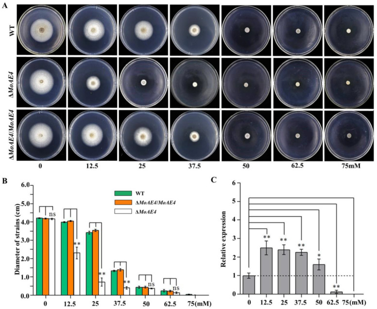
To identify the function of MoAE4 in response to NaHCO3, M. oryzae was cultivated under NaHCO3 stress conditions. By using the knockout mutant strain of MoAE4 (ΔMoAE4) and the complemented strain (ΔMoAE4/MoAE4) (Figure S2B–D), the growth of the tested strains was assessed. When cultivated on complete media (CM) plates at 25 °C without NaHCO3, both the ΔMoAE4 and ΔMoAE4/MoAE4 strains grew at a rate similar to that of the wild type, and their colony morphologies exhibited little difference (Figure 3A,B). When subjected with NaHCO3 of different concentrations, all strains including the wild type were repressed in mycelial growth under NaHCO3 stress. Impressively, ΔMoAE4 almost stopped growing at 25 mM of NaHCO3, but the wild type and complementary strains were capable of growing even at the concentration of 37.5 mM (Figure 3A,B), suggesting a role of ΔMoAE4 in tolerance to NaHCO3. The expression patterns in response to NaHCO3 treatment also illustrated this point (Figure 3C).
Figure 3.
NaHCO3 stress assay of the wild type and created strains and expression patterns of MoAE4. (A) NaHCO3 stress assay. ΔMoAE4 strains were more sensitive to NaHCO3 stress than the wild-type strains. The strains were cultured in PDA media at 28 °C under 0–75 mM different concentrations of NaHCO3 and representative colonies were photographed 7 days post-inoculation. (B) The colonies diameter of the wild type, the ΔMoAE4 and ΔMoAE4/MoAE4 mutant strains following treatments under different concentrations of NaHCO3. (C) Transcription abundance of MoAE4 under different concentrations of NaHCO3. Data represent the means ± standard deviation (SD) from three independent experiments in which triplicate plates were examined for each strain in each experiment. ns p > 0.05. * p < 0.05. ** p < 0.01.
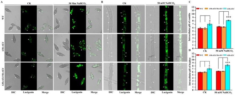
To determine the HCO3− transport activity of MoAE4, lucigenin (bis-N-methylacridinium nitrate), a chemiluminescent probe, was used as to detect the existence of anion radicals in cells under alkaline conditions [33,34]. After treated with NaHCO3 solution (50 mM), the tested strains incubated with 10 mM lucigenin were observed with a 470 nm fluorescence microscope. As expected, lucigenin probed HCO3− with green fluorescence signals was accumulated in vacuoles of the wild type and ΔMoAE4/MoAE4 strains; however, in ΔMoAE4, green fluorescence signals were only detected in cytoplasm (Figure 4A,B). Accordingly, intracellular pH value increased significantly in ΔMoAE4 (Figure 4C,D and Figure S3). These results indicated that MoAE4 functions as a bona fide HCO3− transporter.
Figure 4.
The fluorescence observations and intracellular pH value of the wild type, the ΔMoAE4 and ΔMoAE4/MoAE4 mutant strains. (A) The green fluorescence signals in hyphae. The light (left), fluorescence confocal (middle), and merge (right) microscope observations of strains hypha. Bar = 10 µm. (B) The green fluorescence signals in conidia. The light (left), fluorescence confocal (middle), and merge (right) microscope observations of strains connidia. Bar = 10 µm. (C) The intracellular pH value of conidia. ΔMoAE4 strains were obviously higher than the wild-type strains under 50 mM sodium bicarbonate. (D) The intracellular pH value of the strains in hyphae. Under 50 mM sodium bicarbonate conditions, the intracellular pH of ΔMoAE4 strains remained high in hyphae. The data represent means ± standard deviations (SD) of three experiments. * p < 0.05. *** p < 0.001.
3.4. MoAE4 Is Important for Conidiation and Appressorium Development
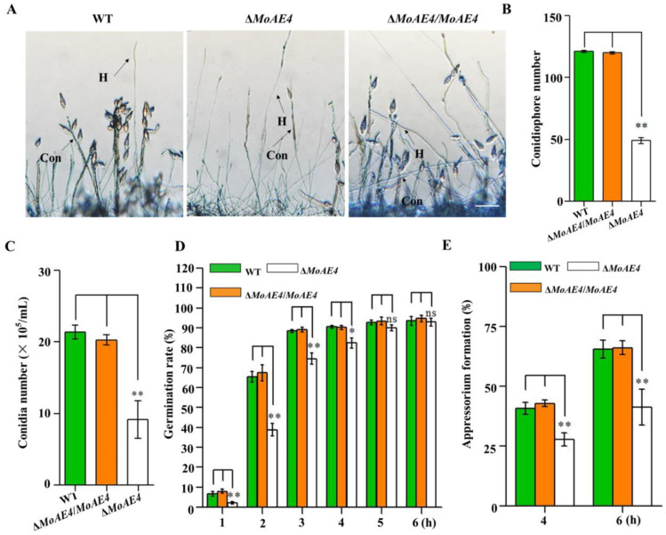
Conidiation and appressorium formation were analyzed among the ΔMoAE4, ΔMoAE4/MoAE4, and wild type strains. The sparse conidiophores with less conidia were observed in the deletion mutant ΔMoAE4, however, both the ΔMoAE4/MoAE4 and wild type produced thick conidiophores and more conidia (Figure 5A–C and Figure S4).
Figure 5.
Conidium and appressorium development analysis of the wild type and created strains. (A) Conidiophores stained with lactophenol cotton blue. The conidiophores of the wild type, the ΔMoAE4 and ΔMoAE4/MoAE4 strains induced for 48 h were stained with lactophenol cotton blue, and observed and counted under a light microscope at room temperature. The hyphae are stained blue, whereas the conidiophore stalks are in gray. Bar = 50 μm. Con, conidiophores; H, hyphae. (B) Statistical analysis of the conidiophores number of the wild type, the ΔMoAE4 and ΔMoAE4/MoAE4 mutant strains. (C) Statistical analysis of conidial production in the strains. The conidia were harvested from the 3-day-old mycelium grown on OMA media, and counted using a hemocytometer for all the three strains. (D) Conidial germination rate. Conidial germination was measured on a hydrophobic cover slips and was calculated under the microscope at 1, 2, 3, 4, 5, and 6 h post inoculation. (E) Appressorial formation rate. Appressorial formation was measured on a hydrophobic cover slips and was calculated under the microscope at 4 and 6 h per inoculation. The analysis was performed using an independent samples t-test. ns p > 0.05. * p < 0.05. ** p < 0.01. Error bars indicate the mean ± SD from three independent experiments.
The conidium germination rate of all strains including the wild type was similar at 4–6 h, although ΔMoAE4 appeared to be a little slow in conidium germination at 1–4 h (Figure 5D and Figure S4B,C). In terms of appressorium formation, ΔMoAE4/MoAE4 had the formation rate similar as the wild type did; but ΔMoAE4 was severely affected (Figure 5E). As conidia and appressoria are essential factors for disease cycle and infection, MoAE4 is proposed to be involved in pathogenesis.
3.5. Requirement of MoAE4 for Pathogenicity in M. oryzae
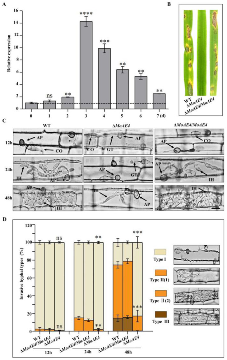
In order to characterize the function of MoAE4 in pathogenic development, pathogenicity assays were carried out using conidia collected from ΔMoAE4, ΔMoAE4/MoAE4, and the wild type. When intact susceptible rice seedlings were spraying-inoculated, at 7 days post inoculation (dpi), some acute expansive disease lesions were observed in rice leaves by the wild type and ΔMoAE4/MoAE4; but no lesions were formed in rice leaves by the ΔMoAE4 (Figure 6). Similarly, when drop-inoculation was assayed, only the wild type and complementary strains still showed pathogenicity.
Figure 6.
Pathogenesis analysis of the wild type and created strains. (A) Transcription abundance of MoAE4 during disease development. (B) Spray-inoculation assay. (C) Rice leaf sheath infection assay. Scale bar = 10 μm. IH, infectious hyphae; CO, Conidium; GT, Germination tube; AP, appressorium. (D) The infection rate was calculated according to the number of type I to type III events. The infection status of more than 100 germinated conidia per leaf sheath was scored at 12, 24, and 48 h post inoculation. Type I, conidia with mature appressoria; Type II, primary hyphae formed, infectious hyphae extended and branched in one cell; Type III, infectious hyphae crossing to neighboring cells. Values represent the averages of five measurements ± standard deviation. The statistical analysis was performed using a one-way ANOVA with Tukey’s multiple comparison test. The averages were taken from the quadruplicate analysis. Values are based on three biological samples and error bars indicate SD, ns p > 0.05. ** p < 0.01; *** p < 0.001. **** p < 0.0001.
Leaf sheath infection assays were performed to examine the infection effects of the MoAE4 in rice host. At 12 hpi, most mature (black) appressoria have been formed in the wild type and ΔMoAE4/MoAE4, but less in ΔMoAE4. At 24 hpi, invasive hyphae of the wild type and ΔMoAE4/MoAE4 commenced to branch in rice cells, but the primary infectious hyphae were just formed in ΔMoAE4. At 48 hpi, the majority of invasive hyphae of the wild-type and ΔMoAE4/MoAE4 branched and started entering neighboring cells, but the ΔMoAE4 strains did not due to the defects in appressorium formation (Figure 6C,D).
To decipher the exact action of MoAE4 during pathogenic development, we defined the three types of infection hyphae according to their developmental morphologies. Then we quantified the proportion of the three types of infection hyphae based on 100 germinated conidia in the inoculated leaf sheath (Figure 6D). As a result, at 48 hpi more than 60% of inoculated conidia from ΔMoAE4/MoAE4 and wild type formed branched infectious hyphae in one cell, of which about 20% extended to neighboring cells of rice (type II and III), suggesting the requirement of MoAE4 in pathogenesis.
3.6. MoAE4 Is Important for H2O2 Tolerance and Clearance Inside or Outside Cells
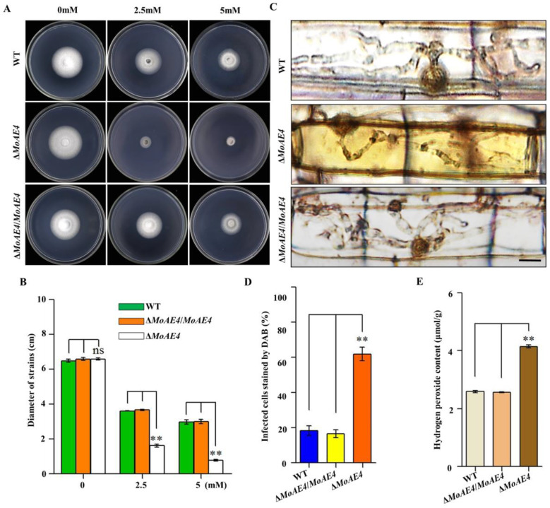
To address the relationship between MoAE4 and endogenous H2O2, the wild type and mutant strains were cultured on CM agar supplemented with 2.5 or 5 mM H2O2 at 28 °C for 5 days. As a result, ΔMoAE4 was markedly inhibited in mycelial growth under H2O2 stress, indicating a role of MoAE4 in oxidative stress tolerance (Figure 7A,B). As rice plant accumulates more H2O2 during pathogen-rice interaction, and MoAE4 expression increases with pathogenic development of M. oryzae, we speculate that MoAE4 is responsible for the clearance of host-derived H2O2 during infection. To test this, DAB staining was used to identify the endogenous ROS accumulated in the cells of rice leaf sheath infected by M. oryzae at 36 hpi (Figure 7C). In the leaf sheaths inoculated with the ΔMoAE4 strains, more than 60% of the infected cells investigated were stained dark brown; in contrast, less than 20% of the infected cells were stained light brown or colorless as ΔMoAE4/MoAE4 and wild type (Figure 7C,D), displaying loss of H2O2 scavenging function in ΔMoAE4. Additionally, endogenous H2O2 was measured in M. oryzae more H2O2 accumulated in ΔMoAE4 than in the wild type and ΔMoAE4/MoAE4 (Figure 7D). These results reveal that MoAE4 is responsible for regulating H2O2 levels exogenous, endogenous, or plant-derived.
Figure 7.
Comparison of oxidative stress, DAB staining, and endogenous H2O2 among the wild type, the ΔMoAE4, and ΔMoAE4/MoAE4 strains. (A) H2O2 stress assay. The strains were cultured in CM media for 7 days at 28 °C with 2.5 or 5 mM H2O2. (B) The colonies diameter of the wild type, ΔMoAE4, and ΔMoAE4/MoAE4 following treatment with 2.5 or 5 mM H2O2. (C) DAB staining of leaf sheath cells of rice infected by wild type, mutant and complementation strains at 36 hpi. Scale bar = 10 μm. (D) Statistical analysis of DAB staining of leaf sheath cells infected by different strains. (E) Endogenous H2O2 assay. The strains of hyphae of Endogenous H2O2 were determined as described in Experimental Procedures. The above experiments were performed in triplicate and repeated three independent times for each strain. Error bars represent the ± SD of three independently repeated samples, ns p > 0.05. ** p < 0.01.
4. Discussion
The family of bicarbonate transport proteins are involved in a wide-range of physiological processes in humans and mammals [1,2,42]. Mutation or dysregulation of these transporters results in physiological diseases in humans [43]. Therefore, bicarbonate transporters have attracted medical attention and have been extensively studied. In fungi, such as the unicellular organism yeast, CO2 can diffuse directly out of yeast cells, so the anion exchanger/carbonic anhydrase system and HCO3− transporter are proposed to be dispensable [32]. Indeed, the AE1 homologue, YNL275w has been verified as an anion transporter just serving for boron detoxification or tolerance in S. cerevisiae [12,13,14]. Bicarbonate gradients modulate growth and colony morphology in A. nidulans [44]. Bioinformatically, other filamentous fungi harbor homologous of mammal HCO3− transporters. In A. nidulans, there are at least five members of SLC family, but the most homologous SbtB still showed boron transporter, not HCO3− transporter [16]. Considering the exclusive activity of BOR1 in fungi and plants, YNL275w and homologues should be grouped into the secondary bicarbonate transporter family specific for boron. The bona fide HCO3− transport function in fungal YNL275w homologues is actually unknown. In this research, we demonstrated that the MoAE4, as a YNL275w homologue, plays a role in HCO3− transport. Importantly, MoAE4 located to cytomembrane and tonoplast promotes conidiation, appressorium formation, and pathogenesis in M. oryzae.
In general, AEs localize to cytoplasmic membrane [10,35]. GFP-tagged Bor1p were detected to localize preferentially to the vacuole and that cells lacking Bor1p have fragmented vacuoles [45]. Recently, kidney anion exchanger 1(kAE1) has been detected on vacuole [46]. These findings suggest Bor1p functions on both cytomembrane and tonoplast. Additionally, in our study, MoAE4 was localized on cytomembrane and tonoplast (Figure 2A; Table S1). In hyphae, MoAE4 protein was concentrated on tonoplast; but in conidia, a strong fluorescence signal was on cytomembrane, suggesting the different subcellular patterns of MoAE4. This may reflect the specific function of MoAE4 in different developmental stages of M. oryzae.
In addition to the homology with BOR1 in sequences and 3-D structures (Figure 1), MoAE4 rescued the defect of ΔScBOR1 in boron tolerance, suggesting an authentic BOR1 homologue. However, MoAE4 also endowed the NaHCO3 tolerance for the mutant strains, for even the wild type failed to grow normally under NaHCO3 stress (Figure 2C). Particularly, loss of MoAE4 resulted in the severe mycelial growth inhibition in ΔMoAE4 compared with the wild type and complementary strains (Figure 3A,B), implying the HCO3− transport activity in MoAE4. Based on the lucigenin fluorescence quenching assay, a great amount of HCO3− was detected in the cytosol of ΔMoAE4, but not in vacuoles of ΔMoAE4; on the contrary, HCO3− was only detected in vacuole in the wild type and ΔMoAE4/MoAE4 (Figure 4), revealing the MoAE4-associated bicarbonate resistant mechanism, by which MoAE4 removes redundant HCO3− from cytosol to vacuole and outside cells.
As a pathogenic fungus, conidiation and appressorium formation are key processes for disease cycle and infection [17,18]. MoAE4 loss resulted in the impaired conidiophore formation and then decreased conidial and appressorial productivity (Figure 5). We speculate that this may be related to the excessively accumulated H2O2 in the MoAE4 deletion mutant (Figure 7D). After all, ΔMoAE4 became sensitive under H2O2 stress (Figure 7B,C). At this point, the reduced pathogenicity in ΔMoAE4 could also be partially explained because ΔMoAE4 was exposed to a high hydrogen peroxide stress both in vivo and in vitro (Figure 7A,E and Figure S6).
CO2, as a labile molecule, is the oxidation waste product of mitochondrial respiration. In humans, redundant CO2 must be released, or the equilibrium with HCO3− + H+ will be disturbed. The ability of HCO3− to undergo pH-dependent conversions is central to its physiological role [1]. CO2 enters the cytoplasm through the membrane and is rapidly hydrated forming carbonic acid (H2CO3). This acid is dissociated into H+ and HCO3− by intracellular carbonic anhydrases [7]. Under normal medium culture conditions, pathogenic fungi such as yeast may not require the anion exchanger/carbonic anhydrase system to help the release of metabolic CO2 [11]. However, the interaction system between host plant and pathogen forms a whole multicellular organism, which should face a challenge in moving membrane impermeant bicarbonate from inside the cell where it is produced to the environment for disposal. In addition, during interactions between plant and pathogen, respiration from both pathogen and plant must be enhanced to produce available energy [24,47,48,49]. Therefore, we propose a pathogenic model mediated by MoAE4/MoCA (carbonic anhydrase) system (Figure 8). Under cultivation conditions, the metabolic CO2 can be released freely from M. oryzae, and both MoAE4 and MoCA genes are at low levels of expression because of the equilibrium of (CO2 + H2O ⇌ HCO3− + H+) (Figure 8A). In the process of invasive hyphae growth, the infected plant cell is a relatively high concentration of CO2 and low concentration of O2 microenvironment; and accordingly, the diffusion of fungal CO2 to the outside (cytosol of plant cell) is hindered. The upregulated MoCA (Figure S5) will increase the concentration of HCO3−, which leads to MoAE4 being upregulated, then MoAE4 transports HCO3− to the vacuole or to plant cells (Figure 8B).
Figure 8.
A pathogenic model mediated by MoAE4/MoCA (carbonic anhydrase) system. (A) M. oryzae is under cultivation conditions. (B) M. oryzae infects the host cells. FM, Fungal Mitochondrion; PM, Plant mitochondrion; V, vacuole; MoCA, M. oryzae carbonic anhydrase.
In the model, MoAE4 maintains the homeostasis of intracellular CO2-HCO3− system, which further ensures the intracellular acid-base balance in cells [50]. According to the importance of the acid-base equilibrium in multiple physiological activities [51], we imply HCO3− regulated by MoAE4 is a pathogenic signal for blast disease development. Actually, in our recent research, a low dose of sodium carbonate can induce the expression of a large number of disease-related genes in wild type, not in the MoAE4 deletion mutant (unpublished data), revealing a potential drug target for blast disease control. To decipher the regulation mechanism, much work remains to be completed.
5. Conclusions
In the rice blast fungus, MoAE4 exhibits more sequence and structure homology with AE proteins. Additionally, MoAE4, localized on cytomembrane and tonoplast, possesses boron and NaHCO3 tolerance in yeast. Lucigenin fluorescence quenching assay indicated that MoAE4 has HCO3− transport activity. Meanwhile, ΔMoAE4 contained more H2O2 than the wild type and complementary strains did, implying a role of MoAE4 in energy metabolism. Importantly, MoAE4 is involved in conidiation, appressorium formation, and pathogenicity in M. oryzae. Overall, MoAE4, as a cytomembrane and tonoplast HCO3− transporter, promotes pathogenesis of M. oryzae. Based on these results, a pathogenic model mediated by MoAE4 is proposed.
Supplementary Materials
The following are available online at https://www.mdpi.com/article/10.3390/jof7110955/s1, Figure S1. Sequence alignment and prediction of MoAE4 structure. Figure S2. The construction strategies for MoAE4 deletion and complementation strains. Figure S3. The green fluorescence signals detection of intracellular pH in conidia. Figure S4. The loss of the MoAE4 gene has a negative effect on sporulation, appressorial development. Figure S5. Transcription abundance of MoCA during disease development. Figure S6. Standard curve line and the endogenous H2O2 of strains hyphae. Table S1. Identifying sub-cellular location. Table S2. Primers used in this study.
Author Contributions
S.-H.Z.: conceptualization, writing—review and editing the manuscript, supervision, and funding acquisition the research; Y.D.: writing and editing original draft preparation, methodology, conceptualization, and data curation; Y.W.: data curation, investigation, validation, and project administration; P.Z., X.L. (Xinchun Liu), X.L. (Xinrui Li), S.W. and H.L.: formal analysis, and software. All authors have read and agreed to the published version of the manuscript.
Funding
This work was supported by the Natural Science Foundation of China (Grant Nos.31670141 to Y.W. and 31671972 to S.-H.Z.); the Local scientific research of Department of Education of Liaoning Province of China (Grant No.01032920021 to S.-H.Z.); Special talent introduction of Shenyang Agricultural University of China (Grant No. 880420019 to S.-H.Z.); Postdoctoral funding of Shenyang Agricultural University of China (Grant No. 770221003 to Y.D.).
Conflicts of Interest
The authors declare no conflict of interest.
Footnotes
Publisher’s Note: MDPI stays neutral with regard to jurisdictional claims in published maps and institutional affiliations.
References
- 1.Cordat E., Casey J.R. Bicarbonate Transport in Cell Physiology and Disease. Biochem. J. 2009;417:423–439. doi: 10.1042/BJ20081634. [DOI] [PubMed] [Google Scholar]
- 2.Romero M.F., Chen A.P., Parker M.D., Boron W.F. The SLC4 Family of Bicarbonate (HCO3−) Transporters. Mol. Asp. Med. 2013;34:159–182. doi: 10.1016/j.mam.2012.10.008. [DOI] [PMC free article] [PubMed] [Google Scholar]
- 3.Shieh Y.W., Minguez P., Bork P., Auburger J.J., Guilbride D.L., Kramer G., Bukau B. Operon Structure and Cotranslational Subunit Association Direct Protein Assembly in Bacteria. Science. 2015;350:678–680. doi: 10.1126/science.aac8171. [DOI] [PubMed] [Google Scholar]
- 4.Hatae H., Inaka K., Okamura R., Furubayashi N., Kamo M., Kobayashi T., Abe Y., Iwata S., Hamasaki N. Crystallization of Human Erythrocyte Band 3, the Anion Exchanger, at the International Space Station “KIBO”. Anal. Biochem. 2018;559:91–93. doi: 10.1016/j.ab.2018.08.009. [DOI] [PubMed] [Google Scholar]
- 5.Tsuganezawa H., Kobayashi K., Iyori M., Araki T., Koizumi A., Watanabe S.I., Kaneko A., Fukao T., Monkawa T., Yoshida T., et al. A New Member of the HCO3− Transporter Superfamily Is an Apical Anion Exchanger of β-Intercalated Cells in the Kidney. J. Biol. Chem. 2001;276:8180–8189. doi: 10.1074/jbc.M004513200. [DOI] [PubMed] [Google Scholar]
- 6.Ko S.B.H., Luo X., Hager H., Rojek A., Choi J.Y., Licht C., Suzuki M., Muallem S., Nielsen S., Ishibashi K. AE4 Is a DIDS-Sensitive Cl−/HCO3− Exchanger in the Basolateral Membrane of the Renal CCD and the SMG Duct. Am. J. Physiol. Cell Physiol. 2002;283:1206–1218. doi: 10.1152/ajpcell.00512.2001. [DOI] [PubMed] [Google Scholar]
- 7.Peña-Münzenmayer G., Catalán M.A., Kondo Y., Jaramillo Y., Liu F., Shull G.E., Melvin J.E. Ae4 (Slc4a9) Anion Exchanger Drives Cl- Uptake-Dependent Fluid Secretion by Mouse Submandibular Gland Acinar Cells. J. Biol. Chem. 2015;290:10677–10688. doi: 10.1074/jbc.M114.612895. [DOI] [PMC free article] [PubMed] [Google Scholar]
- 8.Peña-Münzenmayer G., George A.T., Shull G.E., Melvin J.E., Catalán M.A. Ae4 (Slc4a9) Is an Electroneutral Monovalent Cation-Dependent Cl−/HCO3− Exchanger. J. Gen. Physiol. 2016;147:423–436. doi: 10.1085/jgp.201611571. [DOI] [PMC free article] [PubMed] [Google Scholar]
- 9.Vera-Sigüenza E., Catalán M.A., Peña-Münzenmayer G., Melvin J.E., Sneyd J. A Mathematical Model Supports a Key Role for Ae4 (Slc4a9) in Salivary Gland Secretion. Bull. Math. Biol. 2018;80:255–282. doi: 10.1007/s11538-017-0370-6. [DOI] [PMC free article] [PubMed] [Google Scholar]
- 10.Coudray N., Seyler S.L., Lasala R., Zhang Z., Clark K.M., Dumont M.E., Rohou A., Beckstein O., Stokes D.L. Structure of the SLC4 Transporter Bor1p in an Inward-Facing Conformation. Protein Sci. 2017;26:130–145. doi: 10.1002/pro.3061. [DOI] [PMC free article] [PubMed] [Google Scholar]
- 11.Zhao R., Reithmeier R.A.F. Expression and Characterization of the Anion Transporter Homologue YNL275w in Saccharomyces cerevisiae. Am. J. Physiol. Cell Physiol. 2001;281:33–45. doi: 10.1152/ajpcell.2001.281.1.C33. [DOI] [PubMed] [Google Scholar]
- 12.Nozawa A., Takano J., Kobayashi M., Von Wirén N., Fujiwara T. Roles of BOR1, DUR3, and FPS1 in Boron Transport and Tolerance in Saccharomyces cerevisiae. FEMS Microbiol. Lett. 2006;262:216–222. doi: 10.1111/j.1574-6968.2006.00395.x. [DOI] [PubMed] [Google Scholar]
- 13.Takano J., Kobayashi M., Noda Y., Fujiwara T. Saccharomyces Cerevisiae Bor1p Is a Boron Exporter and a Key Determinant of Boron Tolerance. FEMS Microbiol. Lett. 2007;267:230–235. doi: 10.1111/j.1574-6968.2006.00556.x. [DOI] [PubMed] [Google Scholar]
- 14.Jennings M.L., Howren T.R., Cui J., Winters M., Hannigan R. Transport and Regulatory Characteristics of the Yeast Bicarbonate Transporter Homolog Bor1p. Am. J. Physiol. Cell Physiol. 2007;293:468–476. doi: 10.1152/ajpcell.00286.2005. [DOI] [PubMed] [Google Scholar]
- 15.Takano J., Noguchi K., Yasumori M., Kobayashi M., Gajdos Z., Miwa K., Hayashi H., Yoneyama T., Fujiwara T. Arabidopsis Boron Transporter for Xylem Loading. Nature. 2002;420:337–340. doi: 10.1038/nature01139. [DOI] [PubMed] [Google Scholar]
- 16.Villarino M., Etxebeste O., Mendizabal G., Garzia A., Ugalde U., Espeso E.A. Boron Tolerance in Aspergillus nidulans Is Sustained by the SltA Pathway through the SLC-Family Transporters SbtA and SbtB. Genes. 2017;8:188. doi: 10.3390/genes8070188. [DOI] [PMC free article] [PubMed] [Google Scholar]
- 17.Howard R.J., Ferrari M.A., Roach D.H., Money N.P. Penetration of Hard Substrates by a Fungus Employing Enormous Turgor Pressures. Proc. Natl. Acad. Sci. USA. 1991;88:11281–11284. doi: 10.1073/pnas.88.24.11281. [DOI] [PMC free article] [PubMed] [Google Scholar]
- 18.Talbot N.J. On the Trail of a Cereal Killer: Exploring the Biology of Magnaporthe grisea. Annu. Rev. Microbiol. 2003;57:177–202. doi: 10.1146/annurev.micro.57.030502.090957. [DOI] [PubMed] [Google Scholar]
- 19.Heath M.C., Valent B., Howard R.J., Chumley F.G. Interactions of Two Strains of Magnaporthe grisea with Rice, Goosegrass, and Weeping Lovegrass. Can. J. Bot. 1990;68:1627–1637. doi: 10.1139/b90-209. [DOI] [Google Scholar]
- 20.Kankanala P., Czymmek K., Valent B. Roles for Rice Membrane Dynamics and Plasmodesmata during Biotrophic Invasion by the Blast Fungus. Plant Cell. 2007;19:706–724. doi: 10.1105/tpc.106.046300. [DOI] [PMC free article] [PubMed] [Google Scholar]
- 21.Hammond-Kosack K.E., Parker J.E. Deciphering Plant-Pathogen Communication: Fresh Perspectives for Molecular Resistance Breeding. Curr. Opin. Biotechnol. 2003;14:177–193. doi: 10.1016/S0958-1669(03)00035-1. [DOI] [PubMed] [Google Scholar]
- 22.Nürnberger T., Brunner F., Kemmerling B., Piater L. Innate Immunity in Plants and Animals: Striking Similarities and Obvious Differences. Immunol. Rev. 2004;198:249–266. doi: 10.1111/j.0105-2896.2004.0119.x. [DOI] [PubMed] [Google Scholar]
- 23.Egan M.J., Wang Z.Y., Jones M.A., Smirnoff N., Talbot N.J. Generation of Reactive Oxygen Species by Fungal NADPH Oxidases Is Required for Rice Blast Disease. Proc. Natl. Acad. Sci. USA. 2007;104:11772–11777. doi: 10.1073/pnas.0700574104. [DOI] [PMC free article] [PubMed] [Google Scholar]
- 24.Grahl N., Puttikamonkul S., Macdonald J.M., Gamcsik M.P., Ngo L.Y., Hohl T.M., Cramer R.A. In Vivo Hypoxia and a Fungal Alcohol Dehydrogenase Influence the Pathogenesis of Invasive Pulmonary Aspergillosis. PLoS Pathog. 2011;7:e1002145. doi: 10.1371/journal.ppat.1002145. [DOI] [PMC free article] [PubMed] [Google Scholar]
- 25.Choi J., Chung H., Lee G.W., Koh S.K., Chae S.K., Lee Y.H. Genome-Wide Analysis of Hypoxia-Responsive Genes in the Rice Blast Fungus, Magnaporthe oryzae. PLoS ONE. 2015;10:e0134939. doi: 10.1371/journal.pone.0134939. [DOI] [PMC free article] [PubMed] [Google Scholar]
- 26.Fernandez J., Marroquin-Guzman M., Wilson R.A. Mechanisms of Nutrient Acquisition and Utilization during Fungal Infections of Leaves. Annu. Rev. Phytopathol. 2014;52:155–174. doi: 10.1146/annurev-phyto-102313-050135. [DOI] [PubMed] [Google Scholar]
- 27.Cui X., Wei Y., Wang Y.H., Li J., Wong F.L., Zheng Y.J., Yan H., Liu S.S., Liu J.L., Jia B.L., et al. Proteins Interacting with Mitochondrial ATP-Dependent Lon Protease (MAP1) in Magnaporthe oryzae Are Involved in Rice Blast Disease. Mol. Plant Pathol. 2015;16:847–859. doi: 10.1111/mpp.12242. [DOI] [PMC free article] [PubMed] [Google Scholar]
- 28.Bertram G., Swoboda R.K., Gooday G.W., Gow N.A.R., Brown A.J.P. Structure and Regulation of the Candida Albicans ADH1 Gene Encoding an Immunogenic Alcohol Dehydrogenase. Yeast. 1996;12:115–127. doi: 10.1002/(SICI)1097-0061(199602)12:2<115::AID-YEA889>3.0.CO;2-E. [DOI] [PubMed] [Google Scholar]
- 29.Li Z., Pei X., Zhang Z., Wei Y., Song Y., Chen L., Liu S., Zhang S.H. The Unique GH5 Cellulase Member in the Extreme Halotolerant Fungus Aspergillus glaucus CCHA Is an Endoglucanase with Multiple Tolerance to Salt, Alkali and Heat: Prospects for Straw Degradation Applications. Extremophiles. 2018;22:675–685. doi: 10.1007/s00792-018-1028-5. [DOI] [PubMed] [Google Scholar]
- 30.Zheng W., Zhou J., He Y., Xie Q., Chen A., Zheng H., Shi L., Zhao X., Zhang C., Huang Q., et al. Retromer Is Essential for Autophagy-Dependent Plant Infection by the Rice Blast Fungus. PLoS Genet. 2015;11:e1005704. doi: 10.1371/journal.pgen.1005704. [DOI] [PMC free article] [PubMed] [Google Scholar]
- 31.Dang Y., Wei Y., Wang Y., Liu S., Julia C., Zhang S.H. Cleavage of PrePL by Lon Promotes Growth and Pathogenesis in Magnaporthe oryzae. Environ. Microbiol. 2020;23:4881–4895. doi: 10.1111/1462-2920.15335. [DOI] [PubMed] [Google Scholar]
- 32.Zhou Z., Li G., Lin C., He C. Conidiophore Stalk-Less1 Encodes a Putative Zinc-Finger Protein Involved in the Early Stage of Conidiation and Mycelial Infection in Magnaporthe oryzae. Mol. Plant-Microbe Interact. 2009;22:402–410. doi: 10.1094/MPMI-22-4-0402. [DOI] [PubMed] [Google Scholar]
- 33.Qi D., Wang D., Zhang C., Tang X., He J., Zhao Y., Deng W., Deng X. Vaspin Protects against LPS-Induced ARDS by Inhibiting Inflammation, Apoptosis and Reactive Oxygen Species Generation in Pulmonary Endothelial Cells via the Akt/GSK-3β Pathway. Int. J. Mol. Med. 2017;40:1803–1817. doi: 10.3892/ijmm.2017.3176. [DOI] [PMC free article] [PubMed] [Google Scholar]
- 34.Oosthuizen M.M.J., Greyling D. Hydroxyl Radical Generation: The Effect of Bicarbonate, Dioxygen and Buffer Concentration on PH-Dependent Chemiluminescence. Redox Rep. 2001;6:105–116. doi: 10.1179/135100001101536111. [DOI] [PubMed] [Google Scholar]
- 35.Schwiening C.J., Boron W.F. Regulation of Intracellular PH in Pyramidal Neurones from the Rat Hippocampus by Na(+)-dependent Cl(-)-HCO3− Exchange. J. Physiol. 1994;475:59–67. doi: 10.1113/jphysiol.1994.sp020049. [DOI] [PMC free article] [PubMed] [Google Scholar]
- 36.Yang L., Zhu L., Xu Y., Zhang H., Ye W., Mao J., Chen L., Wang L. Uncoupling of K+ and Cl− Transport across the Cell Membrane in the Process of Regulatory Volume Decrease. Biochem. Pharmacol. 2012;84:292–302. doi: 10.1016/j.bcp.2012.05.006. [DOI] [PubMed] [Google Scholar]
- 37.Brennan T., Frenkel C. Involvement of Hydrogen Peroxide in the Regulation of Senescence in Pear. Plant Physiol. 2020;59:411–416. doi: 10.1104/pp.59.3.411. [DOI] [PMC free article] [PubMed] [Google Scholar]
- 38.Cui X., Wei Y., Xie X.L., Chen L.N., Zhang S.H. Mitochondrial and Peroxisomal Lon Proteases Play Opposing Roles in Reproduction and Growth but Co-Function in the Normal Development, Stress Resistance and Longevity of Thermomyces lanuginosus. Fungal Genet. Biol. 2017;103:42–54. doi: 10.1016/j.fgb.2017.04.002. [DOI] [PubMed] [Google Scholar]
- 39.Liu X., Qian B., Gao C., Huang S., Cai Y., Zhang H., Zheng X., Wang P., Zhang Z. The Putative Protein Phosphatase MoYvh1 Functions Upstream of MoPdeH to Regulate the Development and Pathogenicity in Magnaporthe oryzae. Mol. Plant-Microbe Interact. 2016;29:496–507. doi: 10.1094/MPMI-11-15-0259-R. [DOI] [PubMed] [Google Scholar]
- 40.Rakotonirainy M.S., Héraud C., Lavédrine B. Detection of Viable Fungal Spores Contaminant on Documents and Rapid Control of the Effectiveness of an Ethylene Oxide Disinfection Using ATP Assay. Luminescence. 2003;18:113–121. doi: 10.1002/bio.710. [DOI] [PubMed] [Google Scholar]
- 41.van den Akker E., Satchwell T.J., Williamson R.C., Toye A.M. Band 3 Multiprotein Complexes in the Red Cell Membrane; of Mice and Men. Blood Cells Mol. Dis. 2010;45:1–8. doi: 10.1016/j.bcmd.2010.02.019. [DOI] [PubMed] [Google Scholar]
- 42.Carignan C.C., Punshon T., Karagas M.R. HHS Public Access. Physiol. Behav. 2016;176:100–106. doi: 10.1007/s00223-017-0311-2.Bicarbonate. [DOI] [Google Scholar]
- 43.Alka K., Casey J.R. Bicarbonate Transport in Health and Disease. IUBMB Life. 2014;66:596–615. doi: 10.1002/iub.1315. [DOI] [PubMed] [Google Scholar]
- 44.Rodríguez-Urra A.B., Jimenez C., Dueñas M., Ugalde U. Bicarbonate Gradients Modulate Growth and Colony Morphology in Aspergillus nidulans. FEMS Microbiol. Lett. 2009;300:216–221. doi: 10.1111/j.1574-6968.2009.01780.x. [DOI] [PubMed] [Google Scholar]
- 45.Decker B.L., Wickner W.T. Enolase Activates Homotypic Vacuole Fusion and Protein Transport to the Vacuole in Yeast. J. Biol. Chem. 2006;281:14523–14528. doi: 10.1074/jbc.M600911200. [DOI] [PubMed] [Google Scholar]
- 46.Sarder H.A.M., Li X., Funaya C., Cordat E., Schmitt M.J., Becker B. Saccharomyces Cerevisiae: First Steps to a Suitable Model System To Study the Function and Intracellular Transport of Human Kidney Anion Exchanger 1. mSphere. 2020;5:e00802-19. doi: 10.1128/mSphere.00802-19. [DOI] [PMC free article] [PubMed] [Google Scholar]
- 47.Rios J.A., de Ávila Rodrigues F., Debona D., Silva L.C. Photosynthetic Gas Exchange in Leaves of Wheat Plants Supplied with Silicon and Infected with Pyricularia oryzae. Acta Physiol. Plant. 2014;36:371–379. doi: 10.1007/s11738-013-1418-3. [DOI] [Google Scholar]
- 48.Domiciano G.P., Cacique I.S., Freitas C.C., Filippi M.C.C., DaMatta F.M., Do Vale F.X.R., Rodrigues F.Á. Alterations in Gas Exchange and Oxidative Metabolism in Rice Leaves Infected by Pyricularia oryzae Are Attenuated by Silicon. Phytopathology. 2015;105:738–747. doi: 10.1094/PHYTO-10-14-0280-R. [DOI] [PubMed] [Google Scholar]
- 49.Torres M.A., Dangl J.L. Functions of the Respiratory Burst Oxidase in Biotic Interactions, Abiotic Stress and Development. Curr. Opin. Plant Biol. 2005;8:397–403. doi: 10.1016/j.pbi.2005.05.014. [DOI] [PubMed] [Google Scholar]
- 50.Massey M.K., Reiterman M.J., Mourad J., Luckie D.B. Is CFTR an Exchanger? Regulation of HCO3− Transport and Extracellular PH by CFTR. Biochem. Biophys. Rep. 2021;25:100863. doi: 10.1016/j.bbrep.2020.100863. [DOI] [PMC free article] [PubMed] [Google Scholar]
- 51.Chen Y., Cann M.J., Litvin T.N., Iourgenko V., Sinclair M.L., Levin L.R., Buck J. Soluble Adenylyl Cyclase as an Evolutionarily Conserved Bicarbonate Sensor. Science. 2000;289:625–628. doi: 10.1126/science.289.5479.625. [DOI] [PubMed] [Google Scholar]
Associated Data
This section collects any data citations, data availability statements, or supplementary materials included in this article.


