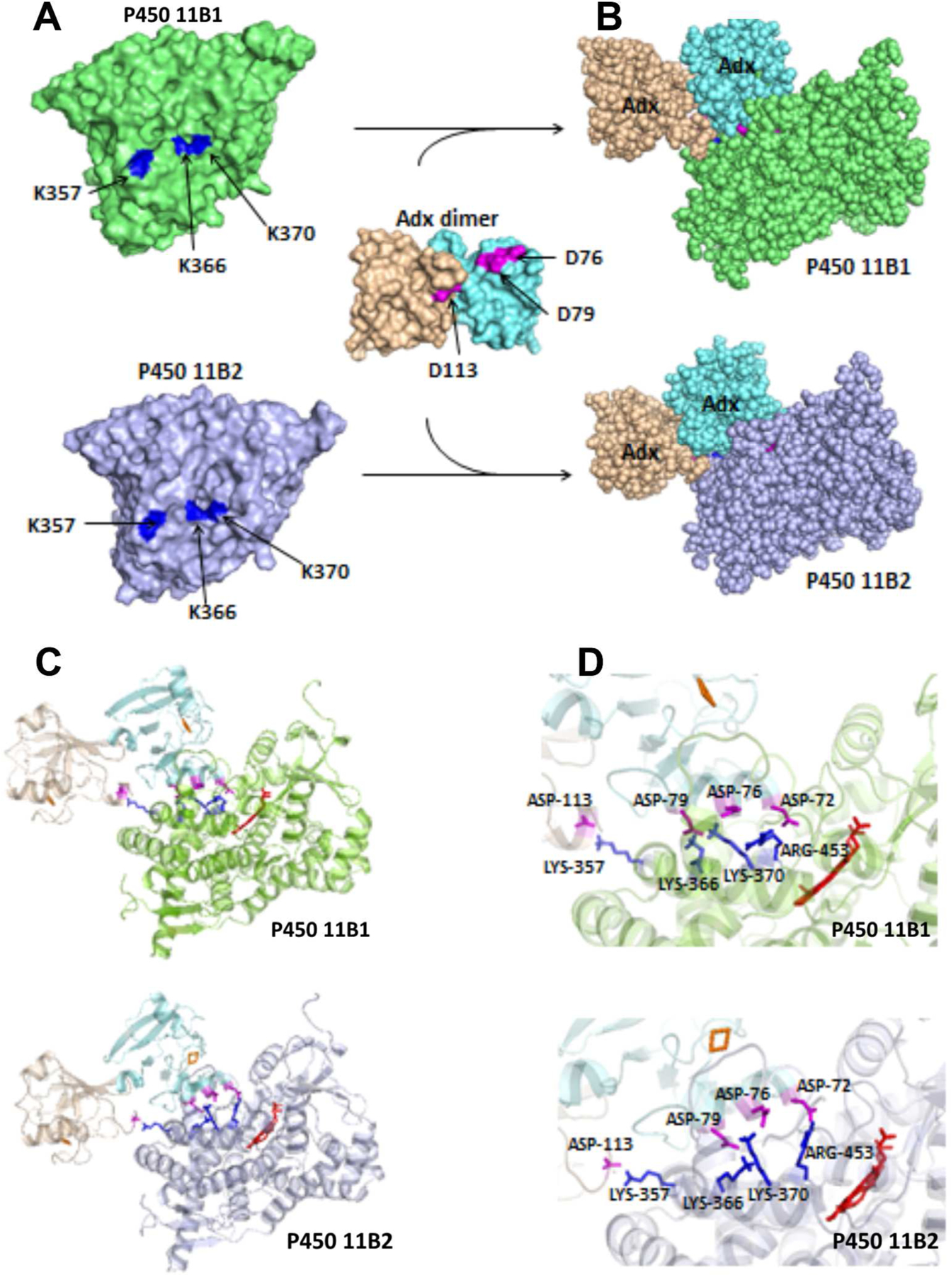
Figure 5.

Docking model of P450 11B1 and P450 11B2 with Adx dimer, based on crystal structures of P450 11B2 (PDB 4DVQ) and Adx (PDB 1CJE). The docking was performed using the programs HADDOCK, with the distance constraints of three intermolecular cross-links, K366 (P450 11B)-D79 (Adx A molecule), K370 (P450 11B)-D76 (Adx A molecule), and K357 (P450 11B)-D113 (Adx B molecule) as described in the Experimental Procedures. (A) The individual proteins are shown as a surface model, and (B) complexes formed are shown as a space-filled model. (C) Ribbon diagram of P450 11B1/11B2 interacting with Adx dimer, and (D) close-up views of the interaction interfaces. The heme in shown in red, and important residues are labeled and shown as sticks on P450 in blue and Adx in magenta.
