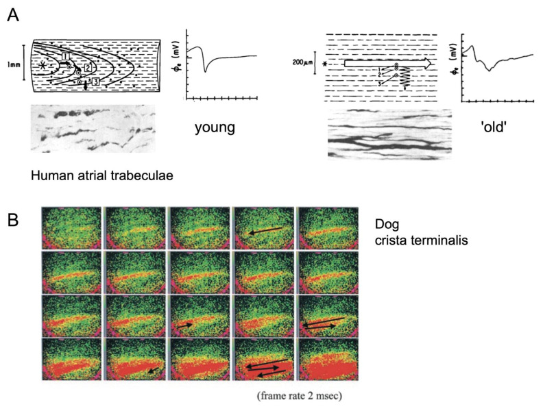
Figure 3.
Non-uniform anisotropy of conduction. (A) Comparison of the conduction patterns and electrograms during pacing in human atrial trabeculae from a young and older individual (top panels). Electrograms recorded at locations transverse to the main wavefront. The corresponding histological illustrations show thickened collagenous septae in the older trabecula, adapted from [12]. (B) Illustration of conduction in the terminal crest of an older dog, showing zig-zag propagation of a narrow activation wave (red), conducting discontinuously in the transverse direction, adapted from [7].

