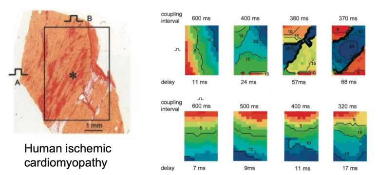
Figure 6.
Impact of fibrotic strands in ventricular cardiomyopathy. In an explanted epicardial tissue slice with long strands of fibrosis, conduction patterns during extrastimulation with progressively shorter intervals were recorded for two different pacing sites (top and bottom row). During pacing at site B, the longitudinal wavefront showed a modest decrease in conduction velocity (bottom row), while at pacing site A, the propagating wavefront showed pronounced transverse conduction block, adapted from [68].

