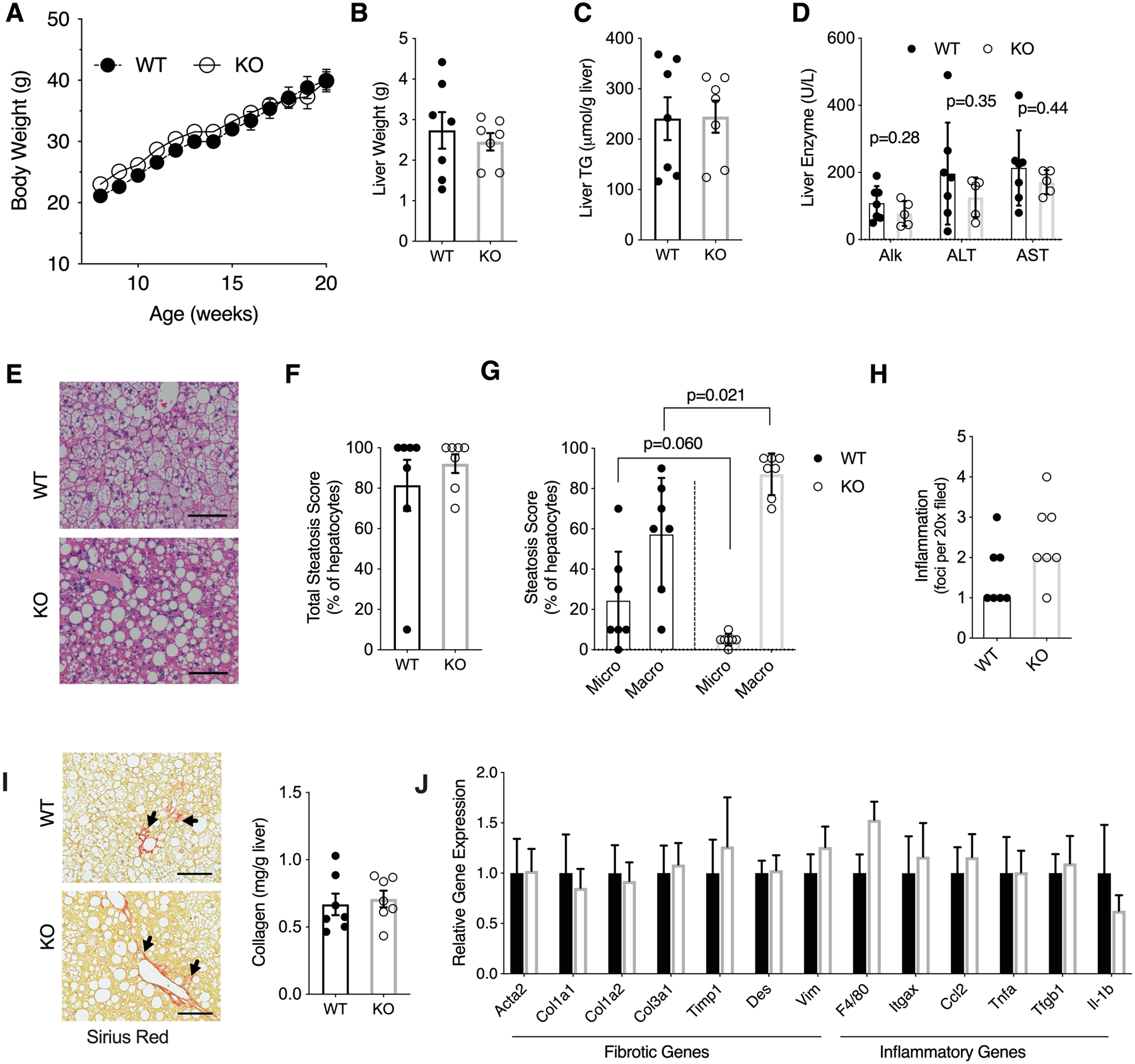
Figure 4. Hsd17b13 deficiency does not protect mice from Western diet (WD) liver injury.

Eight week-old male wild type (WT) and Hsd17b13-knockout (KO) mice were fed with WD for 16 weeks and sacrified at age 24 weeks after a 5 hours fast. (A) Weekly body weight, (B) Liver weight, (C) Liver triglycerides, and (D) Serum liver enzymes. (E) H&E staining of liver sections. Bar indicates 100 μM. (F) Percentage of steatotic hepatocytes. (G) Percentage of hepatocytes with macro- and micro-steatosis. (H) Histological inflammation score. (I) Hepatic collagen content by Sirius Red staining (left) or colorimetric assay (right). Arrow indicates representive positive staining. Bar indicates 100 μM. (J) Hepatic expression level of fibrosis- and inflammation-related genes. N=7 male mice per group. B-J assessed at sacrifice at age 24 weeks. Mean±SEM
