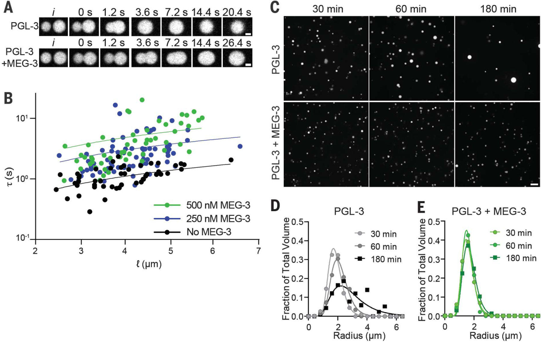
Fig. 2. MEG-3 reduces the surface tension of PGL-3 condensates and prevents coarsening.

(A) Photomicrographs of PGL-3 droplets (3 µM PGL-3 and 80 ng/µl nos-2 RNA) coalescing with or without 0.5 µM MEG-3. (B) Relaxation time (τ) of fusing PGL-3 droplets (as above) is plotted versus length scale (ℓ) with varying concentrations of MEG-3 as indicated. Each dot represents a single fusion event. The linear slope represents the inverse capillary velocity (η/γ). (C) Photomicrographs of a PGL-3 emulsion (max projections) at the indicated time points after assembly. Three micromolar PGL-3 and 80 ng/µl nos-2 RNA were incubated in condensation buffer in the presence or absence of 0.5 µM MEG-3. Scale bar is 5 µm and applies to all images in the set. (D and E) Histograms plotting the size distribution of PGL condensates assembled as in (C). Each data point indicates the fraction of total PGL-3 condensate volume represented by condensates binned by radius from 80 images [as in (C)] collected in four replicates. Lines were fit to a log normal distribution.
