Abstract
In many countries, the 16 years of age threshold is considered to be legally relevant according to the law. This research aims to ascertain the sensitivity and specificity of Olze et al. stages of root pulp visibility (RPV) in a sample of 760 south Indian children aged between 12 and 20 years, with an age threshold of 16 years, using receiver operating characteristic curves and area under the curve (AUC). Spearman’s rho correlation showed a strong positive correlation between the RPV stages and age. No significant difference between the right and left lower first molars was seen. RPV Stage 2 showed the highest AUC in both females (0.813) and males (0.790). The performance of the RPV Stage 2 to discriminate the legal age threshold of 16 years resulted in the sensitivity, specificity and accuracy values of 0.61, 0.96 and 0.77 in males, 0.65, 0.97 and 0.80 in females. It resulted in 3.6% and 2.9% of false positives and 38.5% and 34.5% of false negatives in both sexes. Even though, RPV Stage 2 can discriminate reasonably well between two age categories, due to the high percentage of false negatives we recommend its use in conjunction with other age estimation methods.
Keywords: Dental age estimation;
Keywords: Root pulp visibility;
Keywords: Mandibular first molars, Specificity
Keywords: 16 years;
Keywords: ROC curves
INTRODUCTION
Juvenile crime/ delinquency is a complex problem that continues to have a toll on our society. It involves wrong doing by a child or a young person who is under an age specified by the law. The minimum age of criminal responsibility indicates the lowest age at which children are held responsible for their alleged criminal acts. It has been applied asymmetrically by different countries hugely ranging from as low as 6 up to 18 years of age. (1) The age threshold of 16 years is relevant in many countries like India, China, Maldives, Sri Lanka, Belgium, France, Netherlands, Argentina, UAE etc. It represents the age at which the criminal law is applied to juveniles (minimum of 16 years but less than 18 years) who are accused of heinous crimes and therefore treated as adults. (2, 3) Often sports organizations seek the help of medical and dental experts to estimate the age of players in the 16 year old threshold. (4) Therefore, age estimation methods that have shown reasonable accuracy should be applied to assign an age that the trial or sentencing of the accused will be conducted in the adult or juvenile justice courts.
Currently, age estimation methods are based on the projection radiography of different skeletal elements such as hand and wrist, clavicle, knee and teeth. (5- 10) Depending on the question to be answered, these methods may be more or less useful and reliable. (11) Age estimation using teeth is an accurate, reliable, minimally invasive, well known and widely used historic procedure. (12, 13) For many years, prediction of the legal age thresholds in children and sub-adults was done using the mineralization and maturation of the third molars. (14- 19) In 2010, Olze et al. studied regressive changes in lower third molars with completed root formation in younger age groups. They introduced a 4 stage classification based on the radiographic visibility of the root pulp. The mean age and standard deviation at each stage was used to calculate the likelihood of attaining 18 and 21 years of age. (20) Studies in different racial groups confirmed the radiographic pulp examination as a reliable method and a valuable contribution in forensic age assessment. (21- 24)
The present study is a continuation of previous research (25) that tested the radiographic visibility of the root pulp in the lower first molars for prediction of the attainment of adulthood. As in the previous research, the present study aimed at determining the accuracy of Olze et al (20) classification of root pulp visibility in lower first molars for estimating another key age of medico-legal importance i.e., 16 years. The accuracy of this stage classification was evaluated by Receiver operating characteristic (ROC) analysis, which combines the sensitivity and specificity in a single accuracy measure.
MATERIAL AND METHODS
Sample
A total of 760 orthopantomograms (OPGs) from 380 male and 380 female south Indian subjects with ages ranging 12 to 20 years were collected retrospectively from the digital archives of the Department of Radiology and private dental clinics. Table 1 shows the age and sex distribution of the sample. Ethical clearance was obtained from the Institutional Ethics Committee to conduct the study. The need for obtaining informed consent was waived due to retrospective nature of the study. OPGs belonging to individuals of south Indian origin (confirmed using national identification card), with good diagnostic quality, with intact right and left mandibular first molars were included in the study. OPGs with caries, fillings, endodontically treated first molars, positional anomalies, molars with incomplete root formation or single roots were excluded from the study. Each OPG was assigned a unique identification number (UIN). Details of each individual such as sex, age, UIN, date of birth and date of radiography for each OPG were recorded separately in a Microsoft Excel file. Chronological age of each subject was calculated by subtracting the date of birth from the date of exposure of the radiograph.
Table 1. Age and sex distribution of the sample.
| Age groups | Males | Females | Total |
|---|---|---|---|
| 12-12.9 | 40 | 40 | 80 |
| 13-13.9 | 40 | 40 | 80 |
| 14-14.9 | 50 | 50 | 100 |
| 15-15.9 | 50 | 50 | 100 |
| 16-16.9 | 50 | 50 | 100 |
| 17-17.9 | 50 | 50 | 100 |
| 18-18.9 | 50 | 50 | 100 |
| 19-19.9 | 50 | 50 | 100 |
| Total | 380 | 380 | 760 |
Method
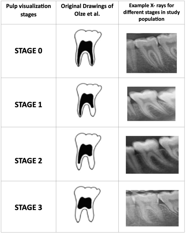
The proportion of obliteration of the root pulp in lower first molars with completed root formation was assessed using the staging system described by Olze et al. (20) Figure 1 shows schematic illustrations of the stage classifications and typical radiographs.
Figure 1.

Schematic representation of root pulp visualization stages proposed by Olze et al., 2010
All OPGs were analyzed a by single examiner, who is a forensic odontologist with 6 years of experience in evaluating radiographic images and in age estimation. The examiner was blinded for subjects age and sex, classified the lower first molars according to the stages of root pulp visibility. To explore the intra- and inter-examiner agreement, 100 OPGs were randomly selected and re-evaluated after three months of the first evaluation.
Statistical analysis
Data was analyzed in IBM SPSS version 20.0 (SPSS Inc., Chicago, IL, USA) with a significance level set at 5% (p<0.05). Cohen’s kappa statistics were performed to calculate the intra- and inter-examiner agreement. Descriptive statistical analysis of the data was expressed in mean, standard deviation (SD), median with lower and upper quartiles, and minimum and maximum of the root pulp visibility of lower first molars considering the 4 stage classification. Correlation between the age and root pulp visibility in lower first molars was evaluated using Spearman rank order correlation (rho). Chi- square test was used to observe the association between age as equal or above 16 years and stages of root pulp visibility.
The performance of the stages of pulp grading was tested by 2×2 contingency table. It generally displays the number of participants who are true positives, true negatives, false positives and false negatives. (26) The performance of each stage was assessed using accurate classification, sensitivity, specificity, positive (LR+) and negative (LR-) likelihood ratios. In the present context, the sensitivity represents the rate of subjects correctly classified as ≥16 years old, while specificity represents the rate of subjects correctly classified as < 16 years. Likelihood ratios combine the sensitivity and specificity into a single value that indicates which cut-off is best in discriminating the age threshold. Values of LR+ > 1, i.e., LR+ between 2 to 5 present small, 5 to 10 present moderate and above 10 present large and often conclusive increases the likelihood of subject to be older than 16 years. LR- between 0.2 and 0.5 present small, 0.2 and 0.1 present moderate, while under 0.1 present large and often conclusive decreases in the likelihood of age being above 16 years. (27)
In the present study, the diagnostic accuracy of each threshold (stages of root pulp visibility) was characterized by calculating diagnostic sensitivity and specificity, which then combined in the ROC plot and by quantifying the areas under the ROC curves (AUC). ROC curve represents a graphical plot which illustrates the performance of a binary classifier system. (28) AUC represents an approximate measurement of the diagnostic accuracy of a quantity. It ranges from 0 to 1, where a value of 0 indicates a perfectly inaccurate test, while a value of 1 perfectly accurate test. (28) An AUC of 0.5 represents no discrimination, 0.7 to 0.8 considered acceptable, 0.8 to 0.9 is excellent and more than 0.9 is considered outstanding. (29)
RESULTS
Kappa statistics revealed the values of 0.87 for intra-examiner and 0.8 for inter-examiner, indicating substantial to almost perfect agreements (Table 2). Table 3 listed the reasons for the exclusion of OPGs that include insufficient image quality or fused roots (1.97% cases) and due to the presence of three rooted molars or multiple root canals (0.65% cases). Total analysed radiographs accounted up to 97.3%. Spearman’s rho correlation showed a strong, positive and statistically significant correlation between the stages and the age for both sexes i.e., 0.823 (p<0.05) and 0.804 (p<0.05) for males and females, respectively.
Table 2. Kappa statistics of the intra- and inter-examiner agreement.
|
Kappa
value |
95% CI | Reliability | ||
|---|---|---|---|---|
| Lower | Upper | |||
| Intra-examiner | 0.874 | 0.832 | 0.904 | Substantial |
| Inter-examiner | 0.803 | 0.776 | 0.839 | Substantial |
CI; Confidence Interval
Table 3. Number of evaluated teeth and the teeth that could not be reliably assessed.
| Tooth | Sex | Number of cases | Evaluated teeth | Insufficient quality image/ fused roots | Three rooted molars/ multiple canals |
|---|---|---|---|---|---|
| 36 | Male | 380 | 368 | 8 | 4 |
| Female | 380 | 372 | 7 | 1 | |
| 46 | Male | 380 | 368 | 9 | 3 |
| Female | 380 | 375 | 5 | 0 |
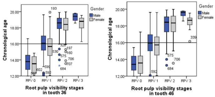
To test the variation between the lower first molars of right and left quadrants, analyses were performed separately for both teeth. In table 4 and 5, descriptive data of each stage of root pulp visibility is displayed for both females and males of both lower first molars. Table 6 and 7 display the proportion of the subjects under and over 16 year age threshold. Figure 2 shows the distribution of the sample according to the chronological age (in years) for different stages of the root pulp visibility in both lower first molars (FDI, 36 & 46).
Table 4. Descriptive statistics of chronological age according to sex and stage of root pulp visibility in tooth 36.
| Stage | N | Min | Max | LQ | Median | UQ | Mean | SD | |
|---|---|---|---|---|---|---|---|---|---|
| Females | 0 | 74 | 12.04 | 14.84 | 12.31 | 13.52 | 14.41 | 13.43 | 0.9 |
| 1 | 162 | 12.06 | 19.93 | 14.53 | 15.67 | 16.65 | 15.56 | 1.5 | |
| 2 | 128 | 13.54 | 19.98 | 17.7 | 18.42 | 19.18 | 18.33 | 1.1 | |
| 3 | 8 | 16.26 | 19.55 | 17.9 | 18.8 | 19.29 | 18.47 | 1.06 | |
| Males | 0 | 99 | 12 | 16.65 | 12.8 | 13.79 | 14.67 | 13.77 | 1.1 |
| 1 | 140 | 12.07 | 19.62 | 15.21 | 16.08 | 16.98 | 15.96 | 1.4 | |
| 2 | 121 | 13.29 | 19.91 | 17.81 | 18.61 | 19.2 | 18.44 | 1.1 | |
| 3 | 8 | 18.59 | 19.81 | 18.75 | 19.42 | 19.77 | 19.32 | 0.5 |
N, Number; Min, Minimum; Max, Maximum; LQ, Lower quartile; UQ, Upper quartile; SD, Standard deviation.
Table 5. Descriptive statistics of chronological age according to sex and stage of root pulp visibility in tooth 46.
| Stage | N | Min | Max | LQ | Median | UQ | Mean | SD | |
|---|---|---|---|---|---|---|---|---|---|
| Females | 0 | 85 | 12.01 | 17.27 | 12.31 | 13.57 | 14.41 | 13.47 | 1.07 |
| 1 | 138 | 12.06 | 19.6 | 14.52 | 15.73 | 16.69 | 15.58 | 1.5 | |
| 2 | 142 | 13.54 | 19.98 | 17.3 | 18.35 | 19.17 | 18.08 | 1.3 | |
| 3 | 10 | 16.15 | 19.53 | 17.99 | 18.67 | 18.95 | 18.37 | 0.9 | |
| Males | 0 | 87 | 12 | 15.41 | 12.69 | 13.41 | 14.39 | 13.55 | 0.9 |
| 1 | 122 | 12.07 | 19.62 | 15.01 | 15.95 | 16.95 | 15.84 | 1.5 | |
| 2 | 154 | 13.29 | 19.91 | 17.25 | 18.44 | 19.1 | 18.03 | 1.3 | |
| 3 | 05 | 18.59 | 19.78 | 18.9 | 19.61 | 19.77 | 19.39 | 0.5 |
N, Number; Min, Minimum; Max, Maximum; LQ, Lower quartile; UQ, Upper quartile; SD, Standard deviation.
Table 6. Stage distribution according to age threshold 16 years for tooth 36.
| Stage | Total (N) | <16 Years | >16 years | |||
|---|---|---|---|---|---|---|
| n | Prop | n | Prop | |||
| Female | 0 | 74 | 74 | 1 | 0 | 0 |
| 1 | 162 | 93 | 0.574 | 69 | 0.426 | |
| 2 | 128 | 5 | 0.390 | 123 | 0.961 | |
| 3 | 08 | 0 | 0 | 08 | 1 | |
| Male | 0 | 99 | 93 | 0.939 | 06 | 0.611 |
| 1 | 140 | 69 | 0.493 | 71 | 0.507 | |
| 2 | 121 | 06 | 0.5 | 115 | 0.95 | |
| 3 | 08 | 0 | 0 | 08 | 1 | |
Prop; Proportion
Table 7. Stage distribution according to age threshold 16 years for tooth 46.
| Stage | Total (N) | <16 Years | >16 years | |||
|---|---|---|---|---|---|---|
| n | Prop | n | Prop | |||
| Female | 0 | 85 | 84 | 0.988 | 1 | 0.012 |
| 1 | 138 | 77 | 0.558 | 61 | 0.442 | |
| 2 | 142 | 14 | 0.099 | 128 | 0.901 | |
| 3 | 10 | 0 | 0 | 10 | 1 | |
| Male | 0 | 87 | 87 | 1 | 0 | 0 |
| 1 | 122 | 62 | 0.508 | 60 | 0.492 | |
| 2 | 154 | 19 | 0.123 | 135 | 0.877 | |
| 3 | 05 | 0 | 0 | 05 | 1 | |
Prop; Proportion
Figure 2.
Box and whisker plots of the stages of root pulp visibility for males and females for tooth 36 (left) and tooth 46 (right)
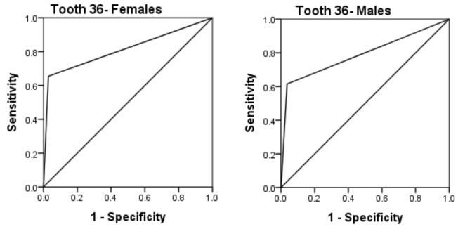
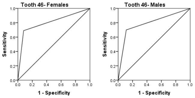
ROC curves were drawn to evaluate the discriminatory ability of the 4 stages and to determine the optimum cut-offs. Figure 3 and 4 show the graphs for females and males for both teeth with respect to the 16 year age threshold. Based on the findings, it was found that the stage 2 of root pulp visibility marked as the optimum cut-off for the 16 year old threshold in both sexes.
Figure 3.
ROC Curves for root pulp visibility stage 2 for 16 year old threshold in females and males for tooth 36
Figure 4.
ROC Curves for root pulp visibility stage 2 for 16 year old threshold in females and males for tooth 46
Table 8 and 9 show the performance measures of the stage 2 root pulp visibility for both sexes in the lower first molars (FDI, 36 & 46). In females, for the lower left first molar (FDI, 36) the values of AUC, sensitivity, specificity, LR+, LR- and accuracy were 0.813, 0.65, 0.97, 22.53, 0.36 and 0.80, respectively. In males these values were 0.790, 0.61, 0.96, 17.22, 0.4 and 0.77, respectively. LR+ value of 22.53 and 17.22 in females and males. This indicates that when the stage 2 of root pulp visibility was scored then a female is 23 times and a male is 17 times more likely to be over than the under 16 years of age threshold. LR- value of 0.36 and 0.4 in females and males indicate that when the stage 2 of pulp visibility was not attained, then both the sexes are approximately 10 times more likely to be under than over 16 years of age. On the other hand, for the lower right first molar (FDI, 46), the values AUC, sensitivity, specificity, LR+, LR- and accuracy 0.805, 0.72, 0.92, 9.02, 0.3 and 0.81 in females and 0.793, 0.70, 0.89, 6.19, 0.34 and 0.79 in males, respectively.
Table 8. Performance measures of pulp visibility stage 2 for legal age threshold over 16 years using tooth 36.
| Measures | Females | Males |
|---|---|---|
| Pulp visibility stage 2 | ||
| AUC | 0.813 (0.768- 0.858) | 0.790 (0.743- 0.837) |
| Sensitivity | 0.65 (0.58- 0.72) | 0.61 (0.54- 0.68) |
| Specificity | 0.97 (0.93- 0.99) | 0.96 (0.92- 0.99) |
| LR+ | 22.53 (9.44- 53.76) | 17.22 (7.79- 38.07) |
| LR- | 0.36 (0.29- 0.43) | 0.4 (0.33- 0.48) |
| Accuracy | 0.80 (0.76- 0.84) | 0.77 (0.73- 0.82) |
Table 9. Performance measures of pulp visibility stage 2 for legal age threshold over 16 years using tooth 46.
| Measures | Females | Males |
|---|---|---|
| Pulp visibility stage 2 | ||
| AUC | 0.805 (0.759- 0.851) | 0.793 (0.746- 0.841) |
| Sensitivity | 0.72 (0.66- 0.78) | 0.70 (0.63- 0.76) |
| Specificity | 0.92 (0.87- 0.96) | 0.89 (0.83- 0.93) |
| LR+ | 9.02 (5.42- 15.01) | 6.19 (4.01- 9.54) |
| LR- | 0.3 (0.24- 0.38) | 0.34 (0.27- 0.42) |
| Accuracy | 0.81 (0.77- 0.85) | 0.79 (0.74- 0.83) |
DISCUSSION
Diagnostic tests play a significant role in modern medicine and have become a critical feature of standard medical practice. (30) Sensitivity and specificity are the measures of the intrinsic diagnostic accuracy, known to be independent of the prevalence of the condition. (31) In the present investigation, we set out to find the optimum cut-off value (i.e., stage of root pulp visibility in lower first molars) that combines the highest possible specificity (important in criminal context) and the highest obtainable sensitivity (percentage of true negatives) for discriminating whether a subject is ≥16 years.
Originally, Olze et al (20) studied the root pulp visibility in the lower third molars for forensic age estimation purposes. However, in the present study we tested this method in the lower first molars. There are two important reasons why the authors in the present study chose to evaluate the lower first molars; firstly, the increased frequency of missing third molars and secondly, the presence of third molars with incomplete root formation. It is reported that the prevalence of third molar agenesis is high and is ranging up to 28%. (32) Timme et al (33) in their study reported higher number of missing third molars (46 to 60%) indicating it as a main limitation of this method. According to the original method, (20) third molars with incomplete roots (not attained Demirjian stage H) could not be included for evaluation. In the present study, when the OPGs of subjects aged 12 - 20 years were evaluated; a very high percentage of third molars (>50%) with incomplete mineralization were seen, indicating that this methodology (20) could not be applied to predict the legal age threshold of 16 years using third molars. Therefore, for the reliability of our results and based on the previous study findings, (25) we decided to study the radiographic visibility of root pulp in lower first molars.
The Study Group on Forensic Age Diagnostics (AGFAD) of the German society of Legal Medicine, recommended the use of multiple methods in combination, for optimal accuracy. (34) The ideal age estimation method is a constant quest for forensic experts. Every method exhibits variations in their accuracy, sensitivity and specificity especially when considered at different age thresholds. (35) Very few authors in the past have tested the age threshold of 16 years. Table 10 summarized the list of studies and their outcome measures. The authors have analysed the maturity of the third molars alone, (17) maturity of the second and third molars combined, (3, 16) Demirjian’s staging system for second and third molars combined, (36) and for all lower seven teeth combined (37) and radiographic examination of root pulp visibility in the lower first molars (present study). Cameriere et al (16) analysed the apical maturity of second and third molars and reported sensitivity and specificity values of 0.79 and 0.81. When Wang et al (3) analysed the same parameters in a southern Chinese sample, similar sensitivity value of 0.78 was reported, however, better specificity value of 0.97 was observed. When the maturity of third molars alone was assessed, sensitivity and specificity values of 0.91 and 0.85 in males and 0.9 and 0.87 in females, were obtained in a south Indian sample. (17) Cardoso et al (36) examined the second and third molars separately, and in combination using the Demirjian staging system. They reported a sensitivity of 0.86 and specificity of 0.77 when the combination of second and third molars were assessed. In the present investigation, we have obtained optimum sensitivity values (0.61 and 0.65) and better specificity values (0.96 and 0.97) in males and females, respectively. Pinchi et al (37) tested the usefulness of Demirjian and Willem age estimation methods for the assessment of the attainment of the 14 years threshold in Italian children. Both methods have resulted in higher sensitivity and lower specificity values. These differences in sensitivity and specificity values might be related to the parameters tested and the existing developmental variations between the populations.
Table 10. Data from previously published studies with different cut-off values tested in various populations for predicting the legal age threshold of 16 years.
|
Author
(Year) |
Parameters
assessed |
Accuracy | Sensitivity | Specificity | AUC |
|---|---|---|---|---|---|
| Pinchi et al (2012) (37) | Maturation of all mandibular teeth excluding third molars | --- | D: 0.80 W: 0.95 |
D: 0.61 W: 0.86 |
--- |
| Cameriere et al (2018) (16) | Combination of second (I2M) and third molar (I3M) maturity indices | 0.80 | 0.79 | 0.81 | 0.890 |
| Cardoso et al (2018) (36) | Demirjian staging system for 2nd & 3rd molars combined |
0.82 | 0.86 | 0.77 | 0.926 |
|
SB Balla et al
(2019)17 |
Third molar maturity index (I3M) |
M: 0.88 F: 0.89 |
M: 0.91 F: 0.90 |
M: 0.86 F: 0.87 |
0.936 |
|
Wang et al
(2020)3 |
Combination of second (I2M) and third molar (I3M) maturity indices | 0.88 | 0.78 | 0.97 | 0.803 (I2M) 0.945 (I3M) |
|
Present study
(2020) |
Olze et al., stages of Root pulp visibility in lower first molars | M: 0.77 F: 0.80 |
M: 0.61 F: 0.65 |
M: 0.96 F: 0.97 |
M: 0.790 F: 0.813 |
AUC, Area under the curve; I2M, Second molar maturity index; I3M, Third molar maturity index; D, Demirjian’s method; W, Willems method
For each threshold, there is a combination of sensitivity and specificity which are then combined in the ROC plot, resulting in the AUC that indicates the performance of the test. (38) In the present investigation, the AUC for stage 2 root pulp visibility was 0.813 for females and 0.790 in males indicating that a randomly selected individual (either male or female) from the older age category (≥16 years) will have a greater grade of root pulp visibility (stage 2 and above) compared to a randomly chosen individual from the younger age category (<16 years) approximately 80% of the time. This indicates that diagnosing age at least 16 years from root pulp visibility stages can discriminate reasonably well between two age categories. Similarly, every threshold has the likelihood ratios of a positive (LR+) and negative (LR-) test result to express the probability of a diagnostic test result. (36) In the present study, the test result of a stage 2 root pulp visibility is more than 23 times in a female and 17 times in a male more likely to occur in an individual at least 16 years otherwise to someone younger than 16. Similarly, LR- of approximately 0.4 in both sexes indicates that an individual marked with early stages of root pulp visibility (stage 0 and 1) is one in ten more likely to be seen if age is at least 16 years than otherwise i.e., if age is in younger category.
Our study also provides the results for the error in discriminating minors (<16 years) and accountable juveniles (≥16 years). When stage 2 root pulp visibility was used as an age marker for 16 years, it resulted in ethically unacceptable (false positive) and technically unacceptable (false negative) errors. The highest error rates were false negatives i.e., 38.5% of males and 34.5% of females who were aged equal or above 16 years was classified as below 16 years. Misclassification, which is important from the medico-legal point of view (false positives) were kept to minimum i.e., 3.6% of males and 2.9% of females.
Each age estimation method must be overviewed in conjunction with its advantages and disadvantages. The advantages of this method are; firstly, it can be applied in subjects without third molars. Secondly, high specificity values were obtained in both sexes, which is extremely important in the criminal law context, which expresses the rate of false positives. (39) Even though, Wang et al (3) obtained higher specificity values using the combination of second and third molar maturity index values, they reported that this methodology could not be used in case the third molars were missing. One possible disadvantage in the present study is the lower sensitivity values in both sexes. However, it is less damaging to classify an individual as being younger than 16 years, when they are not, than classifying a minor as an accountable juvenile. (40) Although this study provides some indication of the quality age predictive model i.e., reasonably well discrimination between two age categories using stage 2 of root pulp visibility in lower first molars, we advise the combination of methods due to the high percentage of false negatives.
CONCLUSIONS
After examining a sample of south Indian children aged between 12 and 20 years using ROC curves, we found that the stage 2 of root pulp visibility can be used to predict the age of 16 years or over. In particular, this study suggests that dental age assessment can be done with reasonable accuracy, especially the prediction of the 16 year old age threshold using teeth other than the third molars. However, due to the high percentage of false negatives, we advise the use of this method in conjunction with other methods. Further research should be done to validate the use of root pulp visibility in teeth other than molars for prediction of the attainment of legal age thresholds in children and sub-adults.
Footnotes
The authors declare that they have no conflict of interest.
REFERENCES
- 1.Cipriani D, Nelken PD. Children’s Rights and the Minimum Age of Criminal Responsibility: A Global Perspective: Ashgate Publishing Limited; 2013. [Google Scholar]
- 2.India to change age of criminal responsibility for minors accused of rape and murder. The Telegraph [Internet]. [Updated 2013 Dec 6; cited 2021 May 25]. Available from: https://www.telegraph.co.uk/news/worldnews/asia/india/ 10499798/India-to-change-age-of-criminal-responsibility-for-minors-accused-of-rape-and-murder.html
- 3.Wang M, Wang J, Pan Y, Fan L, Shen Z, Ji F, et al. Applicability of newly derived second and third molar maturity indices for indicating the legal age of 16 years in the Southern Chinese population. Leg Med (Tokyo). 2020;46:101725. 10.1016/j.legalmed.2020.101725 [DOI] [PubMed] [Google Scholar]
- 4.BCCI Adopts New Age-Verification Method – ESPN CRICINFO. [Internet] [Updated 2012 July 8; cited 2021 May 25]. Available at: https://www.espncricinfo.com/story/india-news-bcci-adopts-new-age-verification-method-571542
- 5.Greulich WW, Pyle SI. Radiographic Atlas of Skeletal Development of the Hand and Wrist: Stanford University Press; 1959. [Google Scholar]
- 6.Schmeling A, Schulz R, Reisinger W, Muhler M, Wernecke KD, Geserick G. Studies on the time frame for ossification of the medial clavicular epiphyseal cartilage in conventional radiography. Int J Legal Med. 2004;118(1):5–8. 10.1007/s00414-003-0404-5 [DOI] [PubMed] [Google Scholar]
- 7.Schulz R, Muhler M, Reisinger W, Schmidt S, Schmeling A. Radiographic staging of ossification of the medial clavicular epiphysis. Int J Legal Med. 2008;122(1):55–8. 10.1007/s00414-007-0210-6 [DOI] [PubMed] [Google Scholar]
- 8.Hackman L, Black S. Age estimation from radiographic images of the knee. J Forensic Sci. 2013;58(3):732–7. 10.1111/1556-4029.12077 [DOI] [PubMed] [Google Scholar]
- 9.Demirjian A, Goldstein H, Tanner JM. A new system of dental age assessment. Hum Biol. 1973;45(2):211–27. [PubMed] [Google Scholar]
- 10.Moorrees CF, Fanning EA, Hunt EE, Jr. Age Variation of Formation Stages for Ten Permanent Teeth. J Dent Res. 1963;42:1490–502. 10.1177/00220345630420062701 [DOI] [PubMed] [Google Scholar]
- 11.Tisè M, Ferrante L, Mora S, Tagliabracci A. A biochemical approach for assessing cutoffs at the age thresholds of 14 and 18 years: a pilot study on the applicability of bone specific alkaline phosphatase on an Italian sample. Int J Legal Med. 2016;130(4):1149–58. 10.1007/s00414-016-1382-8 [DOI] [PubMed] [Google Scholar]
- 12.Ritz-Timme S, Cattaneo C, Collins MJ, Waite ER, Schutz HW, Kaatsch HJ, et al. Age estimation: the state of the art in relation to the specific demands of forensic practice. Int J Legal Med. 2000;113(3):129–36. 10.1007/s004140050283 [DOI] [PubMed] [Google Scholar]
- 13.Lewis JM, Senn DR. Forensic Dental Age Estimation: An Overview. J Calif Dent Assoc. 2015;43(6):315–9. [PubMed] [Google Scholar]
- 14.Mincer HH, Harris EF, Berryman HE. The A.B.F.O. study of third molar development and its use as an estimator of chronological age. J Forensic Sci. 1993;38(2):379–90. 10.1520/JFS13418J [DOI] [PubMed] [Google Scholar]
- 15.Cameriere R, Ferrante L, De Angelis D, Scarpino F, Galli F. The comparison between measurement of open apices of third molars and Demirjian stages to test chronological age of over 18- year olds in living subjects. Int J Legal Med. 2008;122(6):493–7. 10.1007/s00414-008-0279-6 [DOI] [PubMed] [Google Scholar]
- 16.Cameriere R, Velandia Palacio LA, Pinares J, Bestetti F, Paba R, Coccia E, et al. Assessment of second (I2M) and third (I3M) molar indices for establishing 14 and 16 legal ages and validation of the Cameriere’s I3M cut-off for 18 years old in Chilean population. Forensic Sci Int 2018;285:205.e1-.e5. [DOI] [PubMed]
- 17.Balla SB, Chinni SS, Galic I, Alwala AM, Machani P, Cameriere R. A cut-off value of third molar maturity index for indicating a minimum age of criminal responsibility: Older or younger than 16 years? J Forensic Leg Med. 2019;65:108–12. 10.1016/j.jflm.2019.05.014 [DOI] [PubMed] [Google Scholar]
- 18.Sharma P, Wadhwan V. Comparison of accuracy of age estimation in Indian children by measurement of open apices in teeth with the London Atlas of tooth development. J Forensic Odontostomatol. 2020;1(38):39–47. [PMC free article] [PubMed] [Google Scholar]
- 19.Prasad H, Kala N. Accuracy of two dental age estimation methods in the Indian population - A meta-analysis of published studies. J Forensic Odontostomatol. 2019;3(37):2–11. [PMC free article] [PubMed] [Google Scholar]
- 20.Olze A, Solheim T, Schulz R, Kupfer M, Schmeling A. Evaluation of the radiographic visibility of the root pulp in the lower third molars for the purpose of forensic age estimation in living individuals. Int J Legal Med. 2010;124(3):183–6. 10.1007/s00414-009-0415-y [DOI] [PubMed] [Google Scholar]
- 21.Pérez -Mongiovi D, Teixeira A, Caldas IM. The radiographic visibility of the root pulp of the third lower molar as an age marker. Forensic Sci Med Pathol. 2015;11(3):339–44. 10.1007/s12024-015-9688-2 [DOI] [PubMed] [Google Scholar]
- 22.Lucas VS, McDonald F, Andiappan M, Roberts G. Dental Age Estimation-Root Pulp Visibility (RPV) patterns: A reliable Mandibular Maturity Marker at the 18- year threshold. Forensic Sci Int. 2017;270:98–102. 10.1016/j.forsciint.2016.11.004 [DOI] [PubMed] [Google Scholar]
- 23.Akkaya N, Yılancı HÖ, Boyacıoğlu H, Göksülük D, Özkan G. Accuracy of the use of radiographic visibility of root pulp in the mandibular third molar as a maturity marker at age thresholds of 18 and 21. Int J Legal Med. 2019;133(5):1507–15. 10.1007/s00414-019-02036-x [DOI] [PubMed] [Google Scholar]
- 24.Guo YC, Chu G, Olze A, Schmidt S, Schulz R, Ottow C, et al. Application of age assessment based on the radiographic visibility of the root pulp of lower third molars in a northern Chinese population. Int J Legal Med. 2018;132(3):825–9. 10.1007/s00414-017-1731-2 [DOI] [PubMed] [Google Scholar]
- 25.Balla SB, Ankisetti SA, Bushra A, Bolloju VB, Mir Mujahed A, Kanaparthi A, et al. Preliminary analysis testing the accuracy of radiographic visibility of root pulp in the mandibular first molars as a maturity marker at age threshold of 18 years. Int J Legal Med. 2020;134(2):769–74. 10.1007/s00414-020-02257-5 [DOI] [PubMed] [Google Scholar]
- 26.Fletcher RF, Diagnosis S. In: Fletcher RF, S (ed.). Clinical epidemiology. The essentials. Baltimore: Wolters, Kluwer, Lippincott, Williams & Wilkins; 2005. p 35-58. [Google Scholar]
- 27.Grimes DA, Schulz KF. Refining clinical diagnosis with likelihood ratios. Lancet. 2005;365(9469):1500–5. 10.1016/S0140-6736(05)66422-7 [DOI] [PubMed] [Google Scholar]
- 28.Mandrekar JN. Receiver Operating Characteristic Curve in Diagnostic Test Assessment. J Thorac Oncol. 2010;5(9):1315–6. 10.1097/JTO.0b013e3181ec173d [DOI] [PubMed] [Google Scholar]
- 29.Hosmer DW, Lemeshow S. Applied Logistic Regression, 2nd Ed. Chapter 5, John Wiley and Sons, New York, NY (2000), pp. 160- 164. [Google Scholar]
- 30.ES of Radiology . The future role of radiology in healthcare. Insights Imaging. 2010;1(1):2–11. 10.1007/s13244-009-0007-x [DOI] [PMC free article] [PubMed] [Google Scholar]
- 31.Kumar R, Indrayan A. Receiver operating characteristic (ROC) curve for medical researchers. Indian Pediatr. 2011;48(4):277–87. 10.1007/s13312-011-0055-4 [DOI] [PubMed] [Google Scholar]
- 32.Harris EF, Clark LL. Hypodontia: an epidemiologic study of American black and white people. Am J Orthod Dentofacial Orthop. 2008;134(6):761–7. 10.1016/j.ajodo.2006.12.019 [DOI] [PubMed] [Google Scholar]
- 33.Timme M, Timme WH, Olze A, Ottow C, Ribbecke S, Pfeiffer H, et al. The chronology of the radiographic visibility of the periodontal ligament and the root pulp in the lower third molars. Sci Justice. 2017;57(4):257–61. 10.1016/j.scijus.2017.03.004 [DOI] [PubMed] [Google Scholar]
- 34.Schmeling A. R; Rudolf, E; Vieth, V; Geserick, G. Forensic Age Estimation: Methods, Certainty, and the Law. Dtsch Arztebl Int. 2016;113(4):44–50. [DOI] [PMC free article] [PubMed] [Google Scholar]
- 35.Staaf V, Mörnstad H, Welander U. Age estimation based on tooth development: a test of reliability and validity. Scand J Dent Res. 1991;99(4):281–6. 10.1111/j.1600-0722.1991.tb01029.x [DOI] [PubMed] [Google Scholar]
- 36.Cardoso HFV, Caldas IM, Andrade M. Dental and skeletal maturation as simultaneous and separate predictors of chronological age in post-pubertal individuals: a preliminary study in assessing the probability of having attained 16 years of age in the living. Aust J Forensic Sci. 2018;50(4):371–84. 10.1080/00450618.2016.1237548 [DOI] [Google Scholar]
- 37.Pinchi V, Norelli GA, Pradella F, Vitale G, Rugo D, Nieri M. Comparison of the applicability of four odontological methods for age estimation of the 14 years legal threshold in a sample of Italian adolescents. J Forensic Odontostomatol. 2012;30(2):17–25. [PMC free article] [PubMed] [Google Scholar]
- 38.Liversidge HM, Marsden PH. Estimating age and the likelihood of having attained 18 years of age using mandibular third molars. Br Dent J. 2010;209(8):E13. 10.1038/sj.bdj.2010.976 [DOI] [PubMed] [Google Scholar]
- 39.Pinchi V, Pradella F, Vitale G, Rugo D, Nieri M, Norelli G-A. Comparison of the diagnostic accuracy, sensitivity and specificity of four odontological methods for age evaluation in Italian children at the age threshold of 14 years using ROC curves. Med Sci Law. 2016;56(1):13–8. 10.1177/0025802415575416 [DOI] [PubMed] [Google Scholar]
- 40.Garamendi PM, Landa MI, Ballesteros J, Solano MA. Reliability of the methods applied to assess age minority in living subjects around 18 years old. A survey on a Moroccan origin population. Forensic Sci Int. 2005;154:3–12. 10.1016/j.forsciint.2004.08.018 [DOI] [PubMed] [Google Scholar]


