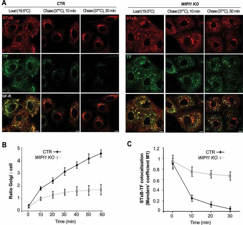
Figure 4.

WIPI1 promotes transfer of STxB from early/recycling endosomes to the Golgi. (A) Confocal microscopy of living HK2 cells. Alexa Fluor 488 TF and Cy3-StxB were bound to control and WIPI1 KO cells for 30 min on ice, internalized by incubating cells at 19.5°C for 45 min (LOAD), and finally chased to the Golgi by shifting cells to 37°C for 0–30 min. Scale bars: 10 μm. (B) Quantification of Golgi localized STxB in images from (A). The ratio of average Golgi-associated fluorescence over the average total cell-associated fluorescence is represented as a function of incubation time at 37°C. Data are means ±s.d. from 3 independent experiments. Number of cells quantified: Control = 250; WIPI1 KO = 245 (C) Colocalization of TF and STxB was quantified in images from (A) at different time points after the shift to 37°C. Values are the mean ± s.d. from 3 independent experiments. Number of cells quantified: Control = 240; WIPI1 KO = 220
