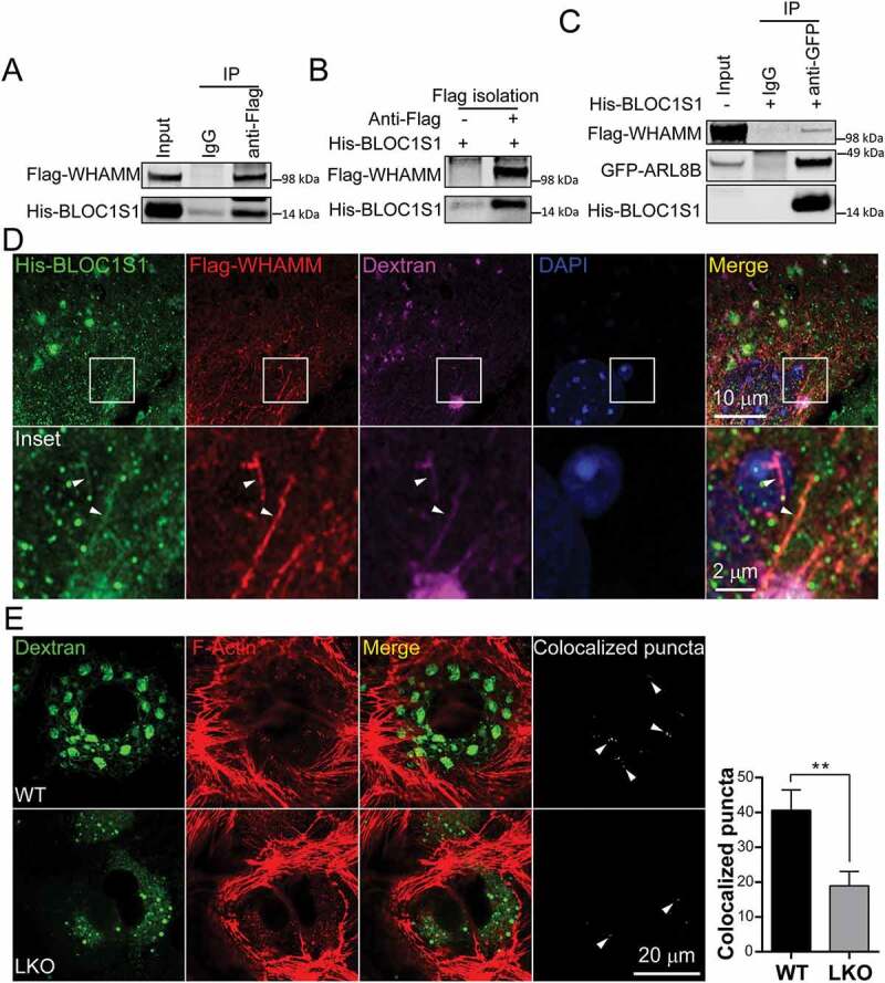
Figure 6.

The BLOC1S1 interacted with and co-localized with WHAMM on lysosomal tubules to regulate actin nucleation on autolysosomes/lysosomes. (A) 293 T cells were co-transfected with Flag-WHAMM and His-BLOC1S1, and then immunoprecipitated with either IgG or anti-Flag antibodies and then subjected to immunoblot analysis using anti-Flag and anti-His antibodies. (B) Flag affinity isolation showed the direct interaction of and Flag-WHAMM with His-BLOC1S1 protein in vitro. 293 T cells were transfected with Flag-WHAMM for 48 h and then immunoprecipitated with either IgG or a Flag antibody conjugated magnetic beads. After washing, the beads were incubated with purified His-BLOC1S1 protein and then subjected to immunoblot analysis. (C) BLOC1S1 concurrently interacted with WHAMM and ARL8B. 293 T cells were co-transfected Flag-WHAMM with GFP-ARL8B for 48 h and then immunoprecipitated with either IgG or GFP antibody conjugated magnetic beads with or without adding His-BLOC1S1 protein and then subjected to immunoblot analysis with antibodies to Flag, His and GFP. (D) WT hepatocytes were co-transfected with His-BLOC1S1 and Flag-WHAMM for 48 h then incubated with fluorescence-labeled dextran (purple). After fixation, the cells were stained using anti-His (green) and anti-Flag (red) antibodies with the nucleus stained by DAPI (blue). The colocalization of WHAMM and BLOC1S1 is show by the arrowhead indicating His-BLOC1S1, Flag-WHAMM and Dextran triple labeled tubules. (E) WT and LKO hepatocytes were co-incubated with Dextran-488 and SiR-actin following 5 h of nutrient depletion, then observed using superresolution confocal microscopy. The colocalization of > 1 μm autolysosomes/lysosomal with actin were extracted by ImagJ and highlighted by arrowheads in far-right panels. The quantitation from n > 14 cells per group are shown in the accompanying histogram
