

Abstract
In this study, we evaluated three PCR methods for epidemiological typing of Francisella tularensis: repetitive extragenic palindromic element PCR (REP-PCR), enterobacterial repetitive intergenic consensus sequence PCR (ERIC-PCR), and random amplified polymorphic DNA (RAPD) assay with both M13 and T3-T7 primers. The analysis was performed with 40 strains of F. tularensis isolated from hares, humans, ticks, and a vole. On the basis of the combination of REP, ERIC, and RAPD fingerprints, F. tularensis strains were divided into 17 genetic groups (designated A to Q), and one Francisella novicida strain was classified in group R. The F. novicida strain is of special concern, since previous genetic methods have been unable to clearly distinguish between F. tularensis and F. novicida. The F. tularensis isolates recovered from hares were included in groups A to J, M, and P; those recovered from humans were included in groups A, D, G, J, L, O, and N; those isolated from ticks were included in groups B and Q; and that recovered from a vole was in group K. The diversities calculated for the 40 F. tularensis isolates, according to Simpson's index, were 0.14 for REP-PCR, 0.52 for ERIC-PCR, 0.39 for RAPD assay with the M13 primer (RAPD/M13-PCR), and 0.65 for RAPD/T3-T7-PCR, and the diversity increased up to 0.90 when ERIC-PCR, RAPD/M13-PCR, and RAPD/T3-T7-PCR were combined. Our results suggest that although limited genetic heterogeneity among F. tularensis strains was observed, this small variation is enough to validate the PCR methods used in this study and their combinations, because they can provide safe, useful, and rapid tools for the typing of F. tularensis.
Tularemia is a clinical syndrome caused by the gram-negative, facultative intracellular bacterium Francisella tularensis. The agent is extremely infectious, and it is thought that as few as 10 organisms can cause infection in humans (2). A severe epidemic tularemia outbreak occurred in 1998 in northwestern Spain (where the disease had never been reported before), with ulceroganglionar, typhoidal, ganglionar, pneumonic, oculoganglionar, and atypical clinical forms diagnosed in humans (1, 5, 11).
There are four recognized biotypes of the bacterium. F. tularensis subsp. tularensis (nearctica, type A) is the common causal agent of tularemia in North America, and it has been recently observed for the first time in Europe (6). This subspecies is highly virulent, causes severe illness in mammals, and is associated mainly with tick-borne tularemia in rabbits. F. tularensis subsp. palaearctica (holarctica, type B) is more widely distributed in nature and is found in Europe, Asia, and to a minor extent in North America. This subspecies is linked to waterborne disease of rodents and hares, and it is considered to be less pathogenic for mammals than F. tularensis subsp. tularensis (15). F. tularensis subsp. mediaasiatica has been found only in central Asia in a part of the former Soviet Union, while F. tularensis subsp. palaearctica japonica has been isolated only in Japan (14).
The correct identification of strains of F. tularensis can be verified by agglutination with specific antisera. However, since strains of Francisella have similar antigenic compositions (to date all of the isolates belong to a unique antigenic group), it is not possible to distinguish the different subspecies or types from each other by this method (12). Subspecies of F. tularensis have been differentiated by biochemical tests (10) and by using probes specific to the 16S rRNAs of each of the two main subspecies (4, 14).
Epidemiological analysis of the 1998 Spanish outbreak was impeded by the lack of a safe, rapid, and easy method to type the isolates. Therefore, there is a need to develop new typing methods for this bacterial species. Repetitive element sequence-based PCR (rep-PCR) is a group of methods which generate DNA fingerprints that allow discrimination between bacterial strains. Two main sets of repetitive elements are used for typing purposes. The repetitive extragenic palindromic (REP) elements are 38-bp sequences consisting of six degenerate positions and a 5-bp variable loop between each side of a conserved palindromic stem. The enterobacterial repetitive intergenic consensus (ERIC) sequences are a second set of DNA sequences which have been successfully used for DNA typing. ERIC sequences are 126-bp elements which contain a highly conserved central inverted repeat and are located in extragenic regions of the bacterial genome (13). On the other hand, the random amplified polymorphic DNA (RAPD) assay is based on the use of short random sequence primers, about 10 to 20 bases in length, which hybridize with sufficient affinity to chromosomal DNA sequences at low annealing temperatures such that they can be used to initiate amplification of regions of the bacterial genome (13).
REP, ERIC, and RAPD sequences have been used as primer binding sites to amplify the genomes of a variety of bacteria by PCR. In this report, rep-PCR and RAPD were used to generate DNA fingerprints of F. tularensis strains isolated from hares, humans, voles, and ticks in Spain and to evaluate the potential of PCR methods for typing F. tularensis isolates of various origins.
MATERIALS AND METHODS
Bacterial strains and culture conditions.
Thirty-five F. tularensis isolates recovered from an epidemic tularemia outbreak that occurred in Castilla y León (northwestern Spain) in 1998, as well as some F. tularensis strains (26, 27, 28, 33, and 40) and Francisella novicida (strain 41) reference strains, were used in this study (Table 1). Of these isolates, 25 were from hares, 8 were from humans, 1 was from a vole, and 1 was from a tick. The geographical origins of these isolates are shown in Table 1. All F. tularensis strains except no. 40 were grown at 37°C for 3 days on modified Thayer-Martin agar plates containing GC medium base (36 g/liter), hemoglobin (10 g/liter), and Vitox supplement (2 vials/liter; Oxoid Ltd., Hampshire, England). F. tularensis no. 40 and F. novicida were grown on Bacto Cystine heart agar (Difco) supplemented with 10% rabbit blood at 37°C for 2 days.
TABLE 1.
Characteristics of F. tularensis strains and the F. novicida strain used in this study and ERIC-PCR, REP-PCR, and RAPD types
| Strain designation
|
Host and geographic origin | REP-PCR type | ERIC-PCR type | RAPD type (M13 primer) | RAPD type (T3-T7 primers) | Global type | |
|---|---|---|---|---|---|---|---|
| This study | Sourcea | ||||||
| 1 | ALG1 | Hare; Valladolid, Spain | 1 | 1 | 1 | 1 | A (1111) |
| 2 | ALG2 | Hare; Valladolid, Spain | 1 | 2 | 1 | 1 | B (1211) |
| 3 | ALG3 | Hare; Valladolid, Spain | 1 | 2 | 1 | 1 | B (1211) |
| 4 | ALG4 | Hare; León, Spain | 1 | 1 | 1 | 2 | C (1112) |
| 5 | ALG5 | Hare; León, Spain | 1 | 1 | 1 | 1 | A (1111) |
| 6 | ALG6 | Hare; Palencia, Spain | 1 | 2 | 1 | 1 | B (1211) |
| 7 | ALG7 | Hare; León, Spain | 1 | 2 | 1 | 1 | B (1211) |
| 8 | ALG8 | Hare; Palencia, Spain | 1 | 2 | 1 | 1 | D (1212) |
| 9 | ALG9 | Hare; Palencia, Spain | 1 | 2 | 1 | 1 | B (1211) |
| 10 | ALG10 | Hare; Valladolid, Spain | 1 | 1 | 1 | 2 | C (1112) |
| 11 | ALG11 | Hare; Valladolid, Spain | 1 | 1 | 1 | 3 | E (1113) |
| 12 | ALG12 | Hare; Zamora, Spain | 1 | 2 | 1 | 1 | B (1211) |
| 13 | ALG13 | Hare; Palencia, Spain | 1 | 1 | 1 | 1 | A (1111) |
| 14 | ALG14 | Hare; Valladolid, Spain | 1 | 2 | 1 | 1 | B (1211) |
| 15 | ALG15 | Hare; Valladolid, Spain | 2 | 1 | 2 | 1 | F (2121) |
| 16 | ALG16 | Hare; Segovia, Spain | 1 | 1 | 1 | 2 | A (1111) |
| 17 | ALG17 | Hare; Palencia, Spain | 1 | 2 | 1 | 4 | G (1214) |
| 18 | ALG18 | Hare; Palencia, Spain | 1 | 1 | 1 | 4 | H (1114) |
| 19 | ALG19 | Hare; Palencia, Spain | 2 | 1 | 2 | 3 | I (2123) |
| 20 | ALG20 | Hare; Palencia, Spain | 1 | 1 | 1 | 3 | E (1113) |
| 21 | ALG21 | Vole (Microtus arvalis); Zamora, Spain | 1 | 1 | 1 | 5 | K (1115) |
| 22 | HLE1 | Human; León, Spain | 1 | 2 | 1 | 3 | J (1213) |
| 23 | HZA1 | Human; Zamora, Spain | 1 | 2 | 1 | 4 | G (1214) |
| 24 | HZA2 | Human; Zamora, Spain | 1 | 2 | 2 | 3 | L (1223) |
| 25 | HZA3 | Human; Zamora, Spain | 1 | 2 | 1 | 2 | D (1212) |
| 26 | CΔPM1 | Hare; strain 130 (= CAPM5536), Czech Republic | 1 | 1 | 3 | 1 | M (1131) |
| 27 | CΔMP2 | Hare; strain 2713 (= CAPM5537), Czech Republic | 1 | 1 | 3 | 1 | M (1131) |
| 28 | CAMP3 | Human; strain SCHU (= CAPM 5600), United States | 3 | 3 | 1 | 6 | N (3316) |
| 29 | ALG24 | Human; Valladolid, Spain | 1 | 1 | 4 | 1 | O (1141) |
| 30 | ALG25 | Human; Valladolid, Spain | 1 | 1 | 4 | 1 | O (1141) |
| 31 | ALG26 | Human; Valladolid, Spain | 1 | 1 | 1 | 1 | A (1111) |
| 32 | ALG27 | Human; Palencia, Spain | 1 | 1 | 1 | 1 | A (1111) |
| 33 | CAPM4 | Hare; strain T-1/59 (= CAPM5151), Czech Republic | 1 | 2 | 3 | 3 | P (1233) |
| 34 | RLAH1 | Hare; Palencia, Spain | 1 | 2 | 1 | 2 | D (1212) |
| 35 | RLAH2 | Hare; Valladolid, Spain | 1 | 2 | 1 | 2 | D (1212) |
| 36 | RLAH3 | Hare; Soria, Spain | 1 | 2 | 1 | 3 | J (1213) |
| 37 | RLAH4 | Hare; Zamora, Spain | 1 | 2 | 1 | 1 | B (1211) |
| 38 | RLAH5 | Hare; Zamora, Spain | 1 | 2 | 1 | 1 | B (1211) |
| 39 | RLAH6 | Tick; Zamora, Spain | 1 | 2 | 1 | 1 | B (1211) |
| 40 | CΔPM5 | Tick; strain 503, former Soviet Union | 1 | 2 | 3 | 1 | Q (1231) |
| 41 | CΔMP6 | F. novicida reference strain ATCC 15482, France | 4 | 4 | 5 | 7 | R (4457) |
ALG, Central Laboratory of Animal Health, Algete, Madrid, Spain; HLE, Department of Medical Microbiology, Hospital Princesa Sofía, Insalud, León, Spain; HZA, Laboratory of Microbiology, Hospital Virgen de la Concha, Insalud, Zamora, Spain; CAPM, Collection of Animal Pathogenic Microorganisms, Brno, Czech Republic; RLAH, Regional Laboratory of Animal Health, León, Spain.
Preparation of genomic DNA.
Chromosomal DNAs from the strains listed in Table 1 were obtained by suspension of log-phase cells in 10 mM Tris-1 mM EDTA and were incubated at 37°C for 2 h in 0.5% sodium dodecyl sulfate and 100 μg of proteinase K per ml. The DNA was then purified by repeated phenol-chloroform extraction, precipitated from the aqueous phase by adding 0.6 volume of isopropanol and 70% ethanol, dried, and resuspended in sterile water.
rep primers and rep-PCR conditions.
For REP-PCR, the primers REP 1R-I (5′-IIIICGICGICATCIGGC-3′) and REP 2-I (5′-ICGICTTATCIGGCCTAC-3′) were used, and for ERIC-PCR, the primers ERIC IR (5′-ATGTAAGCTCCTGGGGATTCA-3′) and ERIC II (5′-AAGTAAGTGACTGGGGTGAGC-3′) were used. The REP 1R-I and REP 2-I primers contain the nucleotide inosine (I) at ambiguous positions in the REP consensus sequence. Inosine can form Watson-Crick base pairs with A, T, G, or C. PCRs were carried out with 5 μl of template DNA per reaction for REP-PCR and ERIC-PCR. Amplification reactions were performed in 50 μl of a solution containing a 1 μM concentration of each of the two opposing primers (Boehringer Mannheim OligoSynthesis, Mannheim, Germany), 0.2 mM each deoxynucleoside triphosphate (Bioline, London, United Kingdom), 2.5 mM MgCl2, 5 μl of 10× amplification buffer [160 mM (NH4)SO4, 670 mM Tris-HCl, pH 8.8 at 25°C], and 4 U of Taq DNA polymerase (Bioline). Amplifications were performed with a DNA thermocycler (GeneAmp PCR System 2400, Perkin-Elmer, Berlin, Germany). For REP-PCR, the temperature profile was 1 cycle at 95°C for 3 min; 30 cycles at 90°C for 30 s, 50°C for 30 s, 45°C for 1 min, and 65°C for 8 min; and 1 cycle at 65°C for 16 min. For ERIC-PCR, the temperature profile was 1 cycle at 95°C for 5 min; 30 cycles at 90°C for 30 s, 50°C for 30 s, 52°C for 1 min, and 72°C for 1 min; and 1 cycle at 72°C for 8 min.
RAPD primers and RAPD-PCR conditions.
For RAPD assay, the primers Universal M13 (5′-TTATGTAAAACGACGGCCAGT-3′), T3 (5′-GCAATTAACCCTCACTAAAG-3′), and T7 (5′-GTAATACGACGCACTATAG-3′) were used. PCRs were carried out as described above. Amplifications were performed under the following conditions: 1 cycle at 94°C for 5 min; 2 cycles at 94°C for 5 min, 40°C for 5 min, and 72°C for 5 min; 35 cycles at 94°C for 1 min, 60°C for 1 min, and 72°C for 1 min; and 1 cycle at 72°C for 5 min.
Reproducibility.
The day-to-day reproducibilities of the above-described REP-PCR and ERIC-PCR were examined by comparing patterns provided by the same strain and method on four different days. One strain was randomly selected from each of the two most significant profiles obtained by every PCR method and then subjected to the whole typing process for four consecutive days. The RAPD assay (with M13 and T3-T7 primers) was further evaluated for reproducibility by testing one randomly selected strain from each of the profiles obtained.
Analysis of PCR products.
rep-PCR (9 μl) and RAPD assay (10 μl) products were analyzed by agarose gel electrophoresis (with 1.1% agarose gels run in 0.5× Tris-borate-EDTA [TBE] buffer). The DNA molecular weight marker X from Boehringer was used as a size standard.
First, DNA fingerprints of the strains were compared for similarity by visual inspection of the band patterns. Two fingerprints were considered different if the presence or absence of at least one band differed in one of the patterns. If the variation was only one band, this result was confirmed by repeating the PCR two more times. Variations in band intensity were not considered to be differences. Bands that were too faint to be interpreted when reproduced were not considered.
Subsequently, Simpson's index of diversity, which is based on the probability that two unrelated strain samples from the test population will be placed into different typing groups, was calculated. This index (D) is given by the following equation:
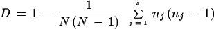 |
where N is the total number of strains in the sample population, s is the total number of types described, and nj is the number of strains belonging to the jth type. Simpson's index of diversity ranges from 0.0 to 1.0, where 1.0 indicates that a typing method is able to distinguish each member of a strain population from all other members of that population and, conversely, 0.0 indicates that all members of a strain population are of an identical type. An index of 0.50 means that if one strain was chosen at random from a strain population, then there would be a 50% probability that the next strain chosen at random was indistinguishable from the first (7, 8).
RESULTS
Molecular typing of Francisella strains by REP-PCR generated from four to eight major amplification products ranging in size from 4 to 0.35 kb. Visual comparison of banding patterns revealed four distinct REP profiles for the 41 strains tested (Fig. 1B). Most of the F. tularensis strains were REP type 1, with six visible bands and an intense band of approximately 1.25 kb in size. Only two of the isolates (strains 15 and 19), recovered from hares, were REP type 2. This profile and type 1 shared REP-PCR amplification bands of approximately 1.25, 0.75, and 0.35 kb in size, but they differed in bands of about 2.5, 1.75, and 0.6 kb. The only reference strain belonging to F. tularensis subsp. tularensis (type A) showed a unique pattern (REP type 3), in which the intense band of approximately 1.25 kb was not present. The F. novicida profile (REP type 4) shared only two bands, approximately 4 and 3.8 kb in size, with F. tularensis profiles. It also had a unique and characteristic band of approximately 1.5 kb in size, but it lacked the band of about 1.25 kb in size (Table 1).
FIG. 1.

(A) ERIC-PCR fingerprints of type 1 (lane 1), type 2 (lane 2), type 3 (lane 3), and type 4 (lane 4). Lanes M, molecular size marker X (Roche Diagnostics). (B) REP-PCR fingerprints of type 1 (lane 2), type 2 (lane 1), type 3 (lane 3), and type 4 (lane 4). Lanes M, molecular size marker X (Roche Diagnostics). (C) RAPD fingerprints, with the M13 primer, of type 1 (lane 1), type 2 (lane 2), type 3 (lane 3), type 4 (lane 4), and type 5 (lane 5). Lanes M, molecular size marker X (Roche Diagnostics). (D) RAPD fingerprints, with T3-T7 primers, of type 1 (lane 1), type 2 (lane 2), type 3 (lane 3), type 4 (lane 4), type 5 (lane 5), type 6 (lane 6), and type 7 (lane 7). Lanes M, molecular size marker X (Roche Diagnostics).
With the ERIC-PCR method, profiles of Francisella strains revealed amplified bands ranging from 3 to 0.9 kb, with various intensities (Fig. 1A). The 41 strains were grouped into four distinct patterns of fingerprints. All of the types shared a very intense band of approximately 3 kb in size. F. novicida (ERIC type 4) did not share any other band, while the American human strain (ERIC type 3) differed from the other F. tularensis strains in one unique band of approximately 1.75 kb in size. In addition, two major common bands of about 2.25 and 1.1 kb were present in all F. tularensis strains tested, and two other bands of approximately 2 and 0.9 kb and two more faint bands of between 1 and 1.6 kb were seen in ERIC types 2 and 3. Of the 25 Spanish isolates recovered from hares, 15 belonged to ERIC type 2 and 10 belonged to ERIC type 1. The distribution of the Spanish hare isolates into two different groups was also observed for the other hare strains isolated in the Czech Republic. The vole and tick isolates were also ERIC type 2. The Spanish human isolates belonged to ERIC profile 1 or 2, while the American human strain was ERIC type 3 (Table 1).
RAPD-PCR produced five distinguishable patterns for the 41 Francisella strains when the M13 primer was used (RAPD/M13). The fragments ranged from 2.2 to 0.35 kb, with various band intensities (Fig. 1C). All strains of F. tularensis (RAPD/M13 types 1 to 4) have common bands of approximately 2.2, 1.6, 1.4, 1.3, 0.9, 0.75, 0.65, 0.45, and 0.35 kb in size. However, F. novicida (RAPD/M13 type 5) shared only bands of approximately 1.4, 1.3, 0.65, and 0.45 kb but showed two unique bands of about 0.67 and 0.5 kb. Additional bands were present only in RAPD/M13 types 2 (0.7 kb), 3 (0.8 kb), and 4 (0.6 kb). RAPD/M13 type 1 included 31 F. tularensis strains and, among them, most of the strains isolated from humans and hares. RAPD/M13 type 2 included the two same hare isolates that were REP type 2 and also human isolate no. 24. RAPD/M13 type 3 included the three hare strains and the tick strain recovered in eastern Europe; RAPD/M13 type 4 was observed for two Spanish human isolates, and RAPD/M13 type 5 was observed for the F. novicida strain (Table 1).
Amplification of genomic DNAs from F. tularensis strains with the T3-T7 primers resulted in six different patterns consisting of four common fragments of approximately 1.5, 0.75, 0.70, and 0.55 kb in size (Fig. 1D). Their differences were in amplification products ranging from 0.95 to 0.55 kb. A seventh profile was observed for the F. novicida strain, which shared with F. tularensis strains only the band of about 0.70 kb but exhibited characteristic main bands of approximately 1.6 and 1.5 kb in size. Hare strains were assigned to RAPD/T3-T7 types 1 to 4 (type 1 included 57.1% of hare strains); human strains were assigned to RAPD/T3-T7 types 1 to 4 and 6 (type 1 included 44.4% of human strains); the two tick strains were assigned to RAPD/T3-T7 type 1; the F. tularensis isolate from a vole was assigned to RAPD/T3-T7 type 5; and the F. novicida strain was assigned to RAPD/T3-T7 type 7 (Table 1).
The day-to-day reproducibilities of the PCRs were examined by comparing patterns amplified on four different days. Excellent reproducibilities were obtained with REP-PCR, ERIC-PCR, and RAPD/M13 assays (Fig. 2 and 3). Although sometimes the major bands produced were less intense and the minor bands were difficult to visualize, their overall positions and whether they were present or absent were highly consistent. For the RAPD assay using the T3-T7 primers, the reproducibility was also high for six of the seven profiles obtained; the exception was type 5 (the F. tularensis strain isolated from a vole), for which one of the bands was not always present (Fig. 4).
FIG. 2.

Reproducibility studies. The day-to-day reproducibilities of the PCR assays were examined by comparing patterns provided by the same strain and method on four different days. Lanes M, molecular size marker X (Roche Diagnostics). (A) ERIC-PCR fingerprints of strains 27 (type 1) (lanes 1, 2, 3, and 4) and 3 (type 2) (lanes 5, 6, 7, and 8). (B) REP-PCR fingerprints of strains 33 (type 1) (lanes 1, 2, 3, and 4) and 19 (type 2) (lanes 5, 6, 7, and 8).
FIG. 3.

Reproducibility studies. The day-to-day reproducibilities of the RAPD/M13 assay were examined by comparing the patterns provided by the five different fingerprints (strain 6, type 1 [lane 1]; strain 19, type 2 [lane 2]; strain 33, type 3 [lane 3]; strain 30, type 4 [lane 4]; and strain 41, type 5 [lane 5]) on four different days (only three days are shown). Lanes M, molecular size marker X (Roche Diagnostics).
FIG. 4.

Reproducibility studies. The day-to-day reproducibilities of the RAPD/T3-T7 assay were examined by comparing the patterns provided by the seven different fingerprints (strain 7, type 1 [lane 1]; strain 10, type 2 [lane 2]; strain 20, type 3 [lane 3]; strain 23, type 4 [lane 4]; strain 21, type 5 [lane 5]; strain 28, type 6 [lane 6]; and strain 41, type 7 [lane 7]) on four different days (panels A to D). Lane M, molecular size marker X (Roche Diagnostics).
When the four assays compared were taken together, the 41 strains listed in Table 1 could be divided into 18 distinct groups, designated A to R. Spanish hare F. tularensis isolates belonged to 10 groups (A to J); Czech hare isolates belonged to groups M and P; Spanish human isolates belonged to groups A, D, G, J, L, O, and N; human strain SCHU belonged to group N; tick isolates belonged to groups B and Q; the vole strain belonged to group K; and the F. novicida strain belonged to group R (Table 1).
Table 2 shows Simpson's index of diversity for the different PCR typing methods used in this study. When the methods were taken separately, RAPD/T3-T7 exhibited the highest discriminating power, with diversity indices (DI) of 0.65 for both the Spanish and the total group of F. tularensis strains. In contrast, the REP-PCR method provided the lowest degree of discrimination (below 0.15). Globally, the highest DI were obtained when ERIC-PCR, RAPD/M13, and RAPD/T3-T7 were combined and when the four methods were taken together (with DI of 0.87 for Spanish strains and of 0.90 for total strains). With the combination of just two methods, ERIC and RAPD/T3-T7, the DI increased to 0.83. The DI for the total F. tularensis strains tested were quite similar to those for the Spanish strains for the different methods compared, with the exception of those obtained for RAPD/M13 and the combination of REP-PCR and RAPD/M13, which were almost twice as high for the total number of strains as for the 35 Spanish isolates.
TABLE 2.
Simpson's index of diversity for the F. tularensis strains used in this study
| Method | No. of different types | No. of strains belonging to the most numerous type | Simpson's index of diversity for:
|
|
|---|---|---|---|---|
| Total strains (n = 40) | Spanish strains (n = 35) | |||
| REP | 3 | 37 | 0.14 | 0.11 |
| ERIC | 3 | 23 | 0.52 | 0.49 |
| RAPD (M13 primer) | 4 | 31 | 0.39 | 0.21 |
| RAPD (T3-T7 primers) | 6 | 22 | 0.65 | 0.65 |
| REP-ERIC | 5 | 23 | 0.55 | 0.51 |
| REP-RAPD (M13 primer) | 6 | 30 | 0.44 | 0.26 |
| REP-RAPD (T3-T7 primers) | 8 | 21 | 0.68 | 0.69 |
| ERIC-RAPD (M13 primer) | 8 | 20 | 0.70 | 0.60 |
| ERIC-RAPD (T3-T7 primers) | 10 | 11 | 0.83 | 0.83 |
| RAPD (M13 primer)-RAPD (T3-T7 primers) | 8 | 16 | 0.81 | 0.75 |
| REP-ERIC-RAPD (M13 primer) | 8 | 20 | 0.70 | 0.60 |
| REP-ERIC-RAPD (T3-T7 primers) | 11 | 11 | 0.84 | 0.85 |
| REP-RAPD (M13 primer)-RAPD (T3-T7 primers) | 9 | 16 | 0.81 | 0.75 |
| ERIC-RAPD (M13 primer)-RAPD (T3-T7 primers) | 17 | 10 | 0.90 | 0.87 |
| REP-ERIC-RAPD (M13 primer)-RAPD (T3-T7 primers) | 17 | 10 | 0.90 | 0.87 |
DISCUSSION
To our knowledge, molecular epidemiological typing for F. tularensis has never been reported before. During the last decade, approaches at the molecular level, such as pulsed-field gel electrophoresis and ribotyping, have been used to assess the relatedness of organisms other than F. tularensis. Despite the broad applicability of these techniques, their use in clinical microbiology laboratories has been limited because they are both time-consuming and labor-intensive. However, the PCR techniques described here have recently been considered easy to perform and to interpret, with high discrimination power, a time of only 1 day to result, moderate cost of equipment, low cost per test, and good (rep) or moderate (RAPD) reproducibility (13).
In addition, molecular typing methods are of special concern when working with pathogenic bacteria, because direct manipulation with live organisms (which takes place when traditional biochemical typing techniques are used) is avoided. On the other hand, although biochemical tests can be used to discriminate among subspecies of F. tularensis strains, such tests have obvious drawbacks for unambiguous classification (14).
In this study, rep-PCR and RAPD assays were used as tools to characterize F. tularensis strains isolated from hares, humans, ticks, and a vole. Both rep-PCR methods, REP- and ERIC-PCR, as well as RAPD assay with the two sets of primers used, showed great possibilities for the identification of F. tularensis isolates. Thus, the banding patterns of F. novicida, an organism closely related to F. tularensis, were completely different from those produced by the four PCR assays compared for the 40 F. tularensis isolates tested in this study. This is of special interest, since previous studies have been unable to clearly discriminate between F. tularensis and F. novicida. Genetic probes have failed so far in this aim (4). Similarly, rep-PCR and RAPD methods were efficient and sensitive molecular typing tools for F. tularensis strains. The lower degree of reproducibility of RAPD compared with rep-PCR has been previously reported for other organisms (9), and it is confirmed in this study for F. tularensis when the T3-T7 primers are used, although the result was only ambiguous for one of the seven profiles obtained. On the other hand, the reproducibilities of both PCR methods were comparable when the M13 primer was used in the RAPD assay.
However, RAPD with the T3-T7 primers provided a higher degree of discrimination by visual analysis within strains than ERIC-PCR and especially than REP-PCR: a DI of 0.65 for RAPD/T3-T7 versus DI of 0.49 and 0.52 for ERIC-PCR and of 0.11 and 0.14 for REP-PCR for Spanish and total strains, respectively, were obtained (Table 2). Some of these values are low, but the combination of some of the PCR methods allow us to achieve DI of 0.8 to 0.9. These results can be considered excellent, especially if both the small area from which the Spanish isolates were recovered and previous information about typing tools for Francisella strains are taken into account (the DI observed for serologic methods is 0, as only one antigenic group has been described so far [3, 12], and DNA methods have been unable to discriminate between F. novicida and F. tularensis [5]). On the other hand, the results in this study were clearly different from those reported by Vila et al. (16), who found that the RAPD assay was less discriminating than rep-PCR methods for typing of the Acinetobacter calcoaceticus-A. baumannii complex.
The existence among the 17 global PCR types of four F. tularensis groups (A, D, G, and J) containing human and hare isolates clearly indicates a common origin for them and confirms that hare F. tularensis strains are infectious for humans. In this respect, it must be pointed out that most of the human infections occurred during the hunting season (hunting is a usual practice among people in Spain). However, it should be noted that the most common profile (group B, containing 25% of the tested strains) was isolated only from hares and a tick, and never from humans. Although the number of isolates in this study is not enough to make epidemiological conclusions, this information should be considered carefully in the future, since it seems to suggest the existence of an F. tularensis group that has a low prevalence in humans but is present in hares and ticks.
Strain no. 39 was recovered from a tick located during the necropsy in the skin of the hare from which strain no. 38 was isolated. The two strains showed identical profiles in the four PCR tests and were included in group B. This finding clearly indicates the existence of tick-borne tularemia in hares. On the other hand, the isolate recovered from a vole was the only strain assigned to group K, and this seems to suggest a completely different origin for this isolate. In any case, as only one strain could be isolated in the outbreak that occurred in Spain, this observation should be validated by the examination of a larger set of vole isolates.
On the other hand, the hare strains were divided into twelve groups (A to J, M, and P) (Table 1). This fact clearly indicates the existence of genetic diversity among F. tularensis strains isolated from this animal. This diversity degree can be considered greater if it is taken into account that 25 of the hare isolates used in this study, recovered from Spain during 1998 in a geographical area of about 11,000 km2, were assigned to 10 different genetic groups (A to J). In the same way, the eight human isolates recovered in Spain were classified into six groups (A, D, G, J, L, and O), while the American human strain showed completely different fingerprints and was assigned to group N. This fact can be easily explained because strain SCHU was the only reference strain used in this study belonging to F. tularensis subsp. tularensis (type A).
Although a high number of groups was obtained, the differences were not more than two or three bands by every PCR method, and, what is more important, many common bands were seen for all of the F. tularensis strains tested. Even without a dendrogram (which was not the purpose of this work), visual inspection of Fig. 1 outlines the possibility of a high clonality among the isolates compared in this study, with several shared bands that would allow, if needed, identification of the genus Francisella or species F. tularensis. Therefore, we can conclude that there is a limited genetic diversity among strains, which is enough for distinguishing subspecies and even different types within subspecies by PCR methods. Our results need further validation by the study of larger amounts of strains isolated in other geographical regions or for longer periods of time. However, because of the high risk assumed when transporting F. tularensis strains and because of the implications of this organism as a biological war agent, we were not able to obtain more isolates from other countries.
In conclusion, all these PCR-based approaches represent useful tools for the epidemiological typing of F. tularensis because of their simplicity and speed. RAPD with T3-T7 primers has the advantage of being more discriminative than rep-PCR. In addition, the results of this study add further evidence for the idea that rep-PCR may be broadly applicable for fingerprinting bacteria which possess repetitive elements such as REP or ERIC sequences.
ACKNOWLEDGMENTS
We thank Laboratorio Central de Sanidad Animal, Ministerio de Agricultura, Pesca y Alimentación, Algete, Madrid, Spain; Servicio de Sanidad Animal and Laboratorio de Sanidad Animal de León, Consejería de Agricultura y Ganadería, Junta de Castilla y León, León, Spain; Laboratorio de Microbiología, Hospital Virgen de la Concha, Insalud, Zamora, Spain; and Departamento de Microbiología Médica, Hospital Princesa Sofía, Insalud, León, Spain; for providing us the F. tularensis isolates.
This work was supported by grant LE 04/00B from the Junta de Castilla y León, Consejería de Educación y Cultura, Spain.
REFERENCES
- 1.Bachiller Luque P, Pérez Castrillón J L, Martín Luquero M, Mena Martín F J, de la Lama López-Areal J, Pérez Pascual P, Mazón M A, Herreros Guilarte V. Preliminary report of an epidemic tularemia outbreak in Valladolid. Rev Clin Esp. 1998;198:789–793. [PubMed] [Google Scholar]
- 2.Bell J F, Owen C R, Larson C C. Virulence of Bacterium tularense: a study of Bacterium tularense in mice, guinea pigs, and rabbits. J Infect Dis. 1955;97:162. doi: 10.1093/infdis/97.2.162. [DOI] [PubMed] [Google Scholar]
- 3.Forsman M, Sandström G, Jaurin B. Identification of Francisella species and discrimination of type A and type B strains of F. tularensis by 16S rRNA analysis. Appl Environ Microbiol. 1990;56:949–955. doi: 10.1128/aem.56.4.949-955.1990. [DOI] [PMC free article] [PubMed] [Google Scholar]
- 4.Forsman M, Sandström G, Sjöstedt A. Analysis of 16S ribosomal DNA sequences of Francisella strains and utilization for determination of the phylogeny of the genus and for identification of strains by PCR. J Syst Bacteriol. 1994;44:38–46. doi: 10.1099/00207713-44-1-38. [DOI] [PubMed] [Google Scholar]
- 5.García Peña F J, Suárez Mayoral P, Cogolludo Cogolludo C, Arriola Garrote C, Anadón Navarro E. An outbreak of tularemia in Castilla-León. First isolation of Francisella tularensis in Spain. Med Vet. 1998;15:418–423. [Google Scholar]
- 6.Gurycova D. First isolation of Francisella tularensis subsp. tularensis in Europe. Eur J Epidemiol. 1998;14:797–802. doi: 10.1023/a:1007537405242. [DOI] [PubMed] [Google Scholar]
- 7.Hunter P R. Reproductibility and indices of discriminatory power of microbial typing methods. J Clin Microbiol. 1990;28:1903–1905. doi: 10.1128/jcm.28.9.1903-1905.1990. [DOI] [PMC free article] [PubMed] [Google Scholar]
- 8.Hunter P R, Gaston M A. Numerical index of the discriminatory ability of typing systems: an application of Simpson's index of diversity. J Clin Microbiol. 1988;26:2465–2466. doi: 10.1128/jcm.26.11.2465-2466.1988. [DOI] [PMC free article] [PubMed] [Google Scholar]
- 9.Liu P Y F, Shi Z Y, Lau Y J, Hu B S, Shyr J M, Tsai W S, Lin Y H, Tseng C Y. Comparison of different PCR approaches for characterization of Burkholderia (Pseudomonas) cepacia isolates. J Clin Microbiol. 1995;33:3304–3307. doi: 10.1128/jcm.33.12.3304-3307.1995. [DOI] [PMC free article] [PubMed] [Google Scholar]
- 10.Marchette N J, Nicholes P S. Virulence and citrulline ureidase activity of Pasteurella tularensis. J Bacteriol. 1961;82:26–32. doi: 10.1128/jb.82.1.26-32.1961. [DOI] [PMC free article] [PubMed] [Google Scholar]
- 11.Montejo M, Pérez-Irezábal J, González de Zárate P, Aguirregengoa K, Vicente J M, Martínez E, Ibarra S, Bereciartúa E, Castell C. Tularemia: report of 16 cases in the Castilla-Leon community. Rev Clin Esp. 1998;198:794–798. [PubMed] [Google Scholar]
- 12.Nutter J E. Antigens of Pasteurella tularensis: preparative procedures. Appl Microbiol. 1971;22:44–48. doi: 10.1128/am.22.1.44-48.1971. [DOI] [PMC free article] [PubMed] [Google Scholar]
- 13.Olive D M, Bean P. Principles and applications of methods for DNA-based typing of microbial organisms. J Clin Microbiol. 1999;37:1661–1669. doi: 10.1128/jcm.37.6.1661-1669.1999. [DOI] [PMC free article] [PubMed] [Google Scholar]
- 14.Sandström G, Sjöstedt A, Forsman M, Pavlovich N V, Mishankin B N. Characterization and classification of strains of Francisella tularensis isolated in the central Asian focus of the Soviet Union and in Japan. J Clin Microbiol. 1992;30:172–175. doi: 10.1128/jcm.30.1.172-175.1992. [DOI] [PMC free article] [PubMed] [Google Scholar]
- 15.Uhari M, Syrjala H, Salminen A. Tularemia in children caused by Francisella tularensis biovar palaearctica. Pediatr Infect Dis J. 1990;9:80–83. doi: 10.1097/00006454-199002000-00003. [DOI] [PubMed] [Google Scholar]
- 16.Vila J, Marcos M A, Jiménez de Anta M T. A comparative study of different PCR-based DNA fingerprinting techniques for typing of the Acinetobacter calcoaceticus-A. baumannii complex. J Med Microbiol. 1996;44:482–489. doi: 10.1099/00222615-44-6-482. [DOI] [PubMed] [Google Scholar]