Abstract
Mutations in HFE cause hereditary hemochromatosis type I hallmarked by increased iron absorption, iron accumulation in hepatocytes and iron deficiency in myeloid cells. HFE encodes an MHC-I like molecule, but its function in immune responses to infection remains incompletely understood. Here, we investigated putative roles of Hfe in myeloid cells and hepatocytes, separately, upon infection with Salmonella Typhimurium, an intracellular bacterium with iron-dependent virulence. We found that constitutive and macrophage-specific deletion of Hfe protected infected mice. The propagation of Salmonella in macrophages was reduced due to limited intramacrophage iron availability for bacterial growth and increased expression of the anti-microbial enzyme nitric oxide synthase-2. By contrast, mice with hepatocyte-specific deletion of Hfe succumbed earlier to Salmonella infection because of unrestricted extracellular bacterial replication associated with high iron availability in the serum and impaired expression of essential host defense molecules such as interleukin- 6, interferon-g and nitric oxide synthase-2. Wild-type mice subjected to dietary iron overload phenocopied hepatocyte-specific Hfe deficiency suggesting that increased iron availability in the serum is deleterious in Salmonella infection and underlies impaired host immune responses. Moreover, the macrophage-specific effect is dominant over hepatocytespecific Hfe-depletion, as Hfe knockout mice have increased survival despite the higher parenchymal iron load associated with systemic loss of Hfe. We conclude that cell-specific expression of Hfe in hepatocytes and macrophages differentially affects the course of infections with specific pathogens by determining bacterial iron access and the efficacy of antimicrobial immune effector pathways. This may explain the high frequency and evolutionary conservation of human HFE mutations.
Introduction
Most patients with hereditary hemochromatosis (HH) show homozygous C282Y missense mutations in the gene HFE.1,2 They are hallmarked by parenchymal iron deposition particularly in hepatocytes, cardiomyocytes and pancreatic acinar cells, leading to organ damage. Conversely, monocytes and macrophages are iron-deficient in type I HH.3-5
An allelic frequency of approximately 5-10% renders the HFE C282Y missense mutation the most common genetic defect in individuals of Northwestern European ancestry. It has been hypothesized that the mutation may protect from iron-deficiency and/or infections; thus conferring an evolutionary advantage to healthy heterozygous carriers.6,7 Several mechanisms by which the HFE protein controls systemic iron balance have been proposed: early studies have shown that HFE, in association with b2-microglobulin, directly interacts with transferrin receptor-1 (TFR1) on the cell surface8 and lowers its affinity for transferrin-bound iron (TBI). Once the iron regulatory hormone hepcidin had been discovered, it became apparent that the HFE C282Y mutation causes systemic hepcidin deficiency and its consequences.9 The HFE mutation disrupts iron-inducible BMP/SMAD (for bone morphogenetic protein/suppressor of mothers against decapentaplegic) signaling and prevents appropriate hepcidin transcription.1,2 The relative lack of hepcidin then causes unrestricted dietary iron absorption by the duodenum and increased iron export from iron-recycling macrophages due to the stabilization of the iron exporter ferroportin (FPN)-1.10,11 As a result, iron accumulates in parenchymal cells where it causes tissue damage by toxic radical formation.12
HFE is an MHC-I like protein, but so far it has remained unclear whether it plays a role in immune function and/or host-pathogen interaction. HFE-deficient monocytes and macrophages are iron poor.3,4,13 Possible explanations include reduced TBI uptake, increased iron export via FPN1 as a consequence of decreased hepcidin levels or increased synthesis of the siderophore-iron binding peptide lipocalin (LCN)-2.3,5,13
For almost all bacteria, iron is essential as it stimulates growth and thus impacts on the course and outcome of many infectious diseases.14 However, iron requirements, iron uptake strategies and proliferation kinetics may greatly vary between bacterial species, possibly explaining species-specific effects on infection outcomes.15 In mice, constitutive Hfe deficiency partially protects from S. enterica Typhimurium (S. Tm.) infection.13 By contrast, Hfe deficient mice are more susceptible to Mycobacterium avium infection.16 Furthermore, human monocyte-derived macrophages from patients with HH limit iron availability for intracellular Mycobacterium tuberculosis, resulting in an improved control of infection.17 On the other hand, individuals with HH type I are highly susceptible to infection with Yersinia species, whose virulence is iron-dependent, as documented by case reports of human subjects and by mouse models.18,19 These diverse outcomes are counterintuitive given that all three pathogens, Salmonella, Mycobacterium and Yersinia, share a predominately intracellular lifestyle pointing to the importance of cell- and tissue- specific iron distribution for susceptibility to these infections.20
Because Hfe exerts contrasting effects in different infectious diseases, we asked whether Hfe plays a cell typespecific role during infection and whether this is linked to alterations of tissue iron distribution or associated with iron-independent effects of Hfe. We herein demonstrate that macrophage-specific deletion of Hfe (LysMCre+ Hfefl/fl) recapitulates the antibacterial phenotype of constitutive Hfe-/- mice in response to S. Tm. infection. By contrast, exclusive deletion of Hfe in hepatocytes (AlfpCre+ Hfefl/fl) is associated with an adverse outcome of S. Tm. infection. These contrasting cell type-specific effects of Hfe-deficiency correlate with bacterial iron availability and anti-microbial effector immune functions. Our findings support the idea that Hfe controls iron concentrations in the microenvironment thus differentially affecting immune effector mechanisms and bacterial growth in intra- and extracellular compartments.
Methods
Salmonella infection in vivo
All infection experiments were performed according to the guidelines of the Medical University of Innsbruck and the Austrian Ministry for Science and Education based on the Austrian Animal Testing Act of 1988 (approvals BMWFW-66.011/0074-C/GT/2007, 66.011/0154- II/3b/2010 and 66.011/0031-WF/V/3b/2015). Male mice were used at 12-16 weeks of age and infected by intraperitoneal (i.p.) injection with 500 colony forming units (CFU) of S. Tm. diluted in 200 mL of phosphate buffered saline (PBS). Unless otherwise specified, S. Tm. Wild-type (WT), strain ATCC 14028s was used for the experiments. Where appropriate, mice were fed an iron adequate control diet (C1000 from Altromin containing 180 mg per g) or an ironenriched diet (C1038 from Altromin supplemented with 25 mg carbonyl iron per g). After 3 weeks, mice were infected by i.p. injection with 500 CFU of S. Tm. diluted in 200 mL of PBS as detailed in the Online Supplementary Methods.
In vitro experiments
The isolation of bone marrow-derived macrophages (BMDM) was performed as detailed in the Online Supplementary Methods.
RNA extraction and quantitative real-time polymerase chain reaction
Preparation of total RNA, reverse transcription and quantification of mRNA expression by quantitative Taqman real-time polymerase chain reaction (qRT-PCR) was performed as described.21 Results were first normalized using the housekeeping gene Hprt and then divided by the means of the control group (WT Hfe+/+ or Cre– mice as appropriate) to obtain expression data that is relative to the respective control group. Sequences of primers and probes are listed in the Online Supplementary Methods.
Measurement of iron and protein concentrations
Measurement of tissue iron concentrations has been described in detail.22 The serum iron concentration was quantified using the QuantiChrom Iron Assay Kit (BioAssay Systems). Intracellular iron concentrations were determined in adherent bone marrow macrophages by atomic absorption spectrometry as described.23 The quantification of protein levels in sera and tissues is detailed in the Online Supplementary Methods.
Statistical analysis
Statistical analysis was carried out using a GraphPad Prism statistical package and Microsoft Excel. We determined significance by unpaired two-tailed Student’s t-test or Mann-Whitney test to assess data, where only two groups existed. For the comparison of organ bacterial loads and mRNA expression, data were log-transformed prior to Student’s t-test. ANOVA with Bonferroni correction was used when more than two groups existed. Survival was compared by log-rank test. Generally, P-values less than 0.05 were considered significant.
Results
Hepatocyte-specific Hfe deletion stimulates extracellular growth of Salmonella Typhimurium
We previously reported that mice lacking Hfe in all cell types (Hfe-/- mice) were partially protected from S. Tm. Infection.13,20
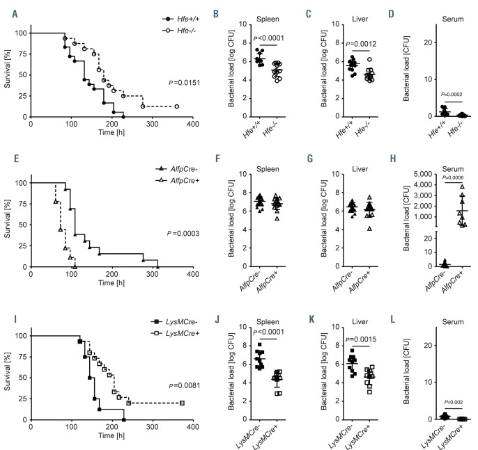
Consistently, we could recapitulate this finding in a different strain of Hfe-/- mice in which exons 3-524 rather than exons 2-313 of Hfe were deleted. We found that also these Hfe-/- mice survived significantly longer (Figure 1A) and carried reduced numbers of bacteria in spleen, liver and serum in response to S. Tm infection when compared to Hfe+/+ littermates (Figure 1B to D). In order to delineate in which cell type the absence of Hfe confers protection from infection, we next analyzed mice with selective Hfe-deficiency in hepatocytes (referred to as AlfpCre+ Hfefl/fl). Previous analyses of the AlfpCre+ Hfefl/fl line showed an iron phenotype comparable to Hfe-/- mice,25 with elevated iron levels in serum and liver and iron deficiency in the spleen.
AlfpCre+ Hfefl/fl and control mice (AlfpCre- Hfefl/fl) were infected with S. Tm. and survival time was monitored for 14 days (336 hours). Unexpectedly and in contrast to the previous findings in Hfe-/- mice, we observed significantly shortened survival in the AlfpCre+ Hfe-/- mice (Figure 1E). Bacterial burden in spleen and liver was not substantially altered, when compared to control mice (Figure 1F and G). By contrast, the number of bacteria circulating in the serum was significantly higher in AlfpCre+ Hfefl/fl mice (Figure 1H). This finding suggested that Hfe-deficiency in hepatocytes does not confer protection against S. Tm. infection related death but even aggravates the infection phenotype.
Macrophage-specific Hfe-deletion phenocopies the protective effect of constitutive Hfe deletion in mice infected with Salmonella Typhimurium
We next tested the response to S. Tm. infection in mice lacking Hfe in myeloid cells (referred to as LysMCre+ Hfefl/fl)25 in comparison to control mice (LysMCre– Hfefl/fl). Interestingly, macrophage-specific Hfe depletion fully recapitulated the protective effect observed in Hfe-/- mice, including prolonged survival (Figure 1I) and reduced bacterial load in spleen, liver and serum (Figure 1J to L). Importantly, the alleles required for tissue-specific recombination to generate the cell type-specific Hfe-depletion models, LysMCre (macrophage-specific Cre-recombinase expression) and AlfpCre (hepatocyte-specific Cre-recombinase expression) alone had no effect on survival and bacterial burden in the spleen, liver and serum (Online Supplementary Figure S1A to D and 26), excluding non-specific effects of the Cre-recombinases. We conclude that the lack of Hfe in myeloid cells is sufficient to protect mice from S. Tm. infection related consequences. This finding demonstrates an important extra-hepatic function of Hfe in vivo.
Salmonella-infection of LysMCre+ Hfefl/fl mice causes iron depletion in macrophages
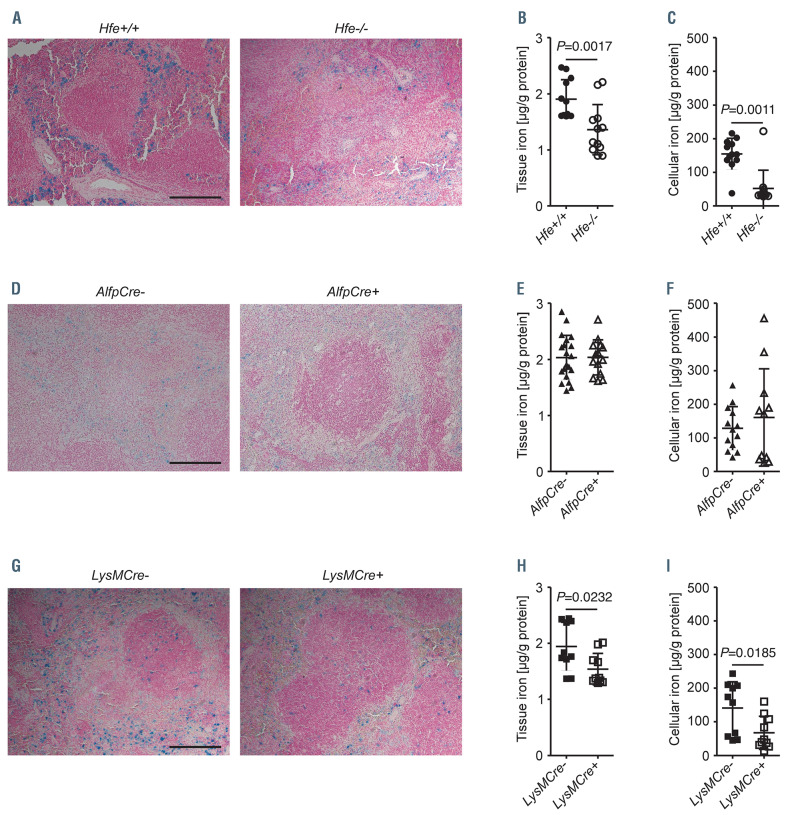
In order to understand the mechanism underlying divergent disease outcomes of S. Tm. infection in AlfpCre+ Hfefl/fl and LysMCre+ Hfefl/fl mice, we analyzed iron-related parameters. Iron localization was detected in tissue sections of Salmonella-infected mice by Prussian blue staining and tissue iron levels were quantified by colorimetric measurement. S. Tm.-infected Hfe-/- (Figure 2A and B) and LysMCre+ Hfefl/fl mice (Figure 2G and H) showed reduced iron levels in the spleen consistent with the protective phenotype observed in these mouse strains. This was not apparent in infected AlfpCre+ Hfefl/fl mice (Figure 2D and E). By contrast, infected Hfe-/- (Online Supplementary Figure S2A and B) and AlfpCre+ Hfefl/fl mice (Online Supplementary Figure S2C and D) showed hepatocellular iron accumulation, while liver iron levels were normal in infected LysMCre+ Hfefl/fl mice (Online Supplementary Figure S2E and F). Importantly, the reduction of splenic iron levels in infected Hfe-/- and LysMCre+ Hfefl/fl mice correlated with diminished intracellular iron levels in bone marrow macrophages (Figure 2C and I), while AlfpCre+ Hfefl/fl bone marrow macrophages had a normal iron content (Figure 2F). This finding suggests that upon Salmonella infection, macrophages lacking Hfe show reduced iron levels.
High serum iron in AlfpCre+ Hfefl/fl mice allows for increased proliferation of Salmonella
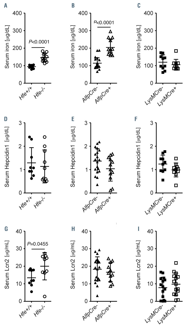
Hfe-/- and AlfpCre+ Hfefl/fl mice infected with S. Tm. WT for 72 hours showed elevated serum iron levels compared to Hfe+/+ or AlfpCre– Hfefl/fl mice, respectively (Figure 3A and B). In contrast, serum iron levels in infected LysMCre+ Hfefl/fl mice were comparable to infected LysMCre– Hfefl/fl mice (Figure 3C). Notably, in the setting of Salmonella infection, serum levels of hepcidin-1 were not different between the mouse strains (Figure 3D to F). Moreover, Salmonella-infected Hfe-/- mice presented with increased serum concentrations of the siderophore-capturing peptide Lcn2 (Figure 3G) while hepatocyte-specific (Figure 3H) or macrophage-specific (Figure 3I) Hfe deletion did not affect serum Lcn2 levels. Thus, the presence of Hfe in hepatocytes is necessary and sufficient to limit serum iron levels both in steady state25 and in response to S. Tm. infection. Moreover, hepcidin-1 induction in response to Salmonella infection is appropriate in mice lacking Hfe. In contrast, the enhanced production of Lcn2 is only observed in the complete absence of Hfe13 suggesting that different cell-types mediate iron- and immune-regulatory effects of Hfe.
Salmonella iron acquisition pathways differently affect extracellular proliferation
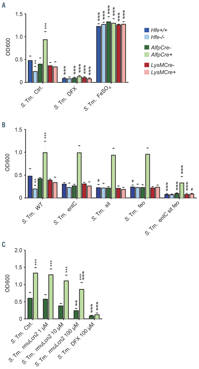
S. Tm. is a bacterial pathogen with dual lifestyle. First, S. Tm. is able to persist and replicate extracellularly, e.g., on contaminated food and surfaces, in the gut lumen and in the serum. Early after the invasion of a murine host, S. Tm. preferentially infects macrophages to propagate intracellularly. We therefore investigated how serum iron availability in Hfe-/-, AlfpCre+ Hfefl/fl and LysMCre+ Hfefl/fl mice may affect bacterial proliferation. We spiked RPMI medium with 10% of serum from uninfected mice of all three strains and inoculated spiked samples with bacteria. We used S. Tm. WT and isogenic mutants lacking either single or all three major bacterial iron uptake systems (enterobactin, feo and sitABCD).27 In addition, we included serum-spiked RPMI treated with 100 mM desferasirox (DFX) to deplete the medium of chelatable iron. Alternatively, we added 100 mM FeSO4 to saturate any iron-binding factors (e.g. transferrin, lactoferrin and Lcn2). S. Tm. WT growth was only inhibited by serum from Hfe-/- mice, possibly due to the presence of high Lcn2.13 In serum from AlfpCre+ Hfefl/fl mice, bacterial growth was strongly enhanced, while it was not affected in serum from LysMCre+ Hfefl/fl (Figure 4A). In addition, growth of S. Tm. WT was strongly restricted by the presence of the iron chelator DFX and enhanced by the addition of FeSO4, independent of the Hfe status of the mice the sera were derived from (Figure 4A). In liquid cultures of iron uptake mutant S. Tm. strains, growth was most pronouncedly inhibited in the case of the triple mutant (entC, feo and sitABCD deletion). Importantly, we saw that the iron-rich serum of AlfpCre+ Hfefl/fl mice facilitated extracellular growth of S. Tm., an effect that was reduced by the lack of all three iron uptake systems (Figure 4B) or abolished by iron chelation (Figure 4A). Notably, the addition of recombinant murine Lcn2 reduced the growth of S. Tm. in a dose-dependent fashion, even though it did not abolish the differences between AlfpCre– Hfefl/fl and AlfpCre+ Hfefl/fl mice (Figure 4C). This suggests that in the presence of high Lcn2 concentrations, iron uptake pathways of Salmonella not targeted by Lcn2, such as the feo and sitABCD systems, are able to compensate. Apparently, feo and sitABCD can maintain a sufficient supply of iron for bacteria when enterobactin incorporation is blocked by Lcn2. Thus, the proliferation advantage of S. Tm. in extracellular compartments of AlfpCre+ Hfefl/fl mice is a specific effect of increased iron availability even though it is not linked to a specific bacterial iron uptake pathway.
Figure 1.
Cell-type specific effect of Hfe deletion on the course of systemic Salmonella infection. Survival (A, E and I) and bacterial load in spleen (B, F and J), liver (C, G and K) and serum (D, H and L) of Hfe-/- (A-D) mice and mice lacking Hfe in hepatocytes (AlfpCre+ Hfefl/fl in E-H) or macrophages (LysMCre+ Hfefl/fl in I-L), respectively, compared to matched controls. Mice were infected with 500 colony forming units (CFU) of S. enterica serovar Typhimurim by intraperitoneal injection and monitored for 14 days (336 hours ). Data represent two independent experiments. Statistics: survival data between control and mutant mice were compared using the Log-rank (Mantel-Cox) Test. n=18 for Hfe+/+, n=16 for Hfe-/-, n=13 for AlfpCre- Hfefl/fl, n=9 for AlfpCre+ Hfefl/fl, n=16 for LysMCre- Hfefl/fl, n=15 for LysMCre+ Hfefl/fl. Log CFU data of tissue bacterial load of randomly selected mice euthanized after 72 hours of Salmonella infection were compared using student t-test. CFU data of serum bacterial load were compared by Mann-Whitney test. n=12 for Hfe+/+, n=12 for Hfe-/-, n=20 for AlfpCre- Hfefl/fl, n=14 for AlfpCre+ Hfefl/fl, n=10 for LysMCre- Hfefl/fl, n=10 for LysMCre+ Hfefl/fl.
Hfe does not affect the phagocytic activity of macrophages
Lower bacterial numbers in Hfe-/- and LysMCre+ Hfefl/fl macrophages could theoretically be explained by altered phagocytosis. Therefore, we next compared the phagocytic capacity of bone marrow-derived macrophages isolated from WT and Hfe-/- mice. However, differences were not detected, suggesting that Hfe in macrophages does not affect the phagocytic capacity of macrophages (Online Supplementary Figure S3).
Cell type-specific Hfe deletions differentially affect iron homeostasis and anti-microbial immune gene and protein expression in spleen and liver
So far, our results indicate that lack of Hfe in macrophages is sufficient to suppress intracellular growth of S. Tm., while hepatocyte-specific Hfe depletion supports iron-dependent extracellular growth of Salmonella. However, the finding that Hfe-/- and AlfpCre+ Hfefl/fl mice show comparable serum iron levels while resulting in contrasting infection outcomes suggested dominant effects of Hfe in myeloid cells. In order to identify the responsible mechanisms, we monitored gene response patterns of iron and immune genes in spleens and livers of S. Tm.-infected mice.
Figure 2.
Reduced iron content in spleen and bone marrow macrophages in the absence of Hfe. Spleen sections of Hfe-/- mice (A), AlfpCre+ Hfefl/fl mice (D) and LysMCre+ Hfefl/fl mice (G) infected for 72 hours with Salmonella were stained by Prussian blue to analyze iron distribution. Scale bars: 200 mM. Iron content in infected spleen (B, E and H) and bone marrow macrophages (C, F and I) was measured and normalized for protein content. Data were compared by Mann-Whitney test. n=12 for Hfe+/+, n=12 for Hfe-/-, n=13-20 for AlfpCre– Hfefl/fl, n=11-14 for AlfpCre+ Hfefl/fl, n=10 for LysMCre– Hfefl/fl, n=10 for LysMCre+ Hfefl/fl.
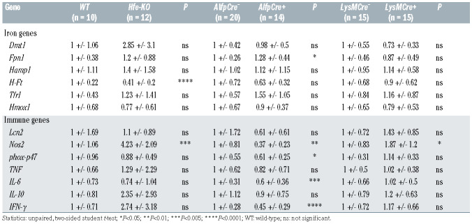
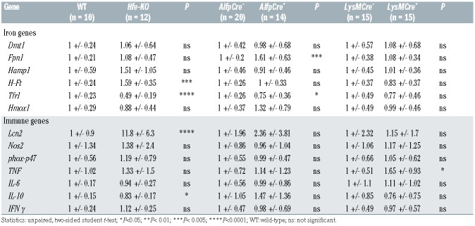
As expected, mRNA expression of ferritin heavy chain (H-Ft) was significantly decreased in the spleen (hallmarked by iron deficiency) and increased in the liver (hallmarked by iron overload) of infected Hfe-/- mice. However, H-Ft remained unchanged in the other infected Hfe-models (Tables 1 and 2). Likewise, in livers of Hfe-/- and AlfpCre+ Hfefl/fl mice, we found significantly reduced expression of Tfr1 (2x and 1.3x in Hfe-/- and AlfpCre+ Hfefl/fl, respectively), consistent with hepatic iron overload (Table 2).
We next studied the expression of central immune genes involved in the control of infection with intramacrophage bacteria. Importantly, mRNA expression of Il-6 and Ifn-g was decreased in the spleen in AlfpCre+ Hfefl/fl, but neither in Hfe-/- nor LysMCre+ Hfefl/fl mice (Table 1). Il-10 was decreased in livers of Hfe-/- mice and Tnf was increased in livers of LysMCre+ Hfefl/fl mice (Table 2).
By contrast, Nos2 (for nitric oxide synthase-2, AKA inducible Nos) expression was increased in mice lacking Hfe either globally or in macrophages, specifically, and decreased in AlfpCre+ Hfefl/fl mice (Table 1). Therefore, reduced macrophage iron levels selectively promote the expression of splenic Nos2, whereas high serum iron has a broader inhibitory effect on antimicrobial host responses in the spleen.
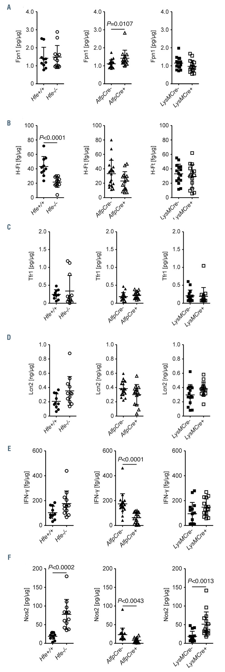
Importantly, the protein levels of iron and immune genes mirrored the mRNA expression levels in both spleen (Figure 5) and liver (Online Supplementary Figure S4). For instance, H-Ft protein expression was lower in the spleen of Hfe-/- mice compared to Hfe+/+ littermates (Figure 5B). Furthermore, splenic Nos2 protein levels were higher in Hfe-/- and LysMCre+ Hfefl/fl mice and lower in AlfpCre+ Hfefl/fl mice as compared to their respective counterparts expressing Hfe (Figure 5F). In addition, H-Ft (Online Supplementary Figure S4B) was higher and Tfr1 protein (Online Supplementary Figure S4C) was lower in both Hfe-/- mice and AlfpCre+ Hfefl/fl mice in comparison to the corresponding controls.
Table 1.
Gene expression in spleen of S. Tm. injected mice, 72 h post infection. Gene expression was normalized to expression of Hprt and is relative to the respective control group (means +/- SD).
Table 2.
Gene expression in livers of S. Tm. injected mice, 72 h post infection. Gene expression was normalized to expression of Hprt and is relative to the respective control group (means +/- SD).
Hepatocyte-specific deletion of Hfe impairs cytokine formation
Finally, we aimed at better understanding why AlfpCre+ Hfefl/fl mice succumb to early death. Based on the result of organ-specific immune gene analyses (Tables 1 and 2), we assessed the levels of these circulating mediators of immunity as well as markers of liver synthesis and damage: C3 is a complement factor produced by hepatocytes and essential for the activation of the membrane attack complex which then destroys bacterial cell walls.
Figure 3.
Serum iron and hepcidin-1 levels are differentially affected by Hfe. Serum iron (A to C), hepcidin-1 (D and F) and Lcn2 (G to I) concentrations of the mice infected for 72 hours were compared by Mann-Whitney test. n=9-12 for Hfe+/+, n=9-12 for Hfe-/-, 20 for AlfpCre– Hfefl/fl, n=11-13 for AlfpCre+ Hfefl/fl, n=10-15 for LysMCre– Hfefl/fl, n=10-15 for LysMCre+ Hfefl/fl.
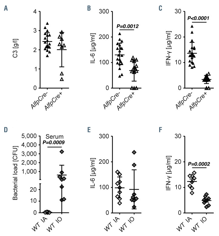
However, C3 levels were not affected by high serum or parenchymal iron (Figure 6A). IL-6, the key cytokine for the initiation of the acute-phase response, was reduced in the serum (Figure 6B) but not in the liver (Online Supplementary Figure 4E) of AlfpCre+ Hfefl/fl mice (Figure 6B). This and the unaltered production of hepcidin-1 (Figure 3E and Table 2) suggest that the acute-phase response is intact in AlfpCre+ Hfefl/fl mice. Moreover, glutamate-pyruvate transaminase (GPT) was reduced in AlfpCre+ Hfefl/fl mice, ruling out increased iron-induced hepatic injury (Online Supplementary Figure S4A). Rather, a specific defect in the pro-inflammatory cytokine output was associated with the poor outcome of Salmonella-infected AlfpCre+ Hfefl/fl mice. Specifically, serum IFN-g concentrations (Figure 6C) were significantly lower in AlfpCre+ Hfefl/fl mice and may have contributed to the insufficient induction of cellular effector mechanisms such as Nos2 in the spleen (Table 1).
Increased serum iron due to dietary iron overload enhances extracellular Salmonella growth and reduces IFN-g levels
In order to further investigate whether the reduced cytokine production in AlfpCre+ Hfefl/fl mice and may have contributed to the insufficient induc mice is linked to increased cellular iron levels, we next maintained WT mice on an iron adequate (IA) or high iron diet for three weeks to induce iron overload (IO) prior to Salmonella infection. We observed an increased bacterial load in the serum (Figure 6D), spleen and liver (Online Supplementary Figure S5B and C) along with unaltered IL-6 (Figure 6E) but reduced IFN-g concentrations in the serum (Figure 6F). This finding suggests that reduced levels of IFN-g, the central cytokine orchestrator of immune responses against intracellular bacteria,28 are a direct consequence of increased serum iron.
Figure 4.
Bacterial proliferation is affected by Hfe. RPMI was spiked with 10% sera of naïve mice of the indicated genotypes. Spiked RPMI was inoculated with wild-type (WT) S. enterica Typhimurium (S. Tm.) and its isogenic derivatives mutated in one of three or all three iron uptake systems (entC, sitABCD, feo). Where applicable, deferasirox (DFX), ferrous sulfate (FeSO4) and recombinant murine Lcn2 (rmuLcn2) was added. Liquid cultures were assessed for extracellular bacterial proliferation (G to I) using the optical density at 600 nm (OD600). **P<0.01, ***P<0.001 for the comparison between mouse genotypes, #P<0.05, ##P<0.01 and ###P<0.001 for the comparison to solvent (Ctrl) or the S. Tm. WT strain as applicable. n=4-6 independent experiments.
Figure 5.
Cell-type specific effects of Hfe on protein expression in the spleen. Spleen homogenates were prepared to quantify the expression of iron and immune relevant proteins by enzyme-linked immunosorbent assay. Protein levels of Fpn1 (A), H-Ft (B), Tfr1 (C), Lcn2 (D), IFN-g (E) and Nos2 (F), normalized for total protein content, are depicted as means ± standard deviations. Statistically significant differences as calculated by unpaired, two-sided student t-test are indicated. n=10 for Hfe+/+, n=12 for Hfe-/-, n=20 for AlfpCre– Hfefl/fl, n=14 for AlfpCre+ Hfefl/fl, n=15 for LysMCre– Hfefl/fl, n=15 for LysMCre+ Hfefl/fl.
Discussion
The challenge of mice with the facultative intracellular bacterium S. Tm. uncovered an important extra-hepatic function of Hfe in macrophages and novel cell type-specific roles of Hfe in infection control and immune regulation: mice lacking Hfe either in all cell types or selectively in the myeloid compartment were more resistant to Salmonella infection and protected from early death compared to WT littermates expressing Hfe. Conversely, hepatocyte-specific Hfe deletion was deleterious to the host, triggering early death in response to Salmonella infection. These findings are somewhat unexpected because the primary iron overload patterns of Hfe-/- mice and hepatocyte-specific Hfe knockout mice are alike.25 Mice with myeloid-specific Hfe deletion by contrast, show no apparent iron-phenotype but are resistant to S. Tm. infection much like Hfe-/- mice.13 This suggests that the putative immune-regulatory roles of Hfe in macrophages are partially separated from its iron-regulatory functions or mediated via micro-environmental rather than systemic effects. Further studies using combinations of pathogens and exogenous iron sources will be required to ravel out underlying regulatory networks. However, the lack of Hfe in macrophages is sufficient to explain the improved survival of constitutive Hfe-/- mice infected with Salmonella. This fact may directly be related to the profound tropism of Salmonella for myeloid cells and points to an important Hfe function in macrophages.29,30 We noted that upon Salmonella infection, LysMCre+ Hfefl/fl and Hfe-/- mice have lower iron levels in the spleen. Indeed, it has been suggested that lower levels of iron in macrophages may be protective against pathogens such as Salmonella that propagate within macrophages.31
Surprisingly, we found accelerated death of AlfpCre+ Hfefl/fl mice due to impaired resistance to Salmonella infection, although macrophage iron content was unaffected, and bacterial loads in the spleen and liver were not different as compared to AlfpCre– Hfefl/fl mice. Our data rather indicate that enhanced extracellular bacterial proliferation in the iron-rich serum is deleterious to AlfpCre+ Hfefl/fl mice. Apart from iron-induced bacterial growth, the reduced levels of IFN-g detected in the spleen and serum of infected AlfpCre+ Hfefl/fl mice and of WT mice maintained on a high iron diet may offer a partial explanation. Unlike AlfpCre+ Hfefl/fl mice in steady state,25 we herein exclusively report on the setting of Salmonella infection and observed that Salmonella-infected AlfpCre+ Hfefl/fl mice show normal iron content in the spleen and bone marrow macrophages, suggesting that high serum iron levels impair IFN-g production and its antimicrobial activity as shown in vitro and in vivo.32,33
Figure 6.
Elevated iron levels correlate with reduced IFN-g production and increased bacterial numbers in the serum. Serum complement factor C3 (A), IL-6 (B) and IFN-γ (C) concentrations were analyzed in AlfpCre+ Hfefl/fl mice infected for 72 hours with Salmonella. n=19-20 for AlfpCre– Hfefl/fl, n=8-14 for AlfpCre+ Hfefl/fl. sIndependently, wild-type (WT) mice were fed an iron-adequate (IA) or iron-enriched diet (IO) 3 weeks prior to and during S. enterica Typhimurium (S. Tm.) infection. Serum bacterial load (D), IL-6 (E) and IFN-g (F) concentrations were determined. Statistically significant differences as calculated by Mann-Whitney test are indicated. n= 8-9 for IA, n=8 for IO.
To a large extent, host defense against intracellular microbes relies on direct antimicrobial effector functions of macrophages.28,34 Reactive nitrogen (RNS) and oxygen species, generated by Nos2 and phagocyte oxidase (phox), interfere with bacterial metabolism and exert toxic effects to limit Salmonella replication within macrophages and counteract systemic spread in infected mice.35-38 TNF and IFN-g have partly overlapping functions in the sense that both of them stimulate the expression of Nos2 and the assembly of phox subunits in Salmonella-infected macrophages.36 IL-6 in contrast, is the major cytokine inducer of the acute-phase reaction and centrally involved in the adaptation of iron homeostasis upon inflammatory stress.39 In the setting of Salmonella infection, IL-6 finetunes myeloid cell functions as it promotes bacterial killing but is also associated with alternative macrophage activation.40
Given unaltered bacterial loads in the mononuclear phagocyte system, the high numbers of bacteria found in the serum of AlfpCre+ Hfefl/fl mice may result from enhanced extracellular proliferation rather than differential phagocytosis, which is supported by the enhanced bacterial growth we observed in AlfpCre+ Hfefl/fl serumspiked medium. Hfe-/- mice, in contrast, have reduced numbers of Salmonella in the serum, which may in part be attributable to increased serum Lcn2 concentrations (Figure 3), which are already present in the absence of infection.13,41 Our findings also support the concept that after invasion of myeloid cells, Salmonella has limited access to serum and hepatocellular iron pools. Rather, Salmonella may use intramacrophage iron sources such as ferritin.26
Iron metabolism and immune function have multiple interconnections including effects of iron availability on immune cell differentiation as well as direct effects of iron on cytokine formation and innate immune responses.42,43 In addition, iron genes and their products modulate the body’s response to inflammation.44 Here we extend these observations by demonstrating that the expression of the antimicrobial enzyme Nos2 is partially affected by Hfe: Nos2 expression in the spleen was highest in Hfe-/- mice, moderately increased in the setting of myeloid-specific Hfe deficiency and markedly reduced in mice with hepatocyte-specific Hfe deficiency. This suggests that the transcriptional induction of Nos2 in macrophages may be affected by a paracrine Hfe-dependent pathway. In contrast, when myeloid cells express Hfe and serum iron levels are high because of hepatocyte-specific Hfe deficiency, the induction of Nos2 was severely impaired in the spleen. Iron inhibits Nos2 transcription45 but this regulation fails to explain the reduced Nos2 mRNA and protein levels in the spleens of AlpfCre+ Hfefl/fl mice, which are relatively iron-poor in steady-state and iron-adequate in S. Tm. Infection.25 We propose that an iron-mediated immunederegulation secondary to the low levels of Ifn-g mRNA in spleens of these mice is a possible explanation because IFN-g is a major inducer of Nos2.46 In addition, RNS counteract Salmonella’s virulence and IFN- promotes Salmonella degradation in mouse macrophages.47 These mechanisms are also of central importance for immunity in human subjects because monogenetic defects in the IFN-g pathway result in increased susceptibility to non-typhoid Salmonella and atypical mycobacteria.48 However, additional studies are required to characterize the regulatory networks that appear to link Hfe to IFN-g and Nos2 levels.
Additionally, the observed differences in the expression of immune genes between spleen and liver argue for the involvement of other types of immune, non-parenchymal or stromal cells. In this context, it will be particularly interesting to study the effects of Hfe depletion in lymphocyte subsets in the context of bacteremia because an effect of Hfe on T-cell differentiation has been proposed in different models.49
Salmonella can acquire iron via different pathways. Its major siderophores, enterobactin and salmochelins, bind ferric iron with extremely high affinity, thus initiating its uptake via siderophore receptors. Independently of siderophores, ionic iron is acquired via feo, sitABCD and a less well characterized low affinity iron uptake system.15 It is interesting to note that the entC sit feo triple mutant did grow better in medium spiked with sera of hepatocyte- specific Hfe-deficient mice than in sera of other mice. The growth of the triple mutant was significantly impaired as compared to Salmonella with only single deletions of iron uptake systems, entC, sit or feo, respectively (Figure 3). This suggests that the growth attenuation of this strain was partially conserved in high iron conditions. Moreover, the addition of high concentrations of recombinant murine Lcn2 to spiked sera of AlfpCre+ Hfefl/fl mice and corresponding controls inhibited bacterial growth in liquid cultures but did not abolish the differences between the two genotypes of mice whereas the iron chelator DFX did. These findings suggest that Salmonella is able to circumvent Lcn2’s growth inhibiting effects by iron uptake mechanisms that act independent of enterobactin and are thus resistant to Lcn2. Salmochelins, which are glycosylated enterobactin derivatives to which Lcn2 cannot bind, may one of the ways by which Salmonella resists the immune response.27 However, further studies are required to understand the mechanisms of Salmonella’s metabolic adaptation to iron withdrawal by the host’s immune response.
In conclusion, our study exclusively reports on Hfe’s role in infection to demonstrate a pivotal extra-hepatic function of Hfe and to highlight the importance of intracellular iron levels within macrophages for the control of infections with Salmonella. We found that the lack of Hfe in macrophages increased host resistance to this particular pathogen secondary to reduced intracellular iron availability and increased Nos2 expression. Importantly, this effect was dominant over possible growth-promoting effects that increased serum iron levels may impose. In contrast, serum iron levels determined both, IFN-g levels and extracellular bacterial replication and increased bacteremia preceding early mortality in mice lacking Hfe exclusively in hepatocytes. Our data suggest that the high penetrance of HFE mutations may originate from the immune modulatory effects of HFE enabling a better control of infections with intracellular pathogens.
Supplementary Material
Acknowledgments
The authors would like to thank Sylvia Berger, Ines Brosch, Sabine Engl, Ines Glatz and Markus Seifert for excellent technical support. We also would like to thank Ferric C. Fang, Departments of Laboratory Medicine and Microbiology, University of Washington, for providing Salmonella mutants and intellectual input.
Funding Statement
Funding: This work was supported by grants from the Austrian Research Fund (FWF sponsored doctoral programme W-1253 HOROS to GW and stand-alone project P 33062, to MN), the Christian Doppler Society (to GW), the Tyrolean Research Fund (TWF, to MN) and by the ‘Verein zur Förderung von Forschung und Weiterbildung in Infektiologie und Immunologie an der Medizinischen Universität Innsbruck’. CM was funded through a postdoctoral scholarship from the Medical Faculty of Heidelberg University, Germany and the Virtual Liver Network funding initiative (BMBF). MUM was supported by a grant from the Deutsche Forschungsgemeinschaft (SFB 1036).
References
- 1.Pietrangelo A. Hereditary hemochromatosis-- a new look at an old disease. N Engl J Med. 2004;350(23):2383-2397. [DOI] [PubMed] [Google Scholar]
- 2.Weiss G. Genetic mechanisms and modifying factors in hereditary hemochromatosis. Nat Rev Gastroenterol Hepatol.; 7(1):50-58. [DOI] [PubMed] [Google Scholar]
- 3.Drakesmith H, Sweetland E, Schimanski L, et al. The hemochromatosis protein HFE inhibits iron export from macrophages. Proc Natl Acad Sci U S A. 2002; 99(24):15602-15607. [DOI] [PMC free article] [PubMed] [Google Scholar]
- 4.Cairo G, Recalcati S, Montosi G, Castrusini E, Conte D, Pietrangelo A. Inappropriately high iron regulatory protein activity in monocytes of patients with genetic hemochromatosis. Blood. 1997;89(7):2546-2553. [PubMed] [Google Scholar]
- 5.Montosi G, Paglia P, Garuti C, et al. Wildtype HFE protein normalizes transferrin iron accumulation in macrophages from subjects with hereditary hemochromatosis. Blood. 2000;96(3):1125-1129. [PubMed] [Google Scholar]
- 6.Ramos P, Guy E, Chen N, et al. Enhanced erythropoiesis in Hfe-KO mice indicates a role for Hfe in the modulation of erythroid iron homeostasis. Blood. 2011; 117(4):1379-1389. [DOI] [PMC free article] [PubMed] [Google Scholar]
- 7.Hollerer I, Bachmann A, Muckenthaler MU. Pathophysiological consequences and benefits of HFE mutations: 20 years of research. Haematologica. 2017;102(5):809-817. [DOI] [PMC free article] [PubMed] [Google Scholar]
- 8.Bennett MJ, Lebron JA, Bjorkman PJ. Crystal structure of the hereditary haemochromatosis protein HFE complexed with transferrin receptor. Nature. 2000;403(6765):46-53. [DOI] [PubMed] [Google Scholar]
- 9.Bridle KR, Frazer DM, Wilkins SJD, et al. Disrupted hepcidin regulation in HFE-associated haemochromatosis and the liver as a regulator of body iron homoeostasis. Lancet. 2003;361(9358):669-673. [DOI] [PubMed] [Google Scholar]
- 10.Muckenthaler MU, Rivella S, Hentze MW, Galy B. A Red Carpet for Iron Metabolism. Cell. 2017. Jan 26;168(3):344-361. [DOI] [PMC free article] [PubMed] [Google Scholar]
- 11.Zoller H, Theurl I, Koch RO, McKie AT, Vogel W, Weiss G. Duodenal cytochrome b and hephaestin expression in patients with iron deficiency and hemochromatosis. Gastroenterology. 2003;125(3):746-754. [DOI] [PubMed] [Google Scholar]
- 12.Brissot P, Pietrangelo A, Adams PC, de Graaff B, McLaren CE, Loreal O. Haemochromatosis. Nat Rev Dis Primers. 2018;4:18016. [DOI] [PMC free article] [PubMed] [Google Scholar]
- 13.Nairz M, Theurl I, Schroll A, et al. Absence of functional Hfe protects mice from invasive Salmonella enterica serovar Typhimurium infection via induction of lipocalin-2. Blood. 2009;114(17):3642-3651. [DOI] [PMC free article] [PubMed] [Google Scholar]
- 14.Schaible UE, Kaufmann SH. Iron and microbial infection. Nat Rev Microbiol. 2004;2(12):946-953. [DOI] [PubMed] [Google Scholar]
- 15.Andrews SC, Robinson AK, Rodriguez-Quinones F. Bacterial iron homeostasis. FEMS Microbiol Rev. 2003;27(2-3):215-237. [DOI] [PubMed] [Google Scholar]
- 16.Gomes-Pereira S, Rodrigues PN, Appelberg R, Gomes MS. Increased susceptibility to Mycobacterium avium in hemochromatosis protein HFE-deficient mice. Infect Immun. 2008;76(10):4713-4719. [DOI] [PMC free article] [PubMed] [Google Scholar]
- 17.Olakanmi O, Schlesinger LS, Britigan BE. Hereditary hemochromatosis results in decreased iron acquisition and growth by Mycobacterium tuberculosis within human macrophages. J Leukoc Biol. 2007;81(1):195-204. [DOI] [PubMed] [Google Scholar]
- 18.Frank KM, Schneewind O, Shieh WJ. Investigation of a researcher's death due to septicemic plague. N Engl J Med. 2011; 364(26):2563-2564. [DOI] [PubMed] [Google Scholar]
- 19.Miller HK, Schwiesow L, Au-Yeung W, Auerbuch V. Hereditary Hemochromatosis Predisposes Mice to Yersinia pseudotuberculosis Infection Even in the Absence of the Type III Secretion System. Front Cell Infect Microbiol. 2016;6:69. [DOI] [PMC free article] [PubMed] [Google Scholar]
- 20.Nairz M, Schroll A, Haschka D, et al. Genetic and Dietary Iron Overload Differentially Affect the Course of Salmonella Typhimurium Infection. Front Cell Infect Microbiol. 2017;7:110. [DOI] [PMC free article] [PubMed] [Google Scholar]
- 21.Nairz M, Fritsche G, Crouch ML, Barton HC, Fang FC, Weiss G. Slc11a1 limits intracellular growth of Salmonella enterica sv. Typhimurium by promoting macrophage immune effector functions and impairing bacterial iron acquisition. Cell Microbiol. 2009;11(9):1365-1381. [DOI] [PMC free article] [PubMed] [Google Scholar]
- 22.Sonnweber T, Ress C, Nairz M, et al. Highfat diet causes iron deficiency via hepcidinindependent reduction of duodenal iron absorption. J Nutr Biochem. 2012; 23(12):1600-1608. [DOI] [PubMed] [Google Scholar]
- 23.Theurl I, Hilgendorf I, Nairz M, et al. Ondemand erythrocyte disposal and iron recycling requires transient macrophages in the liver. Nat Med. 2016;22(8):945-951. [DOI] [PMC free article] [PubMed] [Google Scholar]
- 24.Herrmann T, Muckenthaler M, van der Hoeven F, et al. Iron overload in adult Hfedeficient mice independent of changes in the steady-state expression of the duodenal iron transporters DMT1 and Ireg1/ferroportin. J Mol Med (Berl). 2004;82(1):39-48. [DOI] [PubMed] [Google Scholar]
- 25.Vujic Spasic M, Kiss J, Herrmann T, et al. Hfe acts in hepatocytes to prevent hemochromatosis. Cell Metab. 2008; 7(2):173-178. [DOI] [PubMed] [Google Scholar]
- 26.Nairz M, Ferring-Appel D, Casarrubea D, et al. Iron Regulatory Proteins Mediate Host Resistance to Salmonella Infection. Cell Host Microbe. 2015;18(2):254-261. [DOI] [PMC free article] [PubMed] [Google Scholar]
- 27.Crouch ML, Castor M, Karlinsey JE, Kalhorn T, Fang FC. Biosynthesis and IroC-dependent export of the siderophore salmochelin are essential for virulence of Salmonella enterica serovar Typhimurium. Mol Microbiol. 2008;67(5):971-983. [DOI] [PubMed] [Google Scholar]
- 28.Weiss G, Schaible UE. Macrophage defense mechanisms against intracellular bacteria. Immunol Rev. 2015;264(1):182-203. [DOI] [PMC free article] [PubMed] [Google Scholar]
- 29.Vazquez-Torres A, Jones-Carson J, Baumler AJ, et al. Extraintestinal dissemination of Salmonella by CD18-expressing phagocytes. Nature. 1999;401(6755):804-808. [DOI] [PubMed] [Google Scholar]
- 30.Vazquez-Torres A, Vallance BA, Bergman MA, et al. Toll-like receptor 4 dependence of innate and adaptive immunity to Salmonella: importance of the Kupffer cell network. J Immunol. 2004;172(10):6202-6208. [DOI] [PubMed] [Google Scholar]
- 31.Weinberg ED. Iron availability and infection. Biochim Biophys Acta. 2009; 1790(7):600-605. [DOI] [PubMed] [Google Scholar]
- 32.Weiss G, Fuchs D, Hausen A, et al. Iron modulates interferon-gamma effects in the human myelomonocytic cell line THP-1. Exp Hematol. 1992;20(5):605-610. [PubMed] [Google Scholar]
- 33.Mencacci A, Cenci E, Boelaert JR, et al. Iron overload alters innate and T helper cell responses to Candida albicans in mice. J Infect Dis. 1997;175(6):1467-1476. [DOI] [PubMed] [Google Scholar]
- 34.Fang FC. Antimicrobial reactive oxygen and nitrogen species: concepts and controversies. Nat Rev Microbiol. 2004;2(10):820-832. [DOI] [PubMed] [Google Scholar]
- 35.Vazquez-Torres A, Jones-Carson J, Mastroeni P, Ischiropoulos H, Fang FC. Antimicrobial actions of the NADPH phagocyte oxidase and inducible nitric oxide synthase in experimental salmonellosis. I. Effects on microbial killing by activated peritoneal macrophages in vitro. J Exp Med. 2000;192(2):227-236. [DOI] [PMC free article] [PubMed] [Google Scholar]
- 36.Mastroeni P, Vazquez-Torres A, Fang FC, et al. Antimicrobial actions of the NADPH phagocyte oxidase and inducible nitric oxide synthase in experimental salmonellosis. II. Effects on microbial proliferation and host survival in vivo. J Exp Med. 2000;192(2): 237-248. [DOI] [PMC free article] [PubMed] [Google Scholar]
- 37.Richardson AR, Payne EC, Younger N, et al. Multiple targets of nitric oxide in the tricarboxylic acid cycle of Salmonella enterica serovar typhimurium. Cell Host Microbe. 2011;10(1):33-43. [DOI] [PMC free article] [PubMed] [Google Scholar]
- 38.Rosenberger CM, Finlay BB. Macrophages inhibit Salmonella typhimurium replication through MEK/ERK kinase and phagocyte NADPH oxidase activities. J Biol Chem. 2002;277(21):18753-18762. [DOI] [PubMed] [Google Scholar]
- 39.Kim DK, Jeong JH, Lee JM, et al. Inverse agonist of estrogen-related receptor gamma controls Salmonella typhimurium infection by modulating host iron homeostasis. Nat Med. 2014;20(4):419-424. [DOI] [PubMed] [Google Scholar]
- 40.Fuster JJ, Walsh K. The good, the bad, and the ugly of interleukin-6 signaling. EMBO J. 2014;33(13):1425-1427. [DOI] [PMC free article] [PubMed] [Google Scholar]
- 41.Xiao X, Yeoh BS, Vijay-Kumar M. Lipocalin 2: An Emerging Player in Iron Homeostasis and Inflammation. Annu Rev Nutr. 2017;37: 103-130. [DOI] [PubMed] [Google Scholar]
- 42.Ganz T, Nemeth E. Iron homeostasis in host defence and inflammation. Nat Rev Immunol. 2015;15(8):500-510. [DOI] [PMC free article] [PubMed] [Google Scholar]
- 43.Soares MP, Weiss G. The Iron age of hostmicrobe interactions. EMBO Rep.;16(11): 1482-1500. [DOI] [PMC free article] [PubMed] [Google Scholar]
- 44.Wu Q, Shen Y, Tao Y, Wei J, et al. Hemojuvelin regulates the innate immune response to peritoneal bacterial infection in mice. Cell Discov. 2017;3:17028. [DOI] [PMC free article] [PubMed] [Google Scholar]
- 45.Weiss G, Werner-Felmayer G, Werner ER, Grunewald K, Wachter H, Hentze MW. Iron regulates nitric oxide synthase activity by controlling nuclear transcription. J Exp Med. 1994;180(3):969-976. [DOI] [PMC free article] [PubMed] [Google Scholar]
- 46.Bogdan C. Nitric oxide synthase in innate and adaptive immunity: an update. Trends Immunol. 2015;36(3):161-178. [DOI] [PubMed] [Google Scholar]
- 47.McCollister BD, Bourret TJ, Gill R, Jones- Carson J, Vazquez-Torres A. Repression of SPI2 transcription by nitric oxide-producing, IFNgamma-activated macrophages promotes maturation of Salmonella phagosomes. J Exp Med. 2005;202(5):625-635. [DOI] [PMC free article] [PubMed] [Google Scholar]
- 48.Jouanguy E, Doffinger R, Dupuis S, Pallier A, Altare F, Casanova JL. IL-12 and IFN-gamma in host defense against mycobacteria and salmonella in mice and men. Curr Opin Immunol. 1999;11(3):346-351. [DOI] [PubMed] [Google Scholar]
- 49.Cruz E, Melo G, Lacerda R, Almeida S, Porto G. The CD8+ T-lymphocyte profile as a modifier of iron overload in HFE hemochromatosis: an update of clinical and immunological data from 70 C282Y homozygous subjects. Blood Cells Mol Dis. 2006;37(1):33-39. [DOI] [PubMed] [Google Scholar]
- 50.Nairz M, Schleicher U, Schroll A, et al. Nitric oxide-mediated regulation of ferroportin-1 controls macrophage iron homeostasis and immune function in Salmonella infection. J Exp Med. 2013;210(5):855-873. [DOI] [PMC free article] [PubMed] [Google Scholar]
Associated Data
This section collects any data citations, data availability statements, or supplementary materials included in this article.


