Description
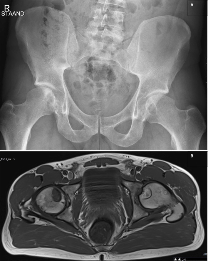
A 38-year-old patient presented with bilateral hip pain of several years’ duration with an increase in severity in the last weeks. The pain was mainly located in the groin radiating to the greater trochanteric region, had no nocturnal component and was associated with a slight functional hip discomfort such as difficulties to get up from the seat and to put on shoes. Conventional anteroposterior radiographs of the right and left hips showed bilateral subchondral cysts in the inferior half of the femoral heads (figure 1A). T1W axial MR image revealed isointense aspect of the cyst on the right side and inhomogeneous hyperintense aspect of the cyst on the left side (figure 1B). As subchondral cysts usually imply the presence of an underlying osteoarthritic disease process, the patient was recommended to consider treatment with total hip prosthesis. From a radiological standpoint, however, the subchondral cysts were not consistent with the typical presentation of osteoarthritis. Herein we discuss, in brief, the differential diagnosis of subchondral cysts, involving four distinct disorders: osteoarthritis (OA), rheumatoid arthritis (RA), calcium pyrophosphate dihydrate deposition disease (CPPD) and osteonecrosis (ON), and we explain why these subchondral cysts should be rather considered in the context of ON.
Figure 1.

(A) Anteroposterior radiographs of the right (and left (hips. Bilateral subchondral cysts in the inferior half of the femoral heads. (B) T1W axial image showing isointense aspect of the cyst on the right side with perilesional discrete isolated bone marrow oedema and inhomogeneous hyperintense aspect of the cyst on the left-side suspect for a mucoid (chronic) component. Both 'lesions' are characterised by intralesional pencil-fine (osseous lamellar) septations and a sharp limited sclerotic peripheral contour delineation. note also the globally preserved sphericity of the femoral heads bilateral with no apparent collapse.
The presence of a subchondral cyst in a weight bearing area, unless otherwise proven, almost always suggests OA, particularly when other characteristics of OA such as narrowing of the joint space, subchondral sclerosis and osteophyte formation are also present. Nevertheless, its presence in patients with OA is only around 30% in comparison with the other three findings, which are observed in more than 90% of patients with OA.1 In this specific case, these three features were not accentuated, and subchondral cysts were not located where we would expect them to be seen in a typical OA, that is, at the superolateral region of the coxofemoral joint. Less frequently, patients with RA may also present with subchondral cysts. These patients, however, usually develop synovial cysts and bursa which relieve the elevated intra-articular pressure eliminating one of the pathophysiologic risk factors for subchondral cyst formation. Given the fact that our patient had no other findings associated with an autoimmune and inflammatory process makes this diagnosis less likely. Finally, subchondral cysts may also be observed in patients with CPPD; but these patients feature usually numerous widespread subchondral cysts which are observed in the context of an advanced degenerative joint process even more accentuated than OA, which was not present in the current case. Consequently, the absence of degenerative, autoimmune and inflammatory findings argues against the diagnosis of OA, RA or CPPD. We therefore concluded a diagnosis by exclusion of the other possibilities and made a tentative diagnosis of ON and recommended follow-up. The difference in the signal intensity between the two cystic structures is due to the different onset of the subchondral cysts and hence different rate of progress.2
The differentiation of a subchondral cyst is not always simple. Its size, location and especially the nature of the accompanying findings help make the differential diagnosis. Treatment of each of the entities discussed above varies, hence a meticulous work-up to avoid misdiagnosis and inappropriate management is important.
Learning points.
Subchondral cysts usually develop in patients with osteoarthritis (OA), rheumatoid arthritis (RA) or calcium pyrophosphate dihydrate deposition disease (CPPD) but may also be observed in patients with osteonecrosis.
The absence of degenerative, autoimmune and inflammatory findings argues against the diagnosis of OA, RA or CPPD.
Treatment of each of these entities varies, hence a meticulous work-up to avoid misdiagnosis and inappropriate management is important.
Footnotes
Contributors: MM and LC contributed to the conception and design of the study. TR participated in reviewing and revising the manuscript. KDS reviewed the manuscript for intellectual content.
Funding: The authors have not declared a specific grant for this research from any funding agency in the public, commercial or not-for-profit sectors.
Case reports provide a valuable learning resource for the scientific community and can indicate areas of interest for future research. They should not be used in isolation to guide treatment choices or public health policy.
Competing interests: None declared.
Provenance and peer review: Not commissioned; externally peer reviewed.
Ethics statements
Patient consent for publication
Consent obtained directly from patient(s).
References
- 1.Landells JW. The bone cysts of osteoarthritis. J Bone Joint Surg Br 1953;35-B:643–9. 10.1302/0301-620X.35B4.643 [DOI] [PubMed] [Google Scholar]
- 2.Wada R, Lambert RGW. Deposition of intraosseous fat in a degenerating simple bone cyst. Skeletal Radiol 2005;34:415–8. 10.1007/s00256-004-0856-9 [DOI] [PubMed] [Google Scholar]


