Abstract
Neonatal lupus is an uncommon entity. The main manifestations are cutaneous and cardiac. It is caused by transplacental passage of maternal antibodies (anti-Ro/SSA or anti-La/SSB), and the diagnosis is made by its detection in the mother or child. The authors present a case of a 4-month-old female infant, with a cutaneous eruption since she was 2 months old. She had no relevant personal or family history. Analytically she had an increase in liver enzymes. The histological aspect of the skin biopsy led to an autoimmunity study on the mother and infant, both of which had positive anti-Ro/SSA antibodies, confirming the diagnosis of neonatal lupus. Cardiological study was normal. The skin lesions resolved during the first year of life. Skin lesions are the most frequent non-cardiac clinical manifestation of neonatal lupus, and they are self-limited. When there is no family history, nor cardiac involvement, the diagnosis can be challenging.
Keywords: infant health, dermatology, rheumatology
Background
Neonatal lupus is a rare autoimmune disease, due to transplacental passage of maternal antibodies (anti-Ro/SSA, anti-La/SSB or, occasionally, anti-RNP).1–6
The main manifestations are cardiac and cutaneous, although others, like haematologic and hepatic, can also appear.1 2 5 Atrioventricular block is the more common and serious cardiac finding, most of the time irreversible and with high morbidity and mortality.1–4 Non-cardiac findings are transitory, coincident with maternal antibodies’ disappearance, which usually happens until 8 months.1–4
In many cases, up to 60% according to some authors, mothers are asymptomatic and become aware of this situation only due to her child’s manifestations.1–4 6 It is estimated that around 1% of healthy women may have anti-Ro/SSA antibodies without associated disease.1 6 Nevertheless, approximately half of the asymptomatic mothers will have an autoimmune disease in the future.6 7
Neonatal lupus occurs in mothers who have anti-Ro/SSA or anti-La/SSB antibodies in circulation and passively transfer them to the fetus. Yet, it is believed that other factors, not fully established, like different maternal antibodies, such as anti-RNP, or genetic factors of the fetus, may play a role in the development of the disease, because neonatal lupus is, in fact, rare in the offspring of these women.1–4 6 The diagnosis is made by antibodies’ detection in the mother and/or child.1 Although previously stated, a correlation between titers of antibodies and severity of clinical signs, namely complete heart block, is not completely established yet. First, laboratories have different cutoffs values, so titer’s definition may differ between them, and second because high titers alone are not enough for the cardiac complications.1 However, some studies describe more serious findings in the presence of both anti-Ro/SSA and anti-La/SSB antibodies.8
The authors describe a case where the diagnosis of neonatal lupus was difficult because of the sole presence of cutaneous lesions, with no cardiac involvement, and no maternal history of rheumatologic diseases known.
Case presentation
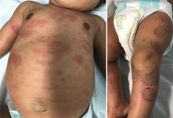
A previously healthy, 4-month-old female infant had a hospital appointment because of a cutaneous eruption that started when she was 2 months old. There were no other symptoms and no relevant personal or family history. Skin lesions were spread all over the body, except the head, which was spared. They were erythematous macules, with rapid progression to well-circumscribed, annular and desquamative plaques (figure 1). The physical examination was otherwise normal. By the time she was assessed, she had already been treated with antifungal therapy, topical corticosteroids and oral antibiotics, without improvement. Analytically she had an increase in liver enzymes (maximum aspartate transaminase 121 UI/L and alanine transaminase 95 UI/L), with no haematological changes. Serologic tests for cytomegalovirus, Epstein-Barr virus, parvovirus and mycoplasma pneumoniae were all negative. A skin biopsy was performed and revealed an interface dermatitis with necrotic keratinocytes, a perivascular/periductal lymphocytic infiltrate and the presence of mucin in the dermis, histological aspects compatible with lupus. An autoimmunity study was carried out on the mother and the infant, both of which had positive anti-Ro/SSA antibodies, which led to the diagnosis of neonatal lupus. Cardiological study, with 12-lead ECG and echocardiogram, was normal.
Figure 1.

Cutaneous lesions at 4 months old.
Outcome and follow-up
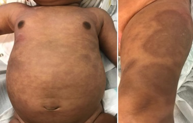
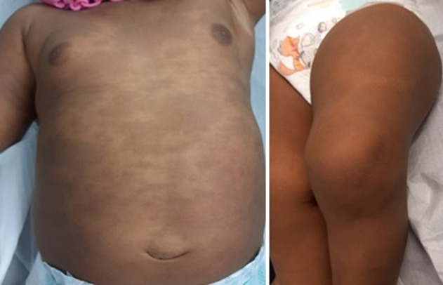
During follow-up, skin lesions slowly resolved (figure 2) and at the age of 12 months they were just hyperpigmented postinflammatory stains (figure 3). Liver enzymes had also gradually decreased to a normal range. At 12 months old she had a negative anti-Ro/SSA antibody.
Figure 2.

Cutaneous lesions at 6 months old.
Figure 3.

Cutaneous lesions at 12 months old.
Throughout this time, the mother has always been asymptomatic. She was referred to a rheumatologist for surveillance.
Discussion
Neonatal lupus is a rare disease, although its real prevalence is unknown,3 and often presents as a clinical challenge. Cardiac and skin manifestations can be isolated or combined, along with other changes, such as haematological, hepatobiliary or neurological.1 6
When there is cardiac involvement, the diagnosis is usually precocious, in utero or in the neonatal period, due to bradycardia. On the other hand, when there is no cardiac disease, clinical findings can appear later and neonatal lupus may be more difficult to diagnose, unless there is some level of suspicion, like a mother’s diagnosis of Sjögren syndrome or Lupus, or the presence of anti-Ro/SSA or anti-La/SSB antibodies in circulation.
Injury to the conducting system is the most common cardiac lesion and can include all degrees of atrioventricular block, with complete or third-degree being the most characteristic.1 6 It usually develops between 18 and 24 weeks of gestation, and clinical findings (bradycardia) depends on the heart block’s severity, with first-degree block being asymptomatic most of the cases and complete one with a typical ventricular rate of 50–80 beats per minute.1 Overall, postnatally, 50%–70% of patients with congenital heart block will need a pacemaker, reaching more than 90% if it is a complete one.4 6 7
Because of the potential severity of the disease and the possibility of prophylaxis, some authors defend a routine screening of maternal antibodies to all pregnant women and, if it is positive, a regular fetal echocardiographic examination from 16 weeks of gestation. Hydroxychloroquine showed to be effective in preventing fetus’ cardiac disease in mothers with anti-Ro/SSA or anti-La/SSB antibodies, which justify a screening as soon as possible.6 7 9 10 Although cardiac manifestations de novo are rare after birth, newborns should be observed cautiously until 28 days of life.6
Cutaneous findings are the most frequent non-cardiac manifestation, and they are present in 4%–16% in offspring of mothers with anti-Ro/SSA or anti-La/SSB antibodies.1 They usually appear from few weeks after birth, until 4 months old.1 6 Their prevalence might be underestimated because they can be mistaken with other skin lesions, like seborrhoeic dermatitis, urticaria, atypical erythema multiforme, tinea corporis, among others.1 3 5 6 Erythematous annular lesions with a slight central atrophy are preferentially located in the head and periorbital area, as they are sun exposed areas.1–4 The rash is self-limited and disappears by 6–8 months, usually with no sequelae, although scars, telangiectasias and hypopigmented areas can persist.1 3 4 11 It is advised to avoid ultraviolet light exposure, because it worsens the lesions.1 2 4 5 11
In our case, the absence of maternal history, cardiac involvement and atypical distribution of skin lesions made the diagnose of lupus neonatal a challenge. As so, the cutaneous lesions motivated a first therapeutic trial with topical antifungal and corticosteroids and oral antibiotic, with no results, which led to a more extensive investigation. The biopsy was the clue to the definitive diagnosis. Skin biopsy is usually not necessary, but, when it is performed, histological findings include keratinocyte degeneration, epidermal atrophy and perivascular lymphocytic infiltrate,3 which are consistent with our results. Direct immunofluorescence demonstrates immunoglobulin and complement deposition at the dermal–epidermal junction.4 Because it is reversible, treatment is not required, although some authors use topical corticosteroids or even systemic if there are severe and persistent lesions.6
Other manifestations of the disease include asymptomatic elevated liver enzymes, mild hepatosplenomegaly, anaemia, neutropenia and thrombocytopenia.1 2 5–7 In most of the cases, there are no need for treatment, although severe ones may need interventive measures, such as systemic corticosteroids, intravenous immunoglobulin or blood/platelets transfusions.6 Our infant only had an elevation of liver enzymes, that resolved in approximately 7 months, which is in accordance with the literature.
The prognosis and severity of neonatal lupus is dictated by the presence of cardiac abnormalities.2 6 9 Children with no cardiac involvement during pregnancy and no evidence of heart block at birth are unlikely to develop cardiac disease later.7 9
The present patient has a good prognosis. Nevertheless, some studies showed that cutaneous neonatal lupus might increase the risk, up to 2% according to some series, of having autoimmune and/or rheumatologic diseases later,1 3 5 6 9 11 so she should have a careful follow-up in life.
For future pregnancies, the mother must have a more intense monitoring, with fetal echocardiographic surveillance, especially during the most vulnerable period (18–24 weeks of pregnancy). Cutaneous neonatal lupus grant a twofold to threefold increased risk of having a new baby with neonatal lupus, including an up to 18% risk of having cardiac involvement, which justify prophylaxis during pregnancy.3–5 9 10 As stated before, hydroxychloroquine have shown some results in doing so. Nevertheless, the efficacy of the drug in cutaneous isolated disease is still not established, although some authors suggested that it might have a protective effect.9
Neonatal lupus’ diagnosis continues to be a clinical challenge, especially if there is no previous family history, nor cardiac involvement. The present case should remind us that it is necessary a high level of suspicion, with a multisystemic and multidisciplinary approach, most of the times using complementary evaluations, in order to diagnose and treat precociously, prenatal or postnatal.
Patient’s perspective.
I did not know what those spots on the skin were and they worried me because we had tried many things without success. I am very glad they disappeared and now I can be better watched if I want to have a new baby.
Learning points.
Neonatal lupus is a rare disease, caused by transplacental passage of maternal antibodies.
When there is cardiac involvement, the diagnosis is usually precocious. When there is not, neonatal lupus may be more difficult to diagnose, unless there is some level of suspicion, like a mother’s diagnosis of a rheumatologic disease.
In this case, the absence of maternal history, cardiac involvement and atypical distribution of skin lesions made the diagnose a challenge, only to be overcome with a skin biopsy.
Footnotes
Contributors: FCC and SF were responsible for child’s clinical evaluation during Paediatric appointment. MP was a Paediatric consultant. SS was a Rheumatology consultant. FCC was responsible for collecting clinical data and drafting the manuscript. SF was responsible for collecting clinical data, revising the manuscript and final approve for submission. SS and MP were responsible for revising the manuscript and final approve for submission.
Funding: The authors have not declared a specific grant for this research from any funding agency in the public, commercial or not-for-profit sectors.
Case reports provide a valuable learning resource for the scientific community and can indicate areas of interest for future research. They should not be used in isolation to guide treatment choices or public health policy.
Competing interests: None declared.
Provenance and peer review: Not commissioned; externally peer reviewed.
Ethics statements
Patient consent for publication
Consent obtained from parent(s)/guardian(s).
References
- 1.Buyon JP. Neonatal lupus: epidemiology, pathogenesis, clinical manifestations, and diagnosis. Available: https://www.uptodate.com/contents/neonatal-lupus-epidemiology-pathogenesis-clinical-manifestations-and-diagnosis?search=Neonatal%20lupus:%20Epidemiology,%20pathogenesis,%20clinicalmanifestaticli,%20and%20diagnosis&source=search_result&selectedTitle=1~34&usage_type=default&displdi_rank=1; [Accessed 13 Dec 2020].
- 2.Coelho R, Ferreira M, Ferreira M, et al. [Neonatal lupus erythematosus]. Acta Med Port 2007;20:229–32. [PubMed] [Google Scholar]
- 3.Teixeira V, Gonçalo M. [Neonatal lupus erythematosus - review of pathophysiology and clinical implications]. Acta Reumatol Port 2012;37:314–23. [PubMed] [Google Scholar]
- 4.Barcelos A, Fernandes B. Cutaneous manifestations of neonatal lupus: a case report. Acta Reumatol Port 2012;37:352–4. [PubMed] [Google Scholar]
- 5.Vega V, Duque M, Don-Pedro D, et al. A neonate with annular cutaneous lesions. Cureus 2020;12:e9138. 10.7759/cureus.9138 [DOI] [PMC free article] [PubMed] [Google Scholar]
- 6.Derdulska JM, Rudnicka L, Szykut-Badaczewska A, et al. Neonatal lupus erythematosus - practical guidelines. J Perinat Med 2021;49:529–38. 10.1515/jpm-2020-0543 [DOI] [PubMed] [Google Scholar]
- 7.Vanoni F, Lava SAG, Fossali EF, et al. Neonatal systemic lupus erythematosus syndrome: a comprehensive review. Clin Rev Allergy Immunol 2017;53:469–76. 10.1007/s12016-017-8653-0 [DOI] [PubMed] [Google Scholar]
- 8.Zuppa AA, Riccardi R, Frezza S, et al. Neonatal lupus: follow-up in infants with anti-SSA/Ro antibodies and review of the literature. Autoimmun Rev 2017;16:427–32. 10.1016/j.autrev.2017.02.010 [DOI] [PubMed] [Google Scholar]
- 9.Braga-Tavares H, Ramos L, Bini-Antunes M. Lúpus eritematoso neonatal. Nascer e Crescer 2009;18:149–51 https://repositorio.chporto.pt/bitstream/10400.16/1252/1/LupusEritematoso_18-3.pdf [Google Scholar]
- 10.Morel N, Georgin-Lavialle S, Levesque K, et al. « Lupus néonatal » : revue de la littérature. La Revue de Médecine Interne 2015;36:159–66. 10.1016/j.revmed.2014.07.013 [DOI] [PubMed] [Google Scholar]
- 11.Buyon JP. Neonatal lupus: management and outcomes. In: Klein-Gitelman M, Triedman JK, Garcia-Prats JA, eds, 2020. https://www.uptodate.com/contents/neonatal-lupus-management-and-outcomes?search=Neonatal%20lupus:%20Epidemiology,%20pathogenesis,%20clinicalmanifestaticli,%20and%20diagnosis&source=search_result&selectedTitle=2~34&usage_type=default&displdi_rank=2


