Abstract
Objective:
To identify esthetic characteristics of the orbital soft tissues of attractive Italian adult women and men.
Materials and Methods:
Three-dimensional computerized digitizers were used to collect the coordinates of facial landmarks in 199 healthy, normal subjects aged 18 to 30 years (71 women, 128 men; mean age, 22 years) and in 126 coetaneous attractive subjects (92 women, 34 men; mean age, 20 years) selected during beauty competitions. From the landmarks, six linear distances, two ratios, six angles, and two areas were calculated. Attractive subjects were compared with normal ones by computing z-scores.
Results:
Intercanthal width was reduced while eye fissure lengths were increased in both genders. Orbital heights (os-or) were increased only in attractive women, with a significant gender-related difference. The inclinations of the eye fissure were increased in attractive subjects, while the inclinations of the orbit were reduced. For several of the analyzed measurements, similar patterns of z-scores were observed for attractive men and women (r = .883).
Conclusion:
Attractive women and men had several specific esthetic characteristics in their orbital soft tissues; esthetic reference values can be used to determine optimal goals in surgical treatment.
Keywords: Face, Attractiveness, Soft tissues
INTRODUCTION
Current scientific literature reports on the esthetic characteristics of the human face are increasing.1–8 For instance, a recent search in the PubMed database using the key word “aesthetics” produced 21,706 entries in the period 1950–2013; using “aesthetics and face,” a total of 3951 entries was obtained; and the combination “aesthetics and (eye or orbital)” produced 987 entries.9 Sixty percent of these papers were published in the current century (2001–2013), thus showing the increasing interest in facial esthetics.
In particular, the eyes and the orbital region play a predominant role in the evaluation and recognition of the craniofacial complex5,10,11 and are among those facial areas that are considered to be at the center of esthetic evaluation.3,5,6,8,12–14 According to the current theories of evolutionary psychology, the esthetic assessment of adult faces depends on various combinations of averageness, symmetry, neoteny (babyness) and youthfulness, and sexual dimorphism.1–4,15
Studies on facial attractiveness focused both on the psychological bases of esthetic perception1,3,16 and on actual measurements computed for faces (or parts of faces) considered more or less attractive.2,4,5,12,17–20 Unfortunately, previous investigations were mostly performed on two-dimensional images (only Farkas19 used three-dimensional measurements), did not report global measures such as facial areas, and scantly assessed men. Most of the preceding studies were made in women, and the assessment of male attractiveness was almost always limited to those parts of the face positively influenced by high testosterone levels (a signal of sexual dimorphism).1,21
In our laboratory, we are currently attempting to measure the facial characteristics of subjects considered attractive by the general public.15,22,23 These subjects' three-dimensional facial soft-tissue dimensions were compared with those collected in healthy subjects of the same gender, age, and ethnicity, and the presence of measurable specific characteristics was assessed. We focused in particular on the orolabial region and on the relationships among the various parts of the face.15,23 In contrast, the eyes and soft-tissue orbital region were almost neglected in our previous studies.
The aim of the current study was to investigate the possible presence of measurable esthetic characteristics in the soft tissue orbital region of adult women and men considered to be attractive (i.e., semi-finalists in beauty competitions).
MATERIALS AND METHODS
Analyzed Subjects
Three-hundred twenty-five white, Italian subjects aged between 18 and 30 years were analyzed. One-hundred ninety-nine (71 women, 128 men; mean age, 22 years) were normal subjects, recruited from subjects and staff attending the university. Part of the data on these subjects has already been published.10
Inclusion Criteria
Inclusion criteria included the following:
Good general health
Clinically normal facial dimensions and proportions (no need for treatment)
No previous history of craniofacial surgery, trauma, or congenital anomalies
One-hundred twenty-six (92 women, 34 men; mean age, 20 years) were attractive subjects, selected by juries of national beauty competitions who were unaware of the scope of the investigation. Attractive subjects were measured during several national beauty competitions that took place in Italy between 2006 and 2008. They were those admitted to the semi-final stage of the beauty competitions and were measured just before the relevant beauty event.15 No information about their clinical or surgical history was available. For attractive subjects, the sample size was higher than that used by Farkas19 in the only previous investigation that analyzed men and women with three-dimensional measurements (171% and 62% higher for women and men, respectively).
All of the analyzed subjects gave their informed consent to the experiment. All procedures were not invasive; did not provoke damage, risks, or discomfort to the subjects; and were approved by the local ethic committee.
Collection of Facial Landmarks
A two-step data collection procedure was used; all mathematical calculations were performed offline, as previously detailed.10,15,23 At first, a set of 50 soft tissue landmarks were located and marked by inspection and palpation on the facial skin of each subject using black eyeliner. Subsequently, the three-dimensional (x, y, z) coordinates of the facial landmarks were obtained using three-dimensional computerized digitizers (an electromagnetic three-dimensional tablet, 3Draw, Polhemus Inc, Colchester, Vt; and an electromechanical instrument, Microscribe G2, Immersion Corporation, San Jose, Calif). An experienced investigator performed all of the procedures. Data collection was performed with the subject sitting in a natural head position in a chair with a backrest. Landmark digitization took approximately 1 minute for each subject. Files of the three-dimensional coordinates were obtained, and original computer programs were used for all subsequent offline calculations. The reproducibility of landmark identification, marker positioning, and data collection procedures was previously reported and found to be reliable.10
Data Analysis
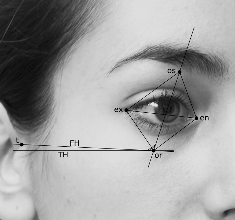
In the present study, from the complete set of 50 landmarks, the following paired soft tissue landmarks were further considered (right and left side noted r and l): exr, exl, exocanthion; enr, enl, endocanthion; orr, orl, orbitale; osr, osl, orbitale superius; tr, tl, tragion (Figure 1).
Figure 1.
Three-dimensional soft tissue facial landmarks used in the current study, identified on an Italian 20-year-old woman: ex indicates exocanthion; en, endocanthion; os, orbitale superius; or, orbitale; t, tragion. Frankfurt plane (FH) and true horizontal (TH) are also traced.
The three-dimensional coordinates of the landmarks obtained on each subject were used to calculate the following measurements10: linear distances (unit: mm): biorbital width (exr-exl), intercanthal width (enr-enl), right and left height of the orbit (os-or), right and left length of the eye fissure (en-ex), ratios (unit: percentage), right and left height of the orbit to length of the eye-fissure ratio (os-or/en-ex × 100); angles (unit: degrees): right and left inclination of the eye fissure (angle of the en-ex line vs the true horizontal, head in natural head position), right and left inclination of the orbit (angle of the os-or line vs the true horizontal, head in natural head position), right and left inclination of the orbit relative to the Frankfurt plane (angle between the os-or and t-or lines); and areas (unit: mm2): right and left external orbital surface area (area of the quadrangle between ex, os, en, and or).
All of the measurements were performed in the three-dimensional space; that is, the position of the landmarks relative to all three planes (frontal, lateral, and horizontal) was considered at the same time (no projections).
Statistical Calculations
For each measurement of the control subjects, descriptive statistics (mean and standard deviation) were computed within gender. Data obtained from the attractive subjects were compared with those collected from normal subjects by computing z-scores.4 The z-score is a measure of the distance between a subject datum and the normal mean expressed in standard deviation units: z-score = (subject value – mean value of the normal group) divided by the standard deviation of the normal group. Positive z-scores indicate that the measurement is larger in the attractive than in the normal population; in contrast, negative z-scores indicate a smaller measurement in the attractive than in the normal population. By definition, the normal population has a mean z-score of 0, with a standard deviation of 1.23 Descriptive statistics of the z-scores were computed within the gender group. The significance of the z-scores was assessed by Student's t-tests (if the attractive subject value is equal to the mean value of the normal group, the z-score is zero; the null hypothesis of the test is that the z-scores are null).
For a global comparison of the facial characteristics of attractive women and men, a correlation analysis between the paired z-scores of the two groups was run: high correlation coefficients indicate very similar patterns.24 In addition, male and female z-scores were compared by independent Student's t-tests. For all comparisons, the significance level was set at 5% (P < .05).
RESULTS
Attractive vs Normal Subjects
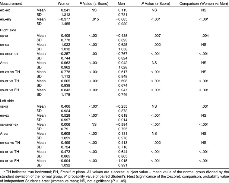
When compared with normal subjects, attractive young women and men had several significant differences in their soft tissue orbital structures. Intercanthal width (enr-enl) was smaller than in normal subjects (Table 1). Orbital heights (os-or) were increased in attractive women but reduced (right side) or unchanged (left side) in men. In both genders, the right and left lengths of the eye fissure (en-ex) were increased. Overall, this produced a larger external orbital surface area in attractive women than in normal ones. The right os-or/en-ex ratio was reduced in both genders, while the left one was reduced only in men.
Table 1.
Three-Dimensional Soft Tissue Orbital Dimensions and Angles in Attractive Subjects as Compared With Normal Subjectsa
The right and left inclinations of the eye fissure (en-ex line vs the true horizontal) were increased in attractive subjects of both genders, while the inclinations of the orbit (os-or line vs the true horizontal and relative to the Frankfurt plane) were reduced: the vertical projections of the orbitale superius and orbitale landmarks were nearer one to the other.
Gender Differences
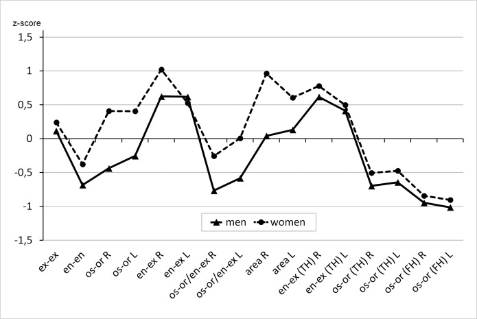
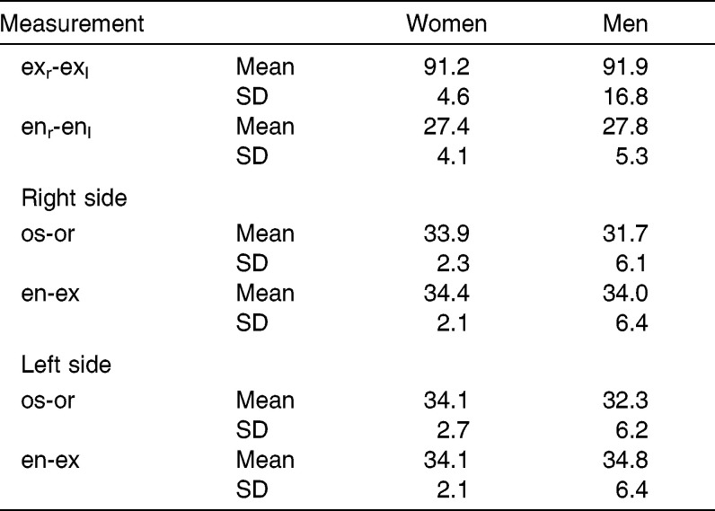
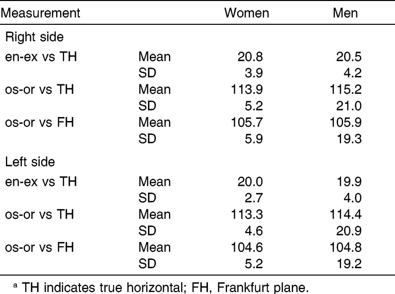
For several of the analyzed measurements, similar patterns of the z-scores were observed for attractive women and men, and the correlation analysis found a very high r value (.883; Figure 2). Most measurements had similar differences in both genders (similar z-scores). Among the differences, there was intercanthal width, os-or/en-ex ratios, and orbital inclinations, all with a larger discrepancy in men than in women. Also, gender differences were observed for orbital heights that were increased in attractive women and decreased in attractive men. Mean values of soft tissue orbital distances and angles in attractive subjects are reported in Table 2 and Table 3.
Figure 2.
Soft tissue orbital measurements in attractive women (interrupted line) and men (continuous line). z-scores were computed using values collected in normal subjects (mean value = 0 for all variables). R indicates right; L, left; TH, true horizontal; FH, Frankfurt plane.
Table 2.
Three-Dimensional Soft Tissue Orbital Linear Distances in Attractive Subjects (in mm)
Table 3.
Three-Dimensional Soft Tissue Orbital Angles in Attractive Subjects (in °)a
DISCUSSION
Facial esthetic characteristics have been analyzed in several studies, but the current study differs from previous investigations in some points. At first, we analyzed men and women considered “beautiful” and “attractive” and selected for the semi-final stage of beauty competitions. They all came from Italy and were admitted to this phase of the competition after a series of selections. Therefore, they should represent what is currently considered “attractive,” “positive,” and “acceptable.”16 The selection was made independently by professionals of the media world (juries of beauty competitions) and not by lay people.5
Indeed, jury ratings were linked to actual facial measurements. Even if the human visual system possesses a better sensitivity than the current anthropometric method,21 health professionals cannot rely only on perception; they need objective data for their diagnosis and treatment.6,8,14 This good agreement may result from the use of three-dimensional, lively stimuli for the current rating procedures, which differ from the methods commonly used in psychological investigations, usually relying on two-dimensional records.4,7,8,15,23
A second difference is that the assessment of attractive adult male faces has been mostly performed using two-dimensional measurements, and three-dimensional analyses seem to have been performed only by Farkas19 for North American white men, and in our laboratory.15,22
The major limitation of the present study is the method used for landmark digitization. We used contact instruments, which digitize only single, selected anatomical landmarks. The instruments not only neglect all surface information comprised between the landmarks but also need a relatively long data collection time (approximately 60 seconds for a set of 50 landmarks), with possible movement artifacts. Nevertheless, we used a carefully controlled protocol to limit this problem and found the method to be reproducible.10,25 Indeed, the performance of contact instruments in clinical practice has been recently assessed and found to be satisfactory.25,26 One additional advantage is that they can be used directly in any environment, for instance, quickly meeting the attractive subjects during beauty competitions, fashion events, or castings. In contrast, optical instruments (laser scans, stereophotogrammetric digitizers) usually necessitate dedicated settings, which may not be organized outside specialized laboratories.26
One of the characteristics of our method is that facial landmarks are identified and marked directly on the face of the subjects before their digitization, thus allowing the use of a wide set of points to calculate measurements in all spatial directions.10,25 Among the others, we could assess the position of landmark orbitale, which has a progressive inferior shift with advancing age,10 thus being a useful indicator of the juvenile characteristics of attractive faces.
A further limitation of the current study is the assessment of a convenience group made by only attractive Italian subjects. The larger number of attractive women depended on the larger number of female beauty competitions organized in Italy. In a different ethnic/sociocultural context, different kinds of attractive faces might be chosen,6,8,12,14 even if the good agreement between the present data and literature reports makes the resulting patterns sufficiently trustworthy for white subjects.
Overall, the soft tissue orbital region in Italian attractive men and women has been found to have specific characteristics. In women, the eye fissure length and the height of the orbit were larger, with a resulting wider soft tissue orbital area; this finding is in good accord with the babyness hypothesis of facial attractiveness.1 In general, attractive women have faces with juvenile characteristics,1 and this is true also for the periorbital area. Aging involves a series of modifications in the periorbital soft tissues, with a resulting perception of a smaller palpebral aperture in older subjects.13 The effect was not so evident in attractive men, who only had larger eye fissure lengths. A similar result was reported by Farkas19 for women. Overall, it is confirmed that the orbital region has a larger importance for attractiveness in women than in men.3,19
The present finding of some degree of hypotelorism contrasts with previous reports of no changes in the intercanthal distance19 or of some degree of hypertelorism.8,12 Both different methods (two-dimensional vs three-dimensional) and ethnicity of the subjects may partly explain the differences. In the current attractive subjects, the orbital height to eye fissure length ratio was reduced, in partial contrast with the data from Farkas.19 The use of different landmarks for the vertical measurement (palpebrale superius and inferius vs orbitale superius and orbitale) can explain the difference.
In good accord with previous studies, performed using both three-dimensional19 and two-dimensional measurements,3,8,12 the inclinations of the eye fissure were increased in attractive subjects. According to Volpe and Ramirez,8 the upward inclination of the intercanthal axis is a specific characteristic of the beautiful eye, suggesting neoteny because of its age-related modifications.3,10,14 In women, makeup can accentuate the medial canthal tilt, thus creating the illusion of more slanted palpebral fissures.3 Also, palpebral fissure inclination is a sexually dimorphic characteristic, with a steeper inclination in women than in men.3,6,13,14 Previous investigations into the esthetic value of this measurement analyzed women only, but we found a similar effect in both genders.
Orbital inclination (os-or line vs the true horizontal and relative to Frankfurt plane) was reduced in both genders, but this measurement was scantily reported by other investigators. Indeed, orbitale landmarks can be difficult to identify on photographs without previous marking.25
Notwithstanding some significant differences between the average z-scores computed in the current attractive men and women, in most occasions the difference was only in the magnitude of the discrepancy relative to their normal peers (z-score value) and not in the direction of the variation (z-score sign). Therefore, the relevant patterns of variation were similar, with a highly significant correlation between the z-scores.24 Pattern profile analysis was devised to identify similar phenotypes in patients with craniofacial malformations, but it is well suitable to appreciate any biological variation.24
The definition of quantitative characteristics of esthetically pleasing faces may help medical practitioners to provide better patient care for both modifications of those facial physiognomies considered as nonattractive (beautification) and variations of those facial features that modify with age (rejuvenation).5,6,8,10,14,27
CONCLUSIONS
Attractive women and men had some specific characteristics in their soft tissue orbital structures that contributed to their general esthetic appearance.
The present data reinforce current esthetic views: the most attractive faces are not average; they combine some averageness with individual features that depart from the population norm.8,12
ACKNOWLEDGMENT
The authors are greatly indebted to all the staff of the Laboratorio di Anatomia Funzionale dell'Apparato Stomatognatico, Università degli Studi di Milano, who participated in data collection and analysis. Dr Patrizia Frangella organized the data collection in attractive subjects. The expert secretarial assistance of Ms Cinzia Lozio is gratefully acknowledged. Financial support was obtained from the University of Milan (FIRST, 2006) and from the Consiglio Direttivo of the Società Italiana Di Ortodonzia (SIDO).
REFERENCES
- 1.Bashour M. History and current concepts in the analysis of facial attractiveness. Plast Reconstr Surg. 2006;118:741–756. doi: 10.1097/01.prs.0000233051.61512.65. [DOI] [PubMed] [Google Scholar]
- 2.Bashour M. An objective system for measuring facial attractiveness. Plast Reconstr Surg. 2006;118:757–774. doi: 10.1097/01.prs.0000207382.60636.1c. [DOI] [PubMed] [Google Scholar]
- 3.Bashour M, Geist C. Is medial canthal tilt a powerful cue for facial attractiveness. Ophthal Plast Reconstr Surg. 2007;23:52–56. doi: 10.1097/IOP.0b013e31802dd7dc. [DOI] [PubMed] [Google Scholar]
- 4.Edler R, Rahim MA, Wertheim D, Greenhill D. The use of facial anthropometrics in aesthetic assessment. Cleft Palate Craniofac J. 2010;47:48–57. doi: 10.1597/08-218.1. [DOI] [PubMed] [Google Scholar]
- 5.Griffin GR, Kim JC. Ideal female brow aesthetics. Clin Plast Surg. 2013;40:147–155. doi: 10.1016/j.cps.2012.07.003. [DOI] [PubMed] [Google Scholar]
- 6.Kunjur J, Sabesan T, Ilankovan V. Anthropometric analysis of eyebrows and eyelids: an inter-racial study. Br J Oral Maxillofac Surg. 2006;44:89–93. doi: 10.1016/j.bjoms.2005.03.020. [DOI] [PubMed] [Google Scholar]
- 7.Sforza C, Laino A, D'Alessio R, et al. Three-dimensional facial morphometry of attractive Italian women. Prog Orthod. 2007;8:282–293. [PubMed] [Google Scholar]
- 8.Volpe CR, Ramirez OM. The beautiful eye. Facial Plast Surg Clin North Am. 2005;13:493–504. doi: 10.1016/j.fsc.2005.06.001. [DOI] [PubMed] [Google Scholar]
- 9.PubMed. Available at: www.pubmed.gov Accessed January, 25, 2013. [Google Scholar]
- 10.Sforza C, Grandi G, Catti F, Tommasi DG, Ugolini A, Ferrario VF. Age- and sex-related changes in the soft tissues of the orbital region. Forensic Sci Int. 2009;185:115.e1–115.e8. doi: 10.1016/j.forsciint.2008.12.010. [DOI] [PubMed] [Google Scholar]
- 11.Sforza C, Elamin F, Tommasi DG, Dolci C, Ferrario VF. Morphometry of the soft tissues of the orbital region in Northern Sudanese subjects. Forensic Sci Int. 2013;228:180.e1–180.e11. doi: 10.1016/j.forsciint.2013.02.003. [DOI] [PubMed] [Google Scholar]
- 12.Husein OF, Sepehr A, Garg R, et al. Anthropometric and aesthetic analysis of the Indian American woman's face. J Plast Reconstr Aesthet Surg. 2010;63:1825–1831. doi: 10.1016/j.bjps.2009.10.032. [DOI] [PubMed] [Google Scholar]
- 13.Pepper JP, Moyer JS. Upper blepharoplasty: the aesthetic ideal. Clin Plast Surg. 2013;40:133–138. doi: 10.1016/j.cps.2012.07.001. [DOI] [PubMed] [Google Scholar]
- 14.Price KM, Gupta PK, Woodward JA, Stinnett SS, Murchison AP. Eyebrow and eyelid dimensions: an anthropometric analysis of African Americans and Caucasians. Plast Reconstr Surg. 2009;124:615–623. doi: 10.1097/PRS.0b013e3181addc98. [DOI] [PubMed] [Google Scholar]
- 15.Sforza C, Laino A, Grandi G, Pisoni L, Ferrario VF. Three-dimensional facial asymmetry in attractive and normal people from childhood to young adulthood. Symmetry. 2010;2:1925–1944. [Google Scholar]
- 16.Orsini MG, Huang GJ, Kiyak HA, et al. Methods to evaluate profile preferences for the anteroposterior position of the mandible. Am J Orthod Dentofacial Orthop. 2006;130:283–291. doi: 10.1016/j.ajodo.2005.01.026. [DOI] [PubMed] [Google Scholar]
- 17.Auger TA, Turley PK. The female soft tissue profile as presented in fashion magazines during the 1900s: a photographic analysis. Int J Adult Orthodon Orthognath Surg. 1999;14:7–18. [PubMed] [Google Scholar]
- 18.Berneburg M, Dietz K, Niederle C, Göz G. Changes in esthetic standards since 1940. Am J Orthod Dentofacial Orthop. 2010;137:450.e1–450.e9. doi: 10.1016/j.ajodo.2009.10.029. [DOI] [PubMed] [Google Scholar]
- 19.Farkas LG. Anthropometry of the attractive North American Caucasian face. In: Farkas LG, editor. Anthropometry of the Head and Face 2nd ed. New York: Raven Press; 1994. pp. 159–179. [Google Scholar]
- 20.Nguyen DD, Turley PK. Changes in the Caucasian male facial profile as depicted in fashion magazines during the twentieth century. Am J Orthod Dentofacial Orthop. 1998;114:208–217. doi: 10.1053/od.1998.v114.a86137. [DOI] [PubMed] [Google Scholar]
- 21.Rhodes G, Yoshikawa S, Palermo R, et al. Perceived health contributes to the attractiveness of facial symmetry, averageness, and sexual dimorphism. Perception. 2007;36:1244–1252. doi: 10.1068/p5712. [DOI] [PubMed] [Google Scholar]
- 22.Bottino A, De Simone M, Laurentini A, Sforza C. A new 3-D tool for planning plastic surgery. IEEE Trans Biomed Eng. 2012;59:3439–3449. doi: 10.1109/TBME.2012.2217496. [DOI] [PubMed] [Google Scholar]
- 23.Sforza C, Laino A, D'Alessio R, Grandi G, Binelli M, Ferrario VF. Soft-tissue facial characteristics of attractive Italian women as compared to normal women. Angle Orthod. 2009;79:17–23. doi: 10.2319/122707-605.1. [DOI] [PubMed] [Google Scholar]
- 24.Garn SM, Smith BH, Lavelle M. Applications of pattern profile analysis to malformations of the head and face. Radiology. 1984;150:683–690. doi: 10.1148/radiology.150.3.6695067. [DOI] [PubMed] [Google Scholar]
- 25.Sforza C, de Menezes M, Ferrario V. Soft- and hard-tissue facial anthropometry in three dimensions: what's new. J Anthropol Sci. 2013;91:159–184. doi: 10.4436/jass.91007. [DOI] [PubMed] [Google Scholar]
- 26.Tartaglia GM, Dolci C, Sidequersky FV, Ferrario VF, Sforza C. Soft tissue facial morphometry before and after total oral rehabilitation with implant-supported prostheses. J Craniofac Surg. 2012;23:1610–1614. doi: 10.1097/SCS.0b013e31825af109. [DOI] [PubMed] [Google Scholar]
- 27.Raschke GF, Rieger UM, Bader RD, Schäfer O, Schultze-Mosgau S. Objective anthropometric analysis of eyelid reconstruction procedures. J Craniomaxillofac Surg. 2013;41:52–58. doi: 10.1016/j.jcms.2012.05.011. [DOI] [PubMed] [Google Scholar]


