Abstract
Objective:
To evaluate the reliability of three-dimensional (3D) landmark identification in cone-beam computed tomography (CBCT) using two different visualization techniques.
Materials and Methods:
Twelve CBCT images were randomly selected. Three observers independently repeated three times the identification of 30 landmarks using 3D reconstructions and 28 landmarks using multiplanar views. The values of the coordinates X, Y, and Z of each point were obtained and the intraclass correlation coefficient (ICC) was calculated.
Results:
The ICC of the 3D visualization was rated >0.90 in 67.76% and 45.56%, and ≤0.45 in 13.33% and 14.46% of the intraobserver and interobserver assessments, respectively. The ICC of the multiplanar visualization was rated >0.90 in 82.16% and 78.56% and ≤0.45 in only 16.7% and 8.33% of the intraobserver and interobserver assessments, respectively. An individual landmark classification was done according to ICC values.
Conclusions:
The frequency of highly reliable values was greater for multiplanar than 3D reconstructions. Overall, lower reliability was found for points on the condyle and higher reliability for those on the midsagittal plane. Depending on the anatomic region, the observer must choose the most reliable type of image visualization.
Keywords: Cone-beam computed tomography, Three-dimensional images, Anatomic reference points, Orthodontics
INTRODUCTION
The cephalometric landmarks located on two-dimensional images have errors that might lead to inaccurate representation of anatomic references, while three-dimensional (3D) analyses seem to offer visualization and identification advantages.1–3 To take full advantage of the information provided by cone-beam computed tomography (CBCT) diagnosis, the development of 3D cephalometric analysis requires appropriate operational definitions of reference points on each of the three planes of space as well as reliable landmark identification and reproducibility.4–6
A previous study7 used only multiplanar (MPR) views and found, in general, an excellent intraobserver and interobserver reproducibility. It concluded that the images from CBCT can provide consistent and reproducible data, but these can be affected by the reference anatomic structure, visualization of anatomic plane, and operator training. The infraorbital landmark is an example of difficult landmark identification because of the curved surface, and according to the authors, it could be better identified using a 3D reconstruction image.
Thus, it is suggested that there are differences in the localization of the structures depending on the image visualized. The software used by the clinical orthodontist must have tools available to facilitate the identification of cephalometric landmarks, and research validation must determine which visualization method is the most reliable for a certain anatomic reference.
This study aimed to assess observer reliability in the identification of anatomic reference points in CBCT images using software features for visualization of multiplanar and 3D reconstructions.
MATERIALS AND METHODS
A total of 12 CBCT images (from 8 female and 4 male patients aged between 20 and 43 years) were randomly selected from 125 orthodontic pretreatment scans from the institution's archive. The inclusion criteria were the presence of complete permanent dentition and no facial growth. Cases presenting skeletal asymmetries, syndromes, poor quality CBCT, or images that did not include all of the cranial structures were excluded. The research protocol was reviewed and approved by the institutional review board.
The CBCTs were obtained with the i-CAT 3D scanner and first processed by the machine software (Xoran Technologies, Ann Arbor, Mich). The acquisition system was calibrated at 120(±5) kV and 3–8(±10%) mA, focusing distance of 0.5 mm, and a source × sensor of 67.5 cm. The dimension of the amorphous silicon-based flat-panel imaging detector with a 1-mm aluminum panel was 20 × 25 cm. The images were acquired at 12 bits in a 360° rotation by using a 20-second cycle, expanded field of view (220 mm), and voxel size of 0.4 mm. The patients were instructed to remain in a natural head position during the scan, with the Frankfort horizontal plane parallel to the ground and in centric occlusion. The images were stored in Digital Imaging and Communications in Medicine (DICOM) format.
Thirty anatomic reference points, previously defined in the three planes of space according to de Oliveira et al.,7 were marked following two different visualization modes available in the InVivo Dental 5.1 software (Anatomage, San Jose, Calif): 3D virtual image model (3D reconstruction) and multiplanar reconstruction of axial, coronal, and sagittal slices. To improve the visualization of the landmarks, tools available in the software such as zoom, rotation, full screen, and contrast settings were used.
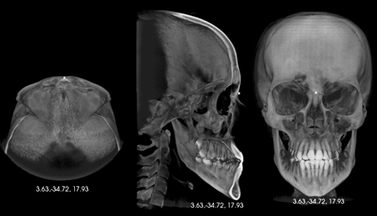
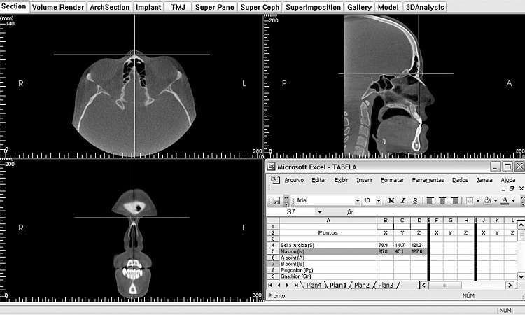
Three operators (students with undergraduate in dentistry, certification in orthodontics, and master's degree in orthodontics) were trained and calibrated to identify 3D landmarks using a set of five CBCT scans not included in this study. Working independently after calibration, they initially used images from a 3D virtual model (Figure 1) for marking 30 reference points on each of the 12 scans at three different time points, with a 1-week interval. By clicking on the image using the marker tool, the coordinate values of X, Y, and Z were provided automatically. Then, the MPR views were analyzed following the same criteria and time interval (Figure 2).
Figure 1.
Identification of the nasion landmark (N) using the visualization of 3D reconstruction images.
Figure 2.
Identification of the nasion landmark (N) using the multiplanar views.
There was consensus among the three operators that the point of the zygomatic-maxillary suture could not be correctly displayed in the MPR views, so it was considered missing data (total of 28 landmarks). In other studies, such as the one by de Oliveira et al.,7 the authors marked this point simultaneously viewing the 3D image reconstruction and the MPR slices, which was not done in the present research.
Landmarks were identified in 12 CBCT images by three observers at three different time intervals using two types of visualization, thus each point was repeated 216 times. Each marking generated three coordinate values (X, Y and Z), thus each point generated a total of 648 values, and 30 points generated a total of 19,440 values.
The intraclass correlation coefficient (ICC) obtained by comparing the values of X, Y, and Z, which indicate the exact location of each point on the axial, coronal and sagittal axes of the skull, was calculated to assess the reliability of the measurements. Mixed-effects models were considered for two-way analysis of variance (ANOVA). The observers were considered as a fixed effect, and the patients (CBCT) as a random effect. The SPSS v14.0 software (Chicago, Ill) was used to calculate the ICC.
RESULTS
Tables of frequencies of the intraobserver and interobserver reliability summarize the results (Tables 1 through 4). The frequency distribution divided the ICC values into above 0.90 (highly reliable), between 0.75 and 0.90 (reliable), between 0.45 and 0.75 (acceptable), and below 0.45 (poor).
Table 1.
Frequency of the Intraobserver Reliability Estimated for the X, Y, and Z Coordinates in the Visualization of 3D Reconstructions
Table 2.
Frequency of the Interobserver Reliability Estimated for the X, Y, and Z Coordinates in the Visualization of 3D Reconstructions
Table 3.
Frequency of the Intraobserver Reliability Estimated for the X, Y, and Z Coordinates in the Visualization of Multiplanar Reconstructions
Table 4.
Frequency of the Interobserver Reliability Estimated for the X, Y, and Z Coordinates in the Visualization of Multiplanar Reconstructions
The frequency of highly reliable values was higher in the intraobserver assessment than in the interobserver in the two types of visualization. Table 1 shows the estimated frequency of intraobserver reliability for the coordinates X, Y, and Z and their total mean values using the 3D reconstruction. The value of ICC was ≥0.90 in 61 assessments (67.76%) with a higher frequency for the coordinate Y (73.30%). The coordinate X (16.70%) showed the lowest reliability, with an ICC of ≤0.45 in 12 assessments (13.33%).
The frequency of interobserver reliability using the 3D rendering is shown in Table 2. The ICC was ≥0.90 in 41 (45.56%) interobserver assessments, and the highest frequency was for the coordinate Z (56.70%). Of the 13 assessments with ICC ≤ 0.45 (14.46% of the total), X and Y (16.70% each) were the coordinates that showed lower reliability.
The intraobserver reliability with the visualization of multiplanar slices is shown in Table 3. The ICC was ≥0.90 in 69 (82.16%) assessments with the highest frequency for the coordinate Y (89.30%). From the total of six assessments (7.16%), the highest frequency of ICC ≤0.45 was for the coordinate X (14.30%).
The frequency of interobserver reliability in multiplanar view is shown in Table 4. The ICC was ≥0.90 in 66 (78.56%) of the interobserver assessments with the highest frequency for the coordinate Y (89.30%). Lower reliability (ICC ≤ 0.45) was more frequent for the coordinate X (14.30%), in a total of seven assessments (8.33%).
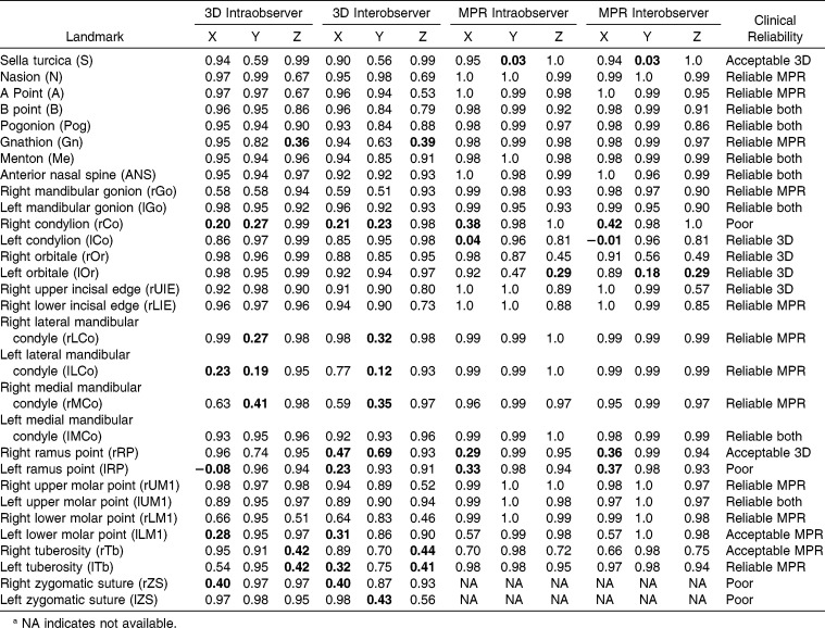
Table 5 shows the estimated reliability by ICC for each landmark and each corresponding coordinate in the intraobserver and interobserver assessments in the visualization of 3D reconstruction and MPR views. Furthermore, it shows the recommendation for clinical use considering the values obtained in the two visualizations, following the classification proposed by the ICC range. The measurements with poor reliability (ICC < 0.45) are in bold.
Table 5.
Reliability Estimated by Intraclass Correlation (ICC) for Each Landmark and Each Coordinate in the Visualization of Three-Dimensional (3D) and Multiplanar (MPR) Reconstructions, and Recommendation for Clinical Use (Values <0.45 Are in Bold)a
The landmarks B, Pg, ME, ANS, rGo, lMCo, and lUM1 showed the best performance in the study, since all values in both visualizations were above 0.75 (reliable), with most being above 0.90 (highly reliable).
All values for one of the visualizations of the landmarks N, A, Gn, rGo, rLIE, rLCo, lLCo, rMCo, rUM1, and lTb (reliable in MPR) and lCo, rOr, lOr, and rUIE (reliable in 3D) were above 0.75 (reliable), and many values of one of the visualizations were above 0.90 (highly reliable).
The values of the landmarks S and rRP (acceptable in 3D) and lLM1 and rTb (acceptable in MPR) were between 0.75 and 0.45 in at least one of the visualizations in at least one axis in the intraobserver or interobserver assessment and should be used with caution.
The values of the landmarks rCo and lRP were <0.45 (poor reliability) in both visualizations in at least one axis in the intraobserver or interobserver assessment, which means either that they are not recommended for clinical use according to the results of the present study or their localization should be modified. The same occurred to the landmarks rZS and eZS, which were not marked in MPR views, but showed poor reliability in 3D image reconstruction.
DISCUSSION
Although some studies3,6–8 have assessed reliability and reproducibility of 3D cephalometric landmarks using CBCT, we found no studies that directly compared visualization of 3D image reconstruction with multiplanar axial, sagittal, and coronal images. Hassan et al.3 found an increase in the precision of identifying landmarks when associated images from MPR views were used with 3D models, but on average double the time was required.
The most widely used software in previous studies was Dolphin 3D (Dolphin Imaging and Management Systems, Chatsworth, Calif), which reinforces the need for resource assessment of other commercial software for clinical use, such as the InVivo.
By working independently after calibration, three observers (one undergraduate, one certification, and one master's student) carried out the landmark identification, unlike other studies2,3 that included only orthodontists. McWilliam and Welander9 affirm that the identification of anatomic landmarks may be related to the level of training of the observers, preferably the most experienced ones. On the other hand, de Oliveira et al.7 conducted a similar experiment to the present study with three observers—one orthodontist, one radiologist, and one undergraduate student. Nevertheless, a larger number of operators with different levels of clinical experience and additional statistical tests could be used in further studies to verify the occurrence of significant differences among operators.
Initially, 30 landmarks were identified on each of the 12 CBCT images, in 3D image visualization at three different time intervals with a 1-week interval between markings, unlike the study of Schlicher et al.8 in which the markings were done at once and Hassan et al.3 in which the second markings were done on the following day. We considered that a longer time interval between the operations is important, so that the anatomic position of the landmarks cannot be memorized. Subsequently, the MPR views were used without the operators having access to 3D visualization, unlike other studies that used MPR images associated with a 3D image.3,7,10–12
In the present study, ICC values were ≤0.45 for 11 landmarks in 3D image visualization, compared with seven points in the MPR image. That is, in general, the identification of landmarks using MPR views proved to be more reliable than the direct marking on the 3D surfaces of the skull. The greater similarity of CBCT images with the commonly used conventional 2D images, especially sagittal images in lateral cephalometry, may have facilitated landmark finding. Additionally, the present study was based on definitions of anatomic location of predefined landmarks7 in MPR images. Furthermore, it might be possible that some structures are not clearly visible in the 3D image reconstructions. It is known that bone density and visibility of the landmark on CBCT is dependent on its location on the skull.10
The frequency of highly reliable values was higher in the intraobserver than in the interobserver assessment in the two types of visualization. The frequency of ICC values in MPR views were >0.75 in 86.92% of the intraobserver assessments, particularly for the coordinate Y (92.9%), while the frequency in interobserver assessment was 84.52%, with frequency of 89.3% for the coordinate Y. These results are similar to those of de Oliveira et al.,7 who found higher frequencies in the intraobserver assessment (91.10%) than in the interobserver assessment (83.2%), but particularly for the coordinate Z (93.33%). The slightly higher results of the previous study may be due to the simultaneous visualization of 3D images and tomographic slices.
In the visualization of the 3D image reconstructions, the ICC value of >0.75 occurred in 74.42% of the intraobserver assessments, also better than the values found in the interobserver assessments (63.66%), particularly for the coordinate Z in both assessments (80.0% and 70.0%, respectively), similar to results found by other studies.2,3,10,12
For the MPR views, intraobserver (86.92%) and interobserver values (84.52%) were less variable when compared with the visualization in 3D image reconstruction (74.42% and 63.66%, respectively). According to Hassan et al.,3 the association between 3D and MPR views increases precision in the identification of cephalometric landmarks.
The sella point (S) showed poor reliability in the MPR views and acceptable reliability in 3D image reconstruction. However, the identification of the landmark S showed high reliability when used for MPR associated with 3D image reconstruction in previous studies.8,12
The identification of the landmarks B, Pg, Me, and SNA showed to be reliable in both image reconstructions. This result is in agreement with Schlicher et al.8 who affirm that the structures in the midsagittal plane are more easily identified because of factors such as ease of identification in the sagittal slice due to similarities with the lateral cephalogram and because they were identified in sequence, with little change in the image position.
On the other hand, the landmark Gn, although it was sagittal, showed low intraobserver and interobserver correlation with respect to the axis Z (sagittal) in the 3D image reconstruction. This result differs from findings of Medelnik et al.,13 which included high dispersion in the localization of Gn for the coordinates X and Y (axial and coronal). A possible explanation, according to Baumrind and Frantz,14 is that reference points located on a prominence or curvature present higher variability compared with the landmarks at defined and plane positions.
The landmarks rUIE, rUM1, and lUM1 showed high reliability in both reconstructions, while the landmarks rTb and lTb showed low reliability in 3D image reconstruction. These results are in agreement with those of Zamora et al.,12 who found high reliability in the identification of central incisors and molars, while the region of tuberosity showed the highest error. The difficulty in locating these anatomic landmarks can be caused by lack of practice since they are not often used in conventional cephalometry.
The left ramus point presented poor reliability in both image reconstructions, while ICC of the homologue was acceptable in 3D image reconstructions. This result differs from a study8 that found more accuracy in identifying landmarks on the left side. Chien et al.15 found low interobserver reliability for the midpoint of the ramus in 3D images when comparing 2D and 3D assessments.
The landmarks rOr and lOr showed high reliability in 3D image reconstruction, agreeing with studies that found no statistically significant differences in intraobserver16 and interobserver17 assessments of this landmark in different sessions. The reliability was poor in MPR views, in agreement with de Oliveira et al.,7 who suggested that the infraorbital landmark, because of its curved surface, could be best identified by 3D image reconstruction. On the other hand, Ludlow et al.18 found accuracy in the identification of landmarks on the orbital anatomy even in reconstructions using axial, coronal, and sagittal slices.
Overall, the landmarks on the condyle presented low reliability in both image reconstructions. Schlicher et al.8 confirmed that landmark identification is difficult in condylar anatomy because of its rounded and irregular structure. Katsavrias and Halazonetis19 found variations in the condylar shape when comparing patients with different malocclusions. This was confirmed when significant individual variability was found in condylar anatomy in different patients using superimposition of 3D CBCT models.20
Based on the results of the present study using commercial software, we suggest that most of the landmarks tested may be used in 3D CBCT cephalometry, where the clinician should consider the type of visualization that generated greater reliability (Table 5).
CONCLUSIONS
The frequency of highly reliable values in the identification of cephalometric landmarks using CBCT was greater in the visualization of multiplanar than in 3D image reconstructions, and it was also higher in intraobserver than in interobserver analyses.
In general, the landmarks on the condyle were those that generated lower reliability; higher reliability was found for those on the midsagittal plane. Depending on the anatomic region, the observer must choose the more reliable type of image visualization.
REFERENCES
- 1.Gribel BF, Gribel MN, Frazäo DC, et al. Accuracy and reliability of craniometric measurements on lateral cephalometry and 3D measurements on CBCT scans. Angle Orthod. 2011;81:26–35. doi: 10.2319/032210-166.1. [DOI] [PMC free article] [PubMed] [Google Scholar]
- 2.Frongia G, Piancino MG, Bracco AA, et al. Assessment of the reliability and repeatability of landmarks using 3-D cephalometric software. Cranio. 2012;30:255–263. doi: 10.1179/crn.2012.039. [DOI] [PubMed] [Google Scholar]
- 3.Hassan B, Nijkamp P, Verheij H, et al. Precision of identifying cephalometric landmarks with cone beam computed tomography in vivo. Eur J Orthod. 2013;35:38–44. doi: 10.1093/ejo/cjr050. [DOI] [PubMed] [Google Scholar]
- 4.Moshiri M, Scarfe WC, Hilgers ML, Scheetz JP, Silveira AM, Farman AG. Accuracy of linear measurements from imaging plate and lateral cephalometric images derived from cone-beam computed tomography. Am J Orthod Dentofacial Orthop. 2007;132:550–560. doi: 10.1016/j.ajodo.2006.09.046. [DOI] [PubMed] [Google Scholar]
- 5.Mozzo P, Procacci C, Tacconi A, Martini PT, Andreis IA. A new volumetric CT machine for dental imaging based on the cone-beam technique: preliminary results. Eur Radiol. 1998;8:1558–1564. doi: 10.1007/s003300050586. [DOI] [PubMed] [Google Scholar]
- 6.Couceiro CP, Vilella OV. 2D/3D Cone-beam CT images or conventional radiography: Which is more reliable. Dental Press J Orthod. 2010;15:72–79. [Google Scholar]
- 7.De Oliveira AEF, Cevidanes LHS, Phillips C, Motta A, Burke B, Tyndall D. Observer reliability of three-dimensional cephalometric landmark identification on cone-beam computerized tomography. Oral Surg Oral Med Oral Pathol Oral Radiol Endod. 2009;107:256–265. doi: 10.1016/j.tripleo.2008.05.039. [DOI] [PMC free article] [PubMed] [Google Scholar]
- 8.Schlicher W, Nielsen Ib, Huang JC, et al. Consistency and precision of landmark identification in three-dimensional cone beam computed tomography scans. Eur J Orthod. 2012;34:263–275. doi: 10.1093/ejo/cjq144. [DOI] [PubMed] [Google Scholar]
- 9.McWilliam JS, Welander U. The effect of image quality on the identification of cephalometric landmarks. Angle Orthod. 1978;48:49–56. doi: 10.1043/0003-3219(1978)048<0049:TEOIQO>2.0.CO;2. [DOI] [PubMed] [Google Scholar]
- 10.Hassan B, Van der Stelt P, Sanderink G. Accuracy of three-dimensional measurements obtained from cone beam computed tomography surface-rendered images for cephalometric analysis: influence of patient scanning position. Eur J Orthod. 2009;31:129–134. doi: 10.1093/ejo/cjn088. [DOI] [PubMed] [Google Scholar]
- 11.Lagravère MO, Low C, Flores-Mir C, et al. Intraexaminer and interexaminer reliabilities of landmark identification on digitized lateral cephalograms and formatted 3-dimensional cone-beam computerized tomography images. Am J Orthod Dentofacial Orthop. 2010;137:598–604. doi: 10.1016/j.ajodo.2008.07.018. [DOI] [PubMed] [Google Scholar]
- 12.Zamora N, Llamas JM, Cibrián R, Gandia JL, Paredes V. A study on the reproducibility of cephalometric landmarks when undertaking a three-dimensional (3D) cephalometric analysis. Med Oral Patol Oral Cir Bucal. 2012;117:678–688. doi: 10.4317/medoral.17721. [DOI] [PMC free article] [PubMed] [Google Scholar]
- 13.Medelnik J, Hertrich K, Steinhäuser-Andresen S, Hirschfelder U, Hofmann E. Accuracy of anatomical landmark identification using different CBCT- and MSCT-based 3D images. An in vitro study [in English, German] J Orofac Orthop. 2011;72:261–278. doi: 10.1007/s00056-011-0032-5. [DOI] [PubMed] [Google Scholar]
- 14.Baumrind S, Frantz R. The reliability of head film measurements. 1. Landmark identification. Am J Orthod. 1971;60:111–127. doi: 10.1016/0002-9416(71)90028-5. [DOI] [PubMed] [Google Scholar]
- 15.Chien PC, Parks ET, Eraso F, Hartsfield JK, Jr, Roberts WE, Ofner S. Comparison of reliability in anatomical landmark identification using two-dimensional digital cephalometrics and three-dimensional cone beam computed tomography in vivo. Dentomaxillofac Radiol. 2009;38:262–273. doi: 10.1259/dmfr/81889955. [DOI] [PubMed] [Google Scholar]
- 16.Park SH, Yu HS, Kim KD, Lee KJ, Baike HS. A proposal for a new analysis of craniofacial morphology by 3-dimensional computed tomography. Am J Orthod Dentofacial Orthop. 2006;129:600.e23–600.e34. doi: 10.1016/j.ajodo.2005.11.032. [DOI] [PubMed] [Google Scholar]
- 17.Olszewski R, Tanesy O, Cosnard G, Zech F, Reychler H. Reproducibility of osseous landmarks used for computed tomography based three-dimensional cephalometric analyses. J Craniomaxillofac Surg. 2010;38:214–221. doi: 10.1016/j.jcms.2009.05.005. [DOI] [PubMed] [Google Scholar]
- 18.Ludlow JB, Gubler M, Cevidanes L, Mold A. Precision of cephalometric landmark identification: cone-beam computed tomography vs conventional cephalometric views. Am J Orthod Dentofacial Orthop. 2009;136:312.e1–312.e10. doi: 10.1016/j.ajodo.2008.12.018. [DOI] [PMC free article] [PubMed] [Google Scholar]
- 19.Katsavrias EG, Halazonetis DJ. Condyle and fossa shape in Class II and Class III skeletal patterns: a morphometric tomographic study. Am J Orthod Dentofacial Orthop. 2005;128:337–346. doi: 10.1016/j.ajodo.2004.05.024. [DOI] [PubMed] [Google Scholar]
- 20.Motta AT, Cevidanes LH, Carvalho FA, Almeida MA, Phillips C. Three-dimensional regional displacements after mandibular advancement surgery: one year of follow-up. J Oral Maxillofac Surg. 2011;69:1447–1457. doi: 10.1016/j.joms.2010.07.018. [DOI] [PMC free article] [PubMed] [Google Scholar]


