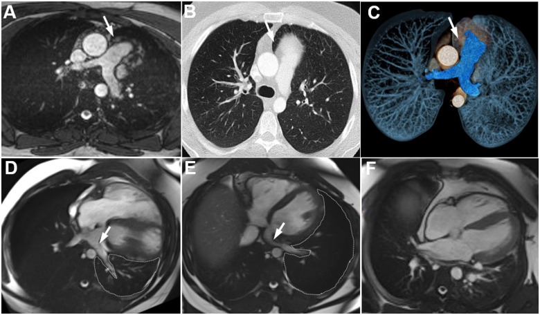
A 27-year-old male was presented at an outpatient cardiology clinic with intermittent but regular chest discomfort and trepopnoea, i.e. dyspnoea when lying in the supine position. However, following a sporting injury with blunt chest trauma several months ago, the symptoms worsened. The electrocardiogram showed a normal sinus rhythm with bradycardia (49 b.p.m.) and right axis deviation (Supplementary material online, Figure S1). Transthoracic echocardiography revealed right ventricular enlargement and paradoxical septal motion (Video 1). Cardiac magnetic resonance (CMR) showed levorotation as well as interposition of lung tissue between the ascending aorta and the main pulmonary artery trunk [Panel A (CMR) and Panels B-C (computed tomography), arrows] suggesting complete left-sided absence of the pericardium. CINE-CMR performed in the lateral decubitus position on both sides revealed impressive cardiac hypermobility (Panel D showing the left side, Video 2; Panel E showing the right side, Video 3). In the left lateral position enlargement of the right ventricle was apparent, as well as a clear displacement of the heart into the left hemithorax with compression of the adjacent lung. However, due to the patient’s symptoms, most likely attributed to motion-dependent cardiac hypermobility with intermittent lung compression (Panels D and E delineations) and constriction of the lower left pulmonary vein between the left atrium and the descending aorta (Panels D and E, arrow), an interdisciplinary decision was made to surgically reconstruct the left pericardium. The post-surgical course showed complete regression of these symptoms (postoperative CMR, Panel F). Accordingly, the thoracic trauma during the soccer game could have sharpened the patient’s awareness of former vague chest discomfort and dyspnoea while moving. In patients with absence of the left pericardium, trepopnoea may worsen when lying on the left side due to increased biventricular volume loading.
The incidence rate of congenital complete unilateral or bilateral pericardial defects is <1 in 10 000 with rare familial occurrence. The majority of these pericardial defects are clinically silent. However, devastating cardiovascular consequences associated with cardiac hypermobility have been reported, including strangulation of myocardial segments or coronary arteries as well as aortic dissection due to persistent strong shear stress of the vascular wall.
Supplementary material
Supplementary material is available at European Heart Journal - Case Reports online.
Consent: The authors confirm that written consent for submission and publication of this case report including images and associated text has been obtained from the patient in line with COPE guidance.
Conflict of interest: None declared.
Funding: None declared.
Supplementary Material
Associated Data
This section collects any data citations, data availability statements, or supplementary materials included in this article.


