Abstract
The tick-borne rickettsia Cowdria ruminantium has been propagated continuously for over 500 days in the Ixodes scapularis tick cell line IDE8 by using the Gardel isolate from bovine endothelial cells as an inoculum. Infection of the tick cells was confirmed by PCR, karyotyping, electron microscopy, and reinfection of bovine cells.
Heartwater is a highly pathogenic, often fatal tick-borne rickettsial disease of domestic ruminants in sub-Saharan Africa and some Caribbean islands (5). Disease control in areas where the disease is endemic depends on intensive acaricide application and, in southern Africa, a live blood vaccine (17). Establishment of Cowdria ruminantium in bovine endothelial cell cultures (2) led to development of inactivated vaccines based on the elementary body (EB) stage of the organism (10, 11, 18), which, although safer than the blood vaccine, are less protective and more expensive to produce and still carry the risks of inducing immunity to bovine products and accidental pathogen transmission. Attempts to grow C. ruminantium in tick cells date back over 25 years (1, 19). Recently, in vitro cultivation of three related tick-borne rickettsiae, Ehrlichia canis, Ehrlichia equi, and Anaplasma marginale, has been achieved (6, 13, 14) in an Ixodes scapularis cell line, IDE8 (12), comprising actively phagocytic hemocyte-like cells with an ability to support growth in vitro of rickettsial species not normally transmitted by this tick in vivo. This paper describes the successful establishment and growth of C. ruminantium in the IDE8 cell line.
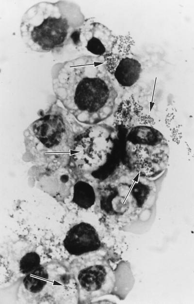
C. ruminantium (Gardel) (20) was maintained in Glasgow minimal essential medium–10% tryptose phosphate broth–10% newborn calf serum in bovine pulmonary artery endothelial cells (BPC) (15). The IDE8 cells were grown in L-15 (Leibovitz) medium–10% tryptose phosphate broth–5% fetal calf serum–0.1% bovine lipoprotein at 30±2°C (12). An IDE8 culture was inoculated with supernatant from an infected BPC culture containing extracellular C. ruminantium EB and immature morulae in association with dying endothelial cells and cell debris, incubated at 37°C overnight, and then returned to 30°C. On day 21 rickettsiae were seen in the extracellular matrix and in vacuoles in a few tick cells. By day 42, 7% of the tick cells were visibly infected, rising to 47% on day 70 (Fig. 1). Subculturing was carried out on day 51, when supernatant was centrifuged at 300 × g for 5 min to remove intact cells, and aliquots were added to each of two IDE8 cultures. Rickettsial inclusions became visible in the IDE8 subcultures within 12 days. A second subculture from the original IDE8 culture was prepared on day 66, when supernatant was transferred directly (without centrifugation and thus containing some intact cells) to a new IDE8 culture; rickettsiae were easily detected in this flask after 9 days. Further subculturing was carried out both as described above and by dividing infected cultures. To date, the infected IDE8 cells have been growing continuously in vitro for over 500 days; they appear healthy, show growth rates comparable to those of uninfected cells, and exhibit very little obvious cytopathic effect, in contrast to E. equi-infected IDE8 cells (13) and C. ruminantium-infected BPC cultures. Infection rates can reach 50% but do not reach the very high levels reported for A. marginale in IDE8 cells (3). Typical spherical EB are not visible in stained preparations.
FIG. 1.
Giemsa-stained cytocentrifuge smear of IDE8 cells on day 70 after inoculation with C. ruminantium, showing rickettsiae within cytoplasmic vacuoles and released from infected cells (arrows). Magnification, ×1,800.
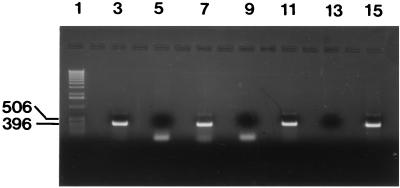
A PCR was carried out (16) on DNA extracted with a commercial kit (Qiagen) from uninfected and rickettsia-infected IDE8 cultures. The primer set consisting of HE1Cow (5′ CAG TTA TTT ATA GCT TCG GCT ATA/G AGT ATC TG) and HE3[S] (5′ GGT ACC GTC ATT ATC TTC CC) was used, designed for specificity at an annealing temperature of 55°C in PCR for amplification of C. ruminantium 16S rRNA gene sequences and an amplicon of 388 bp after 40 thermocycles of PCR. The results confirmed that C. ruminantium genomic DNA was present in the infected IDE8 culture and absent from the uninfected cells (Fig. 2).
FIG. 2.
Agarose gel (1%) electrophoresis of the PCR products amplified from DNA of BPC infected with rickettsiae derived from infected IDE8 cells (lane 3), uninfected IDE8 cells (lane 5), IDE8 cells infected with rickettsiae (lane 7), uninfected BPC (lane 9), BPC infected with C. ruminantium (Gardel) (lane 11), negative control distilled water (lane 13), and positive control C. ruminantium (Gardel) washed EB (lane 15). Molecular weight markers (lane 1) were a 1-kb ladder (Life Technologies).
Karyotyping of IDE8 cells from a culture with a 15% C. ruminantium infection rate was carried out (4) to confirm their tick origin. Chromosome numbers in 100 spreads ranged from 14 to 100, with peaks at 24 (14%) and 26 (14%). Karyotyping done previously on IDE8 cells yielded chromosome numbers ranging from 23 to 30, with a modal number of 28 in >60% of spreads (12). None of the spreads examined in the infected culture had 60 chromosomes, the diploid number for bovine cells (4), indicating that C. ruminantium was growing in IDE8 cells.
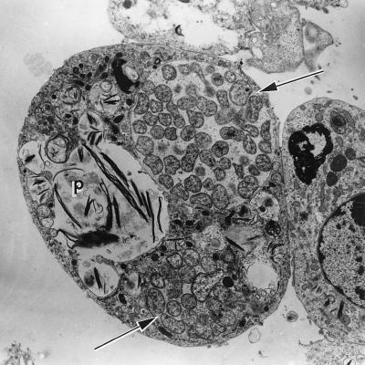
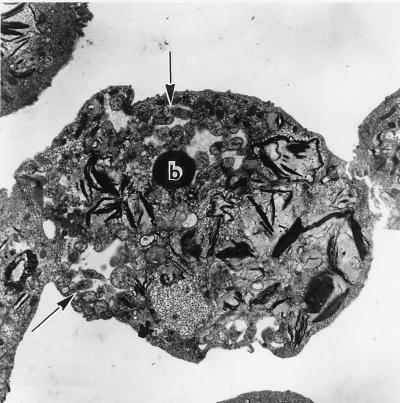
Samples from uninfected and C. ruminantium-infected IDE8 cultures were prepared for transmission electron microscopy by standard techniques. Uninfected IDE8 cells contained vacuoles and inclusions similar to those previously described as phagolysosomes (3, 13, 14). Infected IDE8 cells, however, also had membrane-lined vacuoles containing up to 50 rickettsial organisms 0.4 to 1.5 μm in diameter (Fig. 3 and 4). The organisms had double membranes and were polymorphic, reticulated, and apparently undergoing binary fission, resembling the stages described in nymphal and adult Amblyomma gut epithelium and salivary glands (7, 9). Some rickettsial colonies also contained tiny vesicles visible in the interrickettsial space (Fig. 3), as has been described for A. marginale and E. equi in IDE8 cells (3, 13, 14) or a homogeneous electron-dense inclusion body (Fig. 4) similar to those reported in infected ticks (7, 9). Electron-dense rickettsiae, considered the infective stage for mammalian endothelial cells in vitro (8), were not visible.
FIG. 3.
Electron micrograph of a C. ruminantium-infected IDE8 cell showing reticulated organisms contained within membrane-lined cytoplasmic vacuoles (arrows), small interrickettsial vesicles, and a phagolysosome (p). Magnification, ×5,750.
FIG. 4.
Electron micrograph of a C. ruminantium-infected IDE8 cell showing reticulated organisms contained within cytoplasmic vacuoles (arrows) and an electron-dense inclusion body (b). Magnification, ×5,750.
Infectivity in vitro of the original infected IDE8 culture and cultures with C. ruminantium at passage 2 was demonstrated 68 and 327 days after original inoculation by adding aliquots of supernatant or cell suspension to freshly confluent uninfected BPC; C. ruminantium infection was confirmed by visual examination, preparation and examination of cytocentrifuge smears, PCR (Fig. 2), and subculture onto fresh BPC. Infected IDE8 cultures tested 49, 84, and 348 days after the original inoculation were not infective for BPC.
The lower infection rate for C. ruminantium compared with that for A. marginale in IDE8 cells (3) indicates a need to optimize the culture media and conditions for the tick cell-C. ruminantium system. The apparent absence of EB, the extracellular stage produced in mammalian endothelial cell cultures, from the IDE8 cultures supports the view that development of C. ruminantium in tick cells is significantly different from that in cells of the ruminant host (9). However, since supernatant from infected IDE8 cultures was intermittently infective for BPC, either EB were developing occasionally in these cultures at levels too low to detect visually or EB are not the only stage of C. ruminantium infective for mammalian endothelial cells in vitro.
The successful establishment of C. ruminantium in tick cell culture provides an additional source of material for heartwater vaccination and diagnosis. As the mammalian culture system provided insight into the development of the organism in the ruminant host (8), use of tick cell cultures will lead to increased knowledge and understanding of C. ruminantium development in the vector. In the present-day climate of concern over accidental transmission of viruses and prions of animal origin, such as the bovine spongiform encephalopathy agent, into the food chain and ultimately to humans, the potential advantages of using tick cell lines to replace mammalian cell culture systems for propagation of vector-borne animal pathogens are considerable.
Acknowledgments
This work was supported by grant no. R6566 of the DFID/NRRD Animal Health Research Programme of the United Kingdom Government.
Electron microscopic processing and photography were carried out by Steve Mitchell, EM Unit, Department of Preclinical Veterinary Sciences, Royal (Dick) School of Veterinary Studies, University of Edinburgh, Scotland. C. G. D. Brown's critical comments on the manuscript are appreciated.
REFERENCES
- 1.Andreasen M P. Multiplication of Cowdria ruminantium in monolayer of tick cells. Acta Pathol Microbiol Scand. 1974;82:455–456. doi: 10.1111/j.1699-0463.1974.tb02352.x. [DOI] [PubMed] [Google Scholar]
- 2.Bezuidenhout J D, Paterson C L, Barnard B J H. In vitro cultivation of Cowdria ruminantium. Onderstepoort J Vet Res. 1985;52:113–120. [PubMed] [Google Scholar]
- 3.Blouin E F, Kocan K M. Morphology and development of Anaplasma marginale (Rickettsiales: Anaplasmataceae) in cultured Ixodes scapularis (Acari: Ixodidae) cells. J Med Entomol. 1998;33:656–664. doi: 10.1093/jmedent/35.5.788. [DOI] [PubMed] [Google Scholar]
- 4.Brown C G D. Propagation of Theileria. In: Maramorosch K, Hirumi H, editors. Practical tissue culture applications. New York, N.Y: Academic Press; 1979. pp. 223–254. [Google Scholar]
- 5.Camus E, Barre N, Martinez D, Uilenberg G. Heartwater (cowdriosis): a review. 2nd ed. Paris, France: Office International des Epizooties; 1996. [Google Scholar]
- 6.Ewing S A, Munderloh U G, Blouin E F, Kocan K M, Kurtti T J. Ehrlichia canis in tick cell culture. Proceedings of the 76th Conference of Research Workers in Animal Diseases, Chicago, USA, 13 to 14 November 1995. Ames: Iowa State University Press; 1995. [Google Scholar]
- 7.Hart A, Kocan K M, Bezuidenhout J D, Prozesky L. Ultrastructural morphology of Cowdria ruminantium in midgut epithelial cells of adult Amblyomma hebraeum female ticks. Onderstepoort J Vet Res. 1991;58:187–193. [PubMed] [Google Scholar]
- 8.Jongejan F, Zandbergen T A, Van De Wiel P A, De Groot M, Uilenberg G. The tick-borne rickettsia Cowdria ruminantium has a Chlamydia-like developmental cycle. Onderstepoort J Vet Res. 1991;58:227–237. [PubMed] [Google Scholar]
- 9.Kocan K M, Bezuidenhout J D. Morphology and development of Cowdria ruminantium in Amblyomma ticks. Onderstepoort J Vet Res. 1987;54:177–182. [PubMed] [Google Scholar]
- 10.Mahan S M, Andrew H R, Tebele N, Burridge M J, Barbet A F. Immunisation of sheep against heartwater with inactivated Cowdria ruminantium. Res Vet Sci. 1995;58:46–49. doi: 10.1016/0034-5288(95)90087-x. [DOI] [PubMed] [Google Scholar]
- 11.Martinez D, Maillard J C, Coisne S, Sheikboudou C, Bensaid A. Protection of goats against heartwater acquired by immunisation with inactivated elementary bodies of Cowdria ruminantium. Vet Immunol Immunopathol. 1994;41:153–163. doi: 10.1016/0165-2427(94)90064-7. [DOI] [PubMed] [Google Scholar]
- 12.Munderloh U G, Liu Y, Wang M, Chen C, Kurtti T J. Establishment, maintenance and description of cell lines from the tick Ixodes scapularis. J Parasitol. 1994;80:533–543. [PubMed] [Google Scholar]
- 13.Munderloh U G, Madigan J E, Dumler J S, Goodman J L, Hayes S F, Barlough J E, Nelson C M, Kurtti T J. Isolation of the equine granulocytic ehrlichiosis agent, Ehrlichia equi, in tick cell culture. J Clin Microbiol. 1996;34:664–670. doi: 10.1128/jcm.34.3.664-670.1996. [DOI] [PMC free article] [PubMed] [Google Scholar]
- 14.Munderloh U G, Blouin E F, Kocan K M, Ge N L, Edwards W L, Kurtti T J. Establishment of the tick (Acari: Ixodidae)-borne cattle pathogen Anaplasma marginale (Rickettsiales: Anaplasmataceae) in tick cell culture. J Med Entomol. 1996;33:656–664. doi: 10.1093/jmedent/33.4.656. [DOI] [PubMed] [Google Scholar]
- 15.Mutunga M, Preston P M, Sumption K J. Nitric oxide is produced by Cowdria ruminantium-infected bovine pulmonary endothelial cells in vitro and is stimulated by gamma interferon. Infect Immun. 1998;66:2115–2121. doi: 10.1128/iai.66.5.2115-2121.1998. [DOI] [PMC free article] [PubMed] [Google Scholar]
- 16.Ngumi P N. Characterisation of Cowdria ruminantium (agent of heartwater infection) isolates from Kenya. Ph.D. thesis. Scotland: University of Edinburgh; 1997. [Google Scholar]
- 17.Oberem P T, Bezuidenhout J D. The production of heartwater vaccine. Onderstepoort J Vet Res. 1987;54:485–488. [PubMed] [Google Scholar]
- 18.Tafesse B. Studies on the immunisation of goats and mice against heartwater with inactivated preparation of Cowdria ruminantium (Cowdry 1925). M.S. thesis. Edinburgh, Scotland: University of Edinburgh; 1992. [Google Scholar]
- 19.Uilenberg G. Heartwater (Cowdria ruminantium infection): current status. Adv Vet Sci Comp Med. 1983;27:427–480. [PubMed] [Google Scholar]
- 20.Uilenberg G, Camus E, Barre N. Quelques observations sur une souche de Cowdria ruminantium isolee en Guadeloupe (Antilles francaises) Rev Elev Med Vet Pays Trop. 1985;38:34–42. [PubMed] [Google Scholar]


