PLOS ONE logoLink to PLOS ONE
. 2021 Dec 2;16(12):e0260714. doi: 10.1371/journal.pone.0260714

Patient health records and whole viral genomes from an early SARS-CoV-2 outbreak in a Quebec hospital reveal features associated with favorable outcomes

Bastien Paré 1,2,#, Marieke Rozendaal 1,#, Sacha Morin 3,4, Léa Kaufmann 1,2, Shawn M Simpson 1, Raphaël Poujol 5, Fatima Mostefai 2,5, Jean-Christophe Grenier 5, Henry Xing 1, Miguelle Sanchez 6, Ariane Yechouron 6, Ronald Racette 7, Julie G Hussin 5,8, Guy Wolf 4,9, Ivan Pavlov 7,*, Martin A Smith 1,2,*
Editor: Baochuan Lin10
PMCID: PMC8638998  PMID: 34855869

Abstract

The first confirmed case of COVID-19 in Quebec, Canada, occurred at Verdun Hospital on February 25, 2020. A month later, a localized outbreak was observed at this hospital. We performed tiled amplicon whole genome nanopore sequencing on nasopharyngeal swabs from all SARS-CoV-2 positive samples from 31 March to 17 April 2020 in 2 local hospitals to assess viral diversity (unknown at the time in Quebec) and potential associations with clinical outcomes. We report 264 viral genomes from 242 individuals–both staff and patients–with associated clinical features and outcomes, as well as longitudinal samples and technical replicates. Viral lineage assessment identified multiple subclades in both hospitals, with a predominant subclade in the Verdun outbreak, indicative of hospital-acquired transmission. Dimensionality reduction identified two subclades with mutations of clinical interest, namely in the Spike protein, that evaded supervised lineage assignment methods–including Pangolin and NextClade supervised lineage assignment tools. We also report that certain symptoms (headache, myalgia and sore throat) are significantly associated with favorable patient outcomes. Our findings demonstrate the strength of unsupervised, data-driven analyses whilst suggesting that caution should be used when employing supervised genomic workflows, particularly during the early stages of a pandemic.

Introduction

The first confirmed case of COVID-19 in the province of Quebec, Canada was seen at Verdun Hospital, a 244-bed general adult hospital in Montreal, on February 25, 2020. Community transmission was confirmed in the following weeks, and culminated in a localized outbreak in hospitalized patients. On March 30th, a hospital-wide screening of all admitted asymptomatic patients was performed, 45.2% of whom had detectable levels of SARS-CoV-2 RNA. A policy of universal testing before hospital admission was rapidly established, but did not prevent further smaller localized outbreaks. At the time, there was no publicly available information on SARS-CoV-2 lineage diversity in Quebec. Moreover, reports of asymptomatic and presymptomatic infection and transmission were only beginning to emerge [13]. It has since been established that infected individuals display a range of symptoms of variable clinical severity [47].

In addition to its epidemiological utility, global viral genome sequencing efforts have revealed that SARS-CoV-2 has (and will continue to) quickly evolved, diversified and adapted to the selective pressures of a new mammalian host and large-scale vaccination efforts [8]. The accessibility and affordability of Oxford Nanopore sequencing has facilitated the global adoption of genomic epidemiology during the pandemic. Although once stigmatizing, the error-rate of nanopore sequencing has significantly dropped in the last few years [9, 10], facilitating the reliable generation of consensus sequences–particularly when coupled to expert-backed community-developed analytical pipelines, such as the one rapidly disseminated by the ARTIC Network [11]. The resulting genome consensus is then used for multiple sequence alignment and phylogenetic analysis. Comparing hundreds and thousands of viral genomes and their genetic variants can be a daunting and computationally expensive task. Therefore, genotyping or lineage assignment tools are often used instead of unsupervised approaches for the classification of genomes by leveraging previously-generated phylogenies or lists of curated signature mutations to associate a genome with a pre-defined clade. The most popular lineage assignment tools are Pangolin [12], which uses a supervised learning approach to classify sequences, and Nextstrain/Nextclade [13], which use condensed phylogeny and phylogenetic placement methods, respectively.

An alternative computational strategy for the visualization and interpretation of high-dimensional data, such as that queried when comparing hundreds of viral genomes, is dimensionality reduction. These unsupervised machine learning methods decompose large datasets by projecting dependencies and relationships between data into lower dimensional space (usually 2D). Principal component analysis (PCA), uniform manifold approximation and projection (UMAP) and t-distributed stochastic neighbor embedding (tSNE) are popular dimensionality reduction algorithms commonly used in bioinformatics and genomics [1416]. Potential of heat diffusion for affinity-based transition embedding (PHATE) is a recently developed dimensionality reduction method that was shown to outperform all major dimensionality reduction algorithms at denoising and preserving desirable properties of the surveyed data (Moon et al. 2019). PHATE can be used to extract clusters of data points and as a template to calculate the sample-associated density estimate and relative likelihood, as enabled by the MELD method [17]. Unsupervised learning algorithms can therefore be an efficient alternative to phylogeny for genomic epidemiology, amongst other applications.

In this study, we interrogated the viral genomic diversity and patient outcomes of 242 SARS-CoV-2 infections in a local, first-wave outbreak. We describe the clinical features of this cohort, including symptoms and mortality, and present longitudinal sequencing data from 21 infected individuals. We also compare popular lineage assignment tools with phylogenetic and dimensionality reduction techniques to interrogate the link between viral genomic diversity, symptomology and clinical outcomes.

Results

Clinical observations and outcomes

We retrieved the nasopharyngeal swabs and medical records for 242 individuals (267 samples) who tested positive for the presence of viral nucleic acids between 31 March and 17 April 2020 at Verdun and Notre-Dame hospitals in Montreal, Canada. Table 1 presents general patient data that were included in this study. 163 individuals that participated in this study were female, 79 were male and their age spanned from 2 to 104 years old, with the median age being 50. About half of the participants (134) were hospital employees. 223 individuals presented COVID-19 symptoms before receiving a positive SARS-CoV-2 diagnosis, 21 patients developed symptoms after diagnosis (presymptomatic) and 16 remained asymptomatic. For most patients, symptoms associated with the SARS-CoV-19 infection were the presence of fever, coughing and dyspnea. 87 individuals required hospitalization, of which 23 died during admission. Logistic regression of generic patient data identified age (P>|z| = 0.05520) and the presence of comorbidities (Charlson index >0; P>|z| = 0.00238) over sex, hospital and employee status as the main covariates predictive of mortality, as expected. The Cycle Number (CN) score–a diagnostic measure of viral load from nasopharyngeal samples akin to the cycle threshold (Ct) used in quantitative PCR–was not significantly associated with comorbidities (Wilcoxon rank sum test). However, we observed a slight yet significant positive correlation (0.30, Pearson’s product-moment correlation) between CN and the Charlson comorbidity index. The Charlson comorbidity index is a quantitative metric premised on clinical features and developed to predict the ten-year mortality for a patient who may have a range of comorbid conditions.

Table 1. Cohort summary.

n %
Individuals Total 242
Male 79 32.6
Female 163 66.9
Employee 134 55.4
Hospitalized 87 36
Clinical presentation Symptomatic 205 84.7
Presymptomatic 21 8.7
Asymptomatic 16 6.6
Reason for hospitalization COVID-19 54 62.1
Other 33 37.9
Patient outcome Deceased 23 9.5
Survival or unknown outcome 219 90.5
Age 0–30 27 11.2
31–60 134 55.3
61–104 81 33.5
Charlson comorbidity index 0* 187 77.3
1 7 2.9
2 26 10.7
3 16 6.6
4+ 6 2.5

* Includes individuals with no reported comorbidities.

Viral RNA abundance and bioinformatics parameters affect genome assembly quality

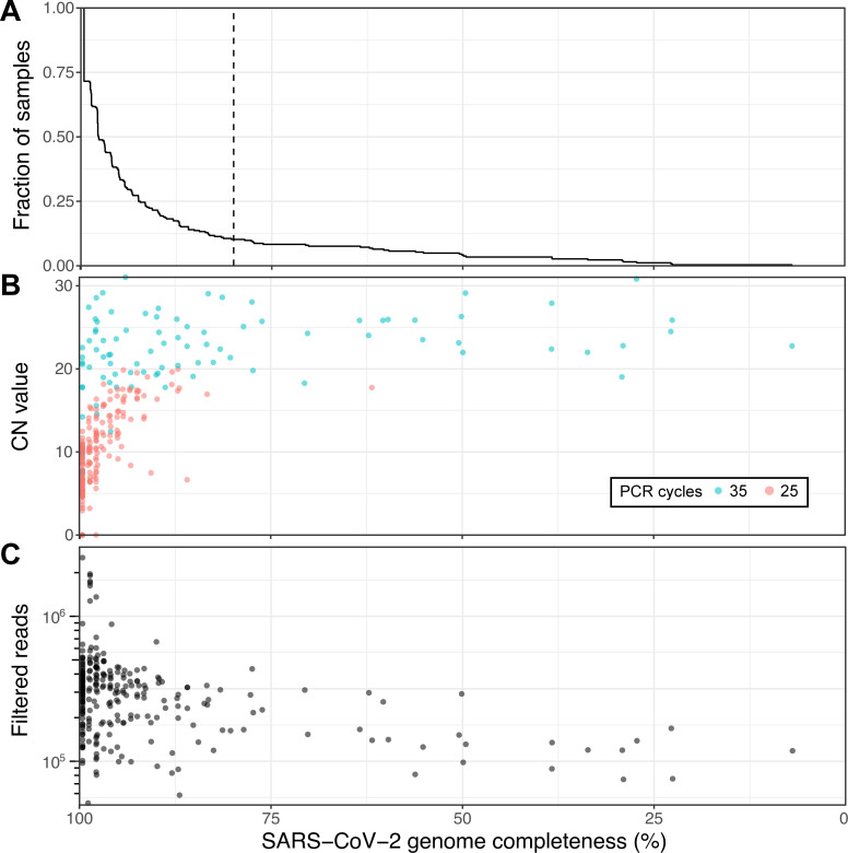
Of the 267 samples, RNA was extracted from 264 samples (240 individuals) and subjected to tiled amplicon sequencing (see Methods), generating a median reference genome coverage (i.e. completeness) of 97.7% from a median of 298,817 filtered reads per sample, including 70 full-length (99.6% complete) genomes (Fig 1 and S1 Table). Of the 12 negative controls, only the sample generated using version 1 of the ARTIC Network protocol generated a false positive genome that could be assigned to a SARS-CoV-2 subclade (S1 Fig, S1 and S2 Tables). A subset of samples (24) with low read coverage was re-sequenced to improve genome completeness and act as a technical replicate (S3 Fig and S4 Table), which recovered 3 samples that were below 80% completeness and another 3 under 90%. Of the 264 sequenced genomes, 207 with at least 90% genome completeness were uploaded to GISAID [18] as soon as the genomes were assembled, including the first publicly disseminated SARS-CoV-2 genomes from Quebec (c.f. submitting laboratory: Smith Laboratory, Centre de Recherche CHU Sainte-Justine).

Fig 1. Viral genome sequencing of 264 SARS-CoV-2 samples with Oxford nanopore.

Fig 1

(A) Cumulative distribution of genome completeness using the ARTIC bioinformatics SOP (see methods). Dashed vertical line corresponds to the 80% completeness threshold used for phylogenetic reconstruction. (B) Relationship between CN score at diagnosis, as measured by the Abbott RealTime M2000rt device (higher CN = lower viral load), and genome completeness. (C) Relationship between number of quality passed reads filtered using the ARTIC bioinformatics SOP and genome completeness.

There is a clear, expected correlation between genome completeness and viral RNA abundance, as measured via the CN score generated during diagnosis (Fig 1B). As the number of PCR cycles used was based on RNA abundance as indicated by the CN score (see Methods) a similar trend in final genome completeness is also observed for this metric. Obtaining full genomes from samples with high CN values required more depth of sequencing given disparities in amplicon coverage, which are exacerbated in samples with lower input RNA. An overview of all amplicons, their abundances and associated negative controls are displayed in S1 Fig. A final set of 237 genomes with at least 80% genome completeness was retained for subsequent analyses.

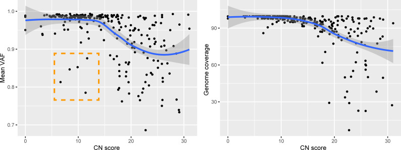
We assessed the impact of two different versions of the ARTIC Network bioinformatics pipeline on consensus genome production: (i) The default version using signal-level correction with Nanopolish and (ii) the experimental version, which uses the Medaka neural network and Longshot variant caller. Excluding regions covered by less than 20 reads, the Nanopolish version generated an average of 2.9 ± 3.5 (standard deviation) ambiguous bases (‘N’) per genome, versus 1.1 ± 2.5 for Medaka, suggesting that the experimental version performs better than the default parameters. Closer inspection of the ambiguous bases in the consensus sequences revealed that these positions were broadly associated with variant allele frequencies (VAF) below ~0.9. We noticed that many of these variant positions were important for SARS-CoV-2 subclade assignment, therefore we replaced ambiguous bases in the consensus sequence with the most dominant variant (for VAF >0.5 only). Interestingly, 5 genomes presented mean VAF (mVAF) scores below 0.9 despite having CN scores <15 and 25 cycles of PCR, suggesting that more than one viral genome haplotype may be present (Fig 2).

Fig 2. Genomic features in relation to RNA abundance at diagnosis.

Fig 2

Average variant allele frequency (VAF) in function of RNA abundance (left). Genome completeness in function of RNA abundance (right). Outliers highlighted in orange. CN = Cycle Number.

Of note, 42/237 genomes with ≥80% completeness had an incomplete S gene and 90/237 had an incomplete N gene. The latter harbors one of the consistently less abundant amplicons from the ARTIC V3 PCR amplification scheme (S1 Fig). However, only 4 unique mutations (7 mutations in total) were observed in 147 genomes with complete N genes, suggesting that the missing sequences are unlikely to contain many mutations. Few mutations were also observed for the S gene; besides the D614G mutation (present in all but one genome), 9 genomes had an A24782G mutation (N1074D substitution) and 7 had a G21641T (A27S substitution).

Unsupervised machine learning outperforms supervised methods at discriminating between viral subclades

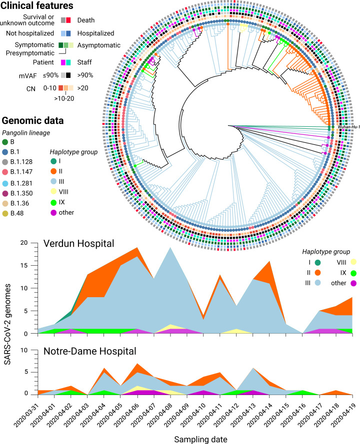
The first batch of 5 SARS-CoV-2 genomes were generated 3 days after receiving all the samples and uploaded to GISAID shortly thereafter. From these first genomes, a preliminary phylogenetic analysis revealed that at least 2 different subclades were present in the Verdun hospital outbreak (not shown). The full phylogenetic relationship of all SARS-CoV-2 genomes with 80% or more completeness is displayed in Fig 3, exposing the presence of a dominant subclade, consistent with suspected nosocomial infection. SARS-CoV-2 lineage classification with Pangolin [12] confirmed that the main cluster mainly consists of subclade B.1, which represents 65.17% (174) of the classified genomes. Other common subclades included B.1.147 (35) and B (35), while a mix of other B lineages was predicted for the remaining samples. NextClade lineage assignment was more conservative, emitting classifications for 205 genomes: 7x 19A, 169x 20A and 29x 20C (S1 Table). When compared to viral diversity from subsequent infections world-wide, only genomes classified as subclade 20C in our samples (corresponding to a subset of the pangolin B.1 classifications) may have persisted and diversified in the human population (S2 Fig).

Fig 3. Genomic and clinical features of 234 SARS-CoV-2 infections.

Fig 3

(Top) Maximum likelihood phylogenetic tree reconstruction of de novo assembled genomes spanning at least 80% of the SARS-CoV-2 reference genome (Wuhan-Hu-1) covered by ≥20 reads and annotated with clinical features of interest. The phylogeny was calculated from a multiple sequence alignment generated with MAFFT [19] using MEGA [20] and visualised with Iroki [21]. Lineage classification performed with Pangolin 2.1.10 [12] (outer circles) and haplotype assignment was performed based on the 20 most common variants in GISAID [18] from the first wave of the pandemic (inner circles). (Bottom) Haplotype diversity over time across two local hospitals. mVAF: Median variant allele frequency, CN: Cycle number.

However, discrepancies were observed between the phylogenetic analysis and Pangolin/NextClade lineage classification, prompting us to employ an alternative genotyping strategy by grouping viral haplotypes based on the 22 most common (allele frequency above 10%) SARS-CoV-2 variants observed in the global community between December 2019 and July 2020, as reported in GISAID on January 20th 2021 (see Methods). The resulting haplotype groups were more consistent with the phylogenetic analysis than the Pangolin/NextClade classifications, prompting us to retain this genotyping strategy in subsequent analyses. A full list of the samples, their assigned subclades, and associated clinical features is listed in S1 Table while the genomic variants used for haplotype assignment are listed in S6 Table. No statistically significant correlation between haplotype assignments and clinical features was observed (Fisher’s exact test, S6 Table).

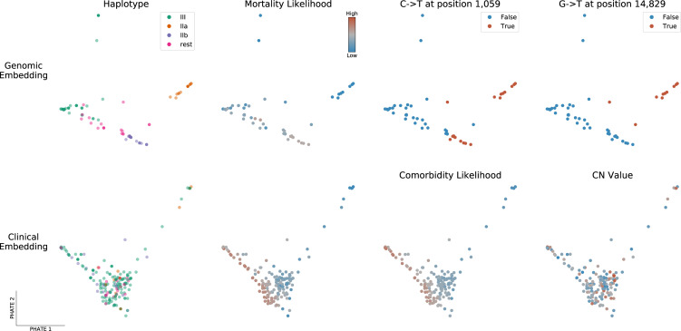
As an alternative to phylogeny, we also applied PHATE [22] to visualize genomic variation and clinical features across all samples (see Fig 4 and Methods). PHATE relies on diffusion geometry to perform nonlinear dimensionality reduction of the data. The resulting representation preserves both local and long-range pairwise similarities, thereby offering a useful way to study how target variables are distributed across the genomic and clinical manifolds.

Fig 4. PHATE embeddings of genomic and clinical features.

Fig 4

Two-dimensional PHATE embeddings of the genomes (Top) and of the clinical features (Bottom). Each marker represents one patient and the embedding location of a given patient indicates feature similarity with surrounding samples as well as dissimilarity with distant ones. The embeddings are unsupervised and the labels of interest are used for coloring only. Specifically, mortality and comorbidity likelihoods were computed using MELD [23], a graph signal processing tool used to smooth a binary variable on the patient-patient graph to determine which regions of its underlying data manifold are enriched or depleted in patients with a specific outcome.

The genomic PHATE embeddings clearly delineate two subclades of haplotype group II, which are supported by the phylogenetic analysis but not by Pangolin lineage assignment (almost all genomes classified as B.1) or NextClade (all 20C). Subclade IIa (16 genomes) likely evolved from IIb (24 genomes) as its members share the same mutations plus 4 additional mutations, 3 of which are non-synonymous: 1150C>T, ORF1a.G295 (synonymous); 4886C>T, ORF1a.P1541S; 14829G>T, ORF1b.M454I; 27964C>T, ORF8.S24L). Interestingly, segregated non-synonymous S gene mutations were observed in both haplogroup IIa/b subclades; 24782A>G (S:N1074D) in 9/16 genomes from IIa and 21641G>T (S: A27S) in 5/24 genomes from IIb. The latter was also present in 2/176 haplogroup III genomes, while few other mutations were observed in the S gene, excluding the T23403G (S:D614G) mutation present in all but one genome. All 9 A24782G mutations in the N gene (see above) were uniquely present in haplogroup IIa genomes, whereas 5/7 G21641T mutations in the S gene were present in haplogroup IIb genomes (the remaining 2 in haplogroup III).

Dimensionality reduction reveals associations between viral subclades, clinical features and patient outcomes

We next queried if there was a link between the observed viral heterogeneity and clinical features. To explore the high-dimensional data, we privileged a dimensionality reduction approach by applying MELD [23] to compute a likelihood gradient across the genomic PHATE embeddings (S4 and S5 Figs). We observed that the haplogroup IIa subclade appeared to be preferentially associated with certain clinical features, prompting us to divide the SARS-CoV-2 genomes into the 4 most dominant haplogroups (IIa, IIb, III and ‘rest’). The PHATE-derived genotype groupings identified that headaches (p-value = 0.01773, Fisher’s exact test), the presence of comorbidities (P = 0.01858) and, to a lesser extent, nausea, vomiting and diarrhea (P = 0.0692) were linked to viral genotypes (S6 Table). The only symptom that was significantly associated with a specific S gene mutation was headaches (mutation S:N1074D, P = 0.01049). Interestingly, patients infected with haplogroup III were significantly more likely to present comorbidities (43/156) than other haplogroups (6/61, P = 0.006173).

Alongside viral genomics, we used PHATE to classify patient samples based on the diverse clinical features surveyed in this cohort (Fig 4, lower panel and S6 and S7 Figs). Coughing and fever are uniformly distributed across the cohort, with the exception of patients requiring breathing assistance for the former. However, patients along the lower-left quadrant are more likely to present severe clinical features (mortality, comorbidities, hospitalization, breathing assistance, etc) whereas those in the upper right were more likely to be employees or present flu-like symptoms (headache, myalgia, sore throat), suggesting that these symptoms are associated with favorable outcomes. Indeed, patients presenting these symptoms were ~10x less likely to die than those that didn’t (odds-ratio 0.0951, P = 9.55x10⁻⁵, Fisher’s exact test). These results indicate that clinical features are more robust indicators of health outcomes than viral genotypes in this patient cohort [2426].

Comparative sampling reveals hospital-acquired transmission of SARS-CoV-2

To validate if the abundance of a particular subclade was due to hospital-acquired transmission or if the observed lineage frequencies were representative of SARS-CoV-2 lineages in the local community, we compared the distribution of viral genotypes between 2 different hospitals (Fig 3, bottom). The nearby Notre-Dame Hospital–which had no reported outbreaks of COVID-19 during the study period–had 29 of 58 (50.0%) infections attributed to haplogroup III, whereas this subclade was assigned to 146 out of 209 infections (69.9%) at Verdun Hospital, consistent with nosocomial transmission.

Longitudinal sequencing of SARS-CoV-2 positive subjects

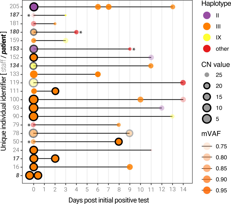
Among this cohort, 21 individuals (mostly hospital employees) were sampled more than once, thus enabling longitudinal analysis (S1 and S7 Tables). Of these, the viral genomes from 13 individuals were assigned a haplotype group for all time points. For all but 3 individuals, the viral load (inversely proportional to the CN value) decreased, while the median genome completeness decreased by 3.4%. As expected, the majority (24/37) of the genomes were composed of haplotype group III. Interestingly, we noticed that some patients presented different viral haplotypes depending on the sampling date (Fig 5). Although most of these occurrences are linked to very high (>25) CN scores–particularly the subsequent samples–some individuals presented more than one viral haplotype with respectable CN scores (individuals 93,100,152,159,205). Only 3 individuals had multiple samples with CN scores below 20, one of which (individual 78, a staff member) produced 2 haplotype III genomes with remarkably lower mVAF values, suggesting possible infection by more than one SARS-CoV-2 subclade.

Fig 5. Longitudinal sequencing of SARS-CoV-2 positive subjects.

Fig 5

Multiple samples for the same individual at different time points are linked by a black line. The size of the points represents the CN score at diagnosis (with black edges corresponding to scores ≤ 20) and the opacity represents the mean variant allele frequency (mVAF). Individual 8 was sampled twice on the same day. Asterisks indicate consensus genomes with <80% completeness.

Discussion

This study provides a description of 264 SARS-CoV-2 genomes associated with 242 infections during one of the first reported local outbreaks in Canada. Quebec is among the provinces most affected by COVID-19 in Canada, with more than 360,000 cases and over 127,000 being confirmed on the island of Montreal alone (as of 2021-05-12). Using nanopore sequencing, we were able to resolve the genetic diversity of SARS-CoV-2 and confirm the presence of multiple subclades of the virus, as well as a dominant subclade indicative of hospital-acquired transmission at the Verdun hospital.

Viral haplotypes and clinical outcomes of SARS-CoV-2

At the beginning of the first wave of the pandemic, reports of asymptomatic infection or transmission were only beginning to emerge. Given that ~15% of individuals were asymptomatic or presymptomatic at the initial diagnosis, we sought to query if the presentation of symptoms (or lack thereof) was preferentially associated with a given viral genotype–a hypothesis that would significantly impact clinical management. The resolved viral haplotypes were largely not associated with clinical outcomes, as confirmed by other studies pertaining to the main viral subclades from the first wave [27, 28]. Medical predispositions and even environmental factors and geographical regions were shown to be important risk factors of severe and deadly cases of SARS-CoV-2 [29, 30]. Notwithstanding, our results suggest that clinical presentations are the predominant prognostic factors to consider when stratifying risk in COVID-19, at least with respect to the viral subclades and patient cohort in question. Interestingly, we report that the presentation of flu-like symptoms (e.g. myalgia, sore throat, headaches, S6 and S7 Figs) appears to be associated with more favorable patient outcomes, potentially indicative of an efficient and protective immune response. Headaches were also reported to be associated with younger age, fewer comorbidities and reduced mortality in a cohort of 379 Spanish COVID-19 patients from March 2020 [31]. Advanced age and the presence of comorbidities were, unsurprisingly, the main correlates of morbidity. The observed correlation between the CN score (viral RNA abundance in nasopharyngeal sample) and the Charlson comorbidity index is counter-intuitive. Possible explanations are the low sample size (55 patients with an index >0) or less productive viral shedding in the nasopharynx in these patients. A less likely explanation could be that these patients present higher levels of anti-SARS-CoV-2 antibodies, which might inhibit viral replication (and, thus, increase the observed CN score) while potentially aggravating their condition through antibody-dependent enhancement [32]. Further serological studies in a larger cohort would be required to confirm this statement.

Genomic diversity of SARS-CoV-2 in an early outbreak

The phylogenetic diversity of SARS-CoV-2 genomes we report in this local outbreak is consistent with the genomic diversity observed across Quebec at the time, specifically subclades B and B.1 [33]. However, we found that different lineage assignment methods we employed (Pangolin, NextClade, phylogeny, dominant haplogroups and PHATE dimensionality reduction) produced disparate results. Pangolin subclades localized to divergent branches of the maximum-likelihood tree we generated from multiple sequence alignments. The latter may be prone to grouping sequences with common amplicon dropouts, a common occurrence in our data, as we sequenced all SARS-CoV-2 positive samples. This composes a unique feature of our study, as current sequencing endeavours ignore samples with lower viral mRNA abundance (i.e. CN >20 or Ct >30). Despite the relatively low abundance of viral RNA in some of the samples, the majority of the consensus genomes we report are over 90% complete. The discrepancies that were observed between the phylogenic analysis and Pangolin classification are very unlikely to be associated with sequencing errors. Even though single-molecule sequencing is generally associated with a high single-read error rate (4–5% in the case of our data), the ARTIC pipeline has integrated error correction methods, based on banded event alignment or deep neural-networks. The fact that the default ARTIC parameter for the number of filtered reads was changed from 200 to 2000 per strand (see Material and Methods section) for our analysis ensures superior consensus accuracy. We can therefore assume that the majority of observed variants are bona fide mutations even though sequencing artifacts could be present in the data regardless of the sequencing technology used.

In April 2020, no publicly available SARS-CoV-2 genomes were available for Quebec. Given the rapid turnaround time of nanopore sequencing, we were able to upload the first 5 SARS-CoV-2 genomes from Quebec to GISAID [18] within a week of receiving the samples from Verdun hospital in a newly established laboratory with limited equipment and reagents. We anticipated that profiling the viral genomic diversity could be a useful epidemiology tool, potentially identifying specific transmission events and targeting specific measures to reduce hospital-acquired infections. However, this proved to be difficult given the relatively low genomic diversity observed in these ‘first wave’ samples and the short timespan between diagnoses. Nonetheless, the relatively higher frequency of the B.1/Haplogroup III in Verdun Hospital versus Notre-Dame Hospital indeed supports the suspected hospital-acquired infections and nosocomial transmission in this establishment. This is also substantiated by the significant enrichment of haplogroup III in patients with comorbidities, suggesting that this viral subclade may have been preferentially transmitted among hospitalized patients potentially sharing the same room or wing of the hospital. Indeed, this was to be anticipated given that the systematic use of personal protective equipment by both employees and patients was not mandated early in the COVID-19 pandemic. This also supports the observed presence of ‘chimeric’ genomes in our cohort, which may represent dual infection events caused by 2 different viral haplotypes, although we cannot rule out the unlikely possibility that this is a consequence of cross-contamination in these samples, despite the use of negative controls.

In contrast to Pangolin lineage assignment, we found that an ad hoc haplotype grouping strategy was globally consistent with the phylogenetic analysis and PHATE clusters. However, both Pangolin and the haplotype grouping failed to identify a significantly divergent subclade of SARS-CoV-2 in this outbreak (IIa/IIb), supporting the use of unsupervised methods for genomic sequence analysis (when possible). Indeed, when considering these additional subclades, we were able to identify a statistically significant association between subclade IIa and the presence of comorbidities and the presentation of headaches, albeit the latter may be a confounding effect of small sample size rather than a consequence of viral evolution itself. Recently, Mostefai et al. successfully used this method on all 2020 SARS-CoV-2 genomes available in GISAID, suggesting it can be used on datasets other than the one presented in this paper, containing more genomes and/or lineages [34].

Recent improvements to SARS-CoV-2 whole genome sequencing by tiled PCR amplicons, including the availability of ligation-based molecular barcodes for 96 samples, and to lineage assignment [12] have greatly facilitated the reliable analysis of SARS-CoV-2 genomes using nanopore sequencing [35]. The experimental and bioinformatics standard operating procedures developed by the ARTIC Network for SARS-CoV-2 [11] have been essential for the rapid and cost-effective sequencing of SARS-CoV-2, which can be performed from start to finish in less than 24h. These standard operating procedures were developed and optimized taking into consideration the best practices or analysis of SARS-CoV-2 that were discussed and published since the beginning of the pandemic [3539].

We found that the medaka version of the ARTIC bioinformatics standard operating procedure provided consensus genomes with less ambiguous bases, as reported by others. However, we believe several improvements can be made to the standard bioinformatics analysis of SARS-CoV-2 nanopore sequencing data. Firstly, much of the ARTIC computational pipeline is single-threaded and could be parallelized to accelerate the bioinformatics turnaround time (particularly for smaller, independent labs with limited computing facilities). Secondly, we believe the default parameters may be a source of technical variation. We found that the quality filtering steps are robust, but the consensus generation steps––which assume a single subclade/haplotype is present––may be a source of artefacts, particularly when confronted to infections with more than one viral subclade or high CN/Ct scores (although many of these issues have been resolved by the global community since the commencement of this study). These might include ‘chimeric’ genomes where amplicon segments might alternate between predominant haplotypes in the consensus, which can explain some of the genomic outliers observed in this study (c.f. Fig 4, top row).

In conclusion, we posit that the rapid nanopore sequencing protocols for SARS-CoV-2, the accessibility of the platform, its low cost and ease of use are significant arguments for the widespread use of this platform for genomic epidemiology in local and global outbreaks. The rise of SARS-CoV-2 variants of concern around the globe combined with the progressive easing of public health restrictions and mass vaccination programs are additional reasons to implement rapid, lightweight genomic surveillance protocols, ideally directly at the point of care or diagnostic laboratory. For instance, performing nanopore sequencing of SARS-CoV-2 samples directly after positive diagnosis in a decentralized manner would (i) save on costs associated with RNA extraction and reverse-transcription, as SARS-CoV-2 sequencing requires the same material as diagnosis by quantitative PCR; (ii) identify genomic variants with a turnaround time of 24-48h; (iii) potentially provide recommendations for clinical management based on the identified variants; and (iv) facilitate effective, variant-focused contact-tracing measures during follow-up with the patient, which is impractical if not impossible to achieve with centralized, off-site next generation sequencing facilities.

Materials and methods

Diagnosis, sample collection and RNA extraction

Consecutive positive samples of SARS-CoV-2 collected from March 30th to April 17th, 2020, were provided by the clinical microbiology laboratory of Verdun Hospital. Those samples were collected from hospitalized patients, patients seen in the emergency department, and healthcare workers. The laboratory also processed samples from the associated Notre-Dame Hospital, a 250-bed general hospital located 10.3 km from Verdun Hospital, and those were included in the analysis.

Standard nasopharyngeal (NP) swabs were collected from patients and suspended in an RNA preservation and lysis solution. RNA extraction was then performed using TRIzol LS following the manufacturer’s protocol (Invitrogen, California, United States of America). The presence of SARS-CoV-2 was detected using the Abbott RealTime SARS-CoV-2 assay (Abbott, Chicago, Illinois, United States of America) on an Abbott RealTime M2000rt, a qualitative multiplex real time PCR device that has FDA emergency use authorization for in vitro diagnostic use. This qualitative test gives a CN value for positive samples that is reminiscent of quantitative PCR values. CN values were manually extracted from digitized copies of the M2000rt reports.

Nanopore sequencing

For each SARS-CoV-2 positive sample, 250 μl aliquots of NP swab were collected and stored at -80°C until RNA extraction. 750 μl of TRIzol LS (Invitrogen, California, United States of America) was added to 250 μl of NP solution and RNA extraction was performed according to the manufacturers’ recommendations. Final RNA pellets were resuspended in 15μl of nuclease-free water (Life Technologies, California, United States of America). For samples with a CN > 20, two aliquots were used for extraction, pellets were suspended in 10 μl of nuclease-free water and then pooled. 11 μl of the RNA was used for subsequent reverse transcription using SuperScript IV reverse transcriptase (Life Technologies, California, United States of America) and random hexamer primers (Life Technologies, California, United States of America).

Library preparation was performed following the Arctic Network nCov19 sequencing protocol version 1 (dx.doi.org/10.17504/protocols.io.bbmuik6w) and using individual V3 PCR primers (Life Technologies, California, United States of America) for samples included in the first sequencing run (e.g. “verd1”). The number of PCR cycles was determined based on the CN values: 25 cycles for a CN ≤ 7; 35 cycles for a CN > 7. For samples with a CN > 20, RNA was extracted from two aliquots, and 35 PCR cycles were used. Library preparation for runs verd2 and verd3 was performed following the similar Oxford Nanopore Technologies (ONT) PCR tiling of COVID-19 virus protocol and using manually pooled, individual ARTIC V3 PCR primers. The rest of the libraries were obtained and barcoded following the Oxford Nanopore Technologies PCR tiling of COVID-19 virus protocol and using the ARTIC nCoV-2019 V3 Panel (10006788, Integrated DNA Technologies, Iowa, United States of America) and ONT Ligation Sequencing Kit (SQK-LSK109, ONT, Oxford, United Kingdom). Two minor changes were made to the protocol, based on comparison with the Arctic Network nCov19 sequencing protocol version 2 (https://dx.doi.org/10.17504/protocols.io.bdp7i5rn): (i) At the reverse transcription step, samples were incubated for 5 minutes at 25°C before incubation at 42°C and (ii) samples were incubated for 10 minutes at both 20°C and 65°C during the end-prep step. Each sample was barcoded using Native Barcoding Expansion 1–12 and 13–24 kits (EXP-NBD104 and EXP-NBD114, ONT, Oxford, United Kingdom) and sequencing performed on FLO-MIN006 (R9.4.1) flow cells using MinION MK1b, MinION MK1c and GridION sequencers (ONT, United Kingdom). Detailed technical information on the sequencing runs is listed in S2 Table.

Data processing and consensus sequence generation

The raw sequencing files (.fast5) were base called offline with the proprietary basecaller Guppy, version 4.4.1 (ONT, Oxford, United Kingdom) using the R9.4.1 high accuracy configuration file. Base called reads were demultiplexed using Guppy with parameters “—require_barcodes_both_ends—arrangements_files barcode_arrs_nb12.cfg barcode_arrs_nb24.cfg". The resulting reads were then size selected between 400 and 700 nt in length and subjected to the ARTIC Network Bioinformatics protocol (https://artic.network/ncov-2019/ncov2019-bioinformatics-sop.html) to generate full-length consensus genomes. Both the “nanopolish” and “medaka” parameters were used with default parameters, with the sole exception of increasing the “—normalise” parameter from 200 to 2000 for both commands.

The medaka consensus genomes were further processed to replace ambiguous (‘N’) bases at positions with lower variant allele frequencies, a consequence of (overly) strict variant filtering in the Medaka pipeline. This was performed by merging the “pass” and “fail” intermediary.vcf files, and filtering variants using the following parameters: (i) supported by at least 20 reads; (ii) not located in masked-regions as determined by the “coverage_mask.txt” files produced by the ARTIC pipeline; and (iii) present at a frequency above 50%. Resulting variants were inserted in the consensus sequence. The associated scripts can be found at https://github.com/TheRealSmithLab/Verdun. The coverage mask files were used to calculate genome completeness and to retain sequences for subsequent phylogenetic analyses.

Phylogenetics

Assembled SARS-CoV-2 genome sequences with 80% or more completeness and the Wuhan-Hu-1 isolate reference genome (Genbank reference MN908947.3) were submitted to a multiple sequence alignment with MAFFT v7.475 using parameters “—maxiterate 500”. The resulting multiple sequence alignment was used to generate a maximum-likelihood phylogeny using MEGA X [20]. The evolutionary history was inferred by using the Maximum Likelihood method and Tamura-Nei model [40]. The tree with the highest log likelihood was retained. Initial tree(s) for the heuristic search were obtained automatically by applying Neighbor-Joining and BioNJ algorithms to a matrix of pairwise distances estimated using the Tamura-Nei model, and then selecting the topology with superior log likelihood value. The tree is drawn to scale, with branch lengths measured in the number of substitutions per site. This analysis involved 235 nucleotide sequences. Codon positions included were 1st+2nd+3rd+Noncoding. All positions with less than 90% site coverage were eliminated, i.e. fewer than 10% alignment gaps, missing data, and ambiguous bases were allowed at any position (partial deletion option).

Lineage calling

Lineage classification was performed using the Phylogenetic Assignment of Named Global Outbreak LINeages (Pangolin) software package (version 2.1.10) proposed by [12], using default parameters, as well as using the Nextstrain: real-time tracking of pathogen evolution (SARS-CoV-2 pipeline:2021/09/15). Nextstrain was built with a multiple input build-config (no filtering) using our sequences and metadata along with GISAID sequences and metadata (on 2021/09/16) [13].

Haplotype grouping

A subset of 20 positions were selected to define viral haplotypes that represent large groups of sequences defined based on the worldwide most common genetic variants (using GISAID consensus sequences as of January 14th 2021) [34]. The 20 nucleotide positions are: 241, 313, 1059, 1163, 3037, 7540, 8782, 14408, 14805, 16647, 18555, 22992, 23401, 23403, 25563, 26144, 28144, 28881, 28882, 28883. These 20 mutations were selected because they exceeded 10% in variant allele frequency in at least one of the months of the first wave of the pandemic (January to July 2020) in GISAID consensus sequences. Further details on these haplotypes can be found in Mostefai et al. 2021. The definition of haplotypes based on these 20 mutations found in samples from this study, and their corresponding NextStrain clades, is reported in S5 Table. We note that haplotype III and IX are grouped within the same Nexstrain clade, despite the fact that they differ at position 25563, and both differ from haplotype II (NextStrain clade 20C) at position 14408.

PHATE embeddings

PHATE embeddings [22] were computed independently for viral variants (257 features) and symptoms (43 features). In both cases, the input data for a given patient consisted of a binary encoding, with a 1-value for the presence of a given variant or symptom, and a 0-value for the absence thereof. PHATE is then applied to find a low-dimensional representation that preserves the geometry of the high-dimensional samples. The overall structure of the embeddings was relatively robust to the choice of hyperparameters. We therefore used the default parameters for PHATE, except for the diffusion-time parameter t, which was set to 30 for both the genotype and clinical data to display cleaner branches. Similarly, we set the knn value of MELD [23] (i.e. the number of considered neighbors) to 5 to be consistent with the PHATE default and the beta parameter (i.e. the amount of smoothing to apply) to 20 to avoid over-smoothing. We used PHATE 1.0.4 and MELD 1.0.0. Source code is respectively available at https://github.com/KrishnaswamyLab/PHATE and https://github.com/KrishnaswamyLab/MELD.

Clinical data extraction

Data from patient files were extracted and entered in a database using a standardized case report form by an experienced research assistant (RR) and cross checked in full by IP.

Ethics statement

Human research ethics approval for this study (MP-21-2021-2938) was provided by the CHU Sainte-Justine Research Centre ethics committee (FWA00021692) designated by the Quebec provincial government. Due to the retrospective nature of the study, and the absence of risk for participants, the need for consent was waived by the ethics committee, both for inclusion in the study, and for access to medical records for the purpose of data extraction. All patient samples have been de-identified.

Supporting information

S1 Fig. Amplicon coverage for all samples.

Amplicon coverage for each sample, including the controls, was calculated using bedtools using a 90% overlap between the query and the target (ARTIC Network amplicon coordinates) as well as 80% overlap between the target and the query.

(EPS)

S2 Fig. Nextstrain genomic epidemiology of SARS-CoV-2.

Time-resolved phylogenetic tree of all genomes reported in our cohort (red dots) in the context of subsequent viral evolution (subsampled genomes from Nexstrain) during the COVID-19 pandemic (as of September 2021) produced via the Nextstrain SARS-CoV-2 pipeline.

(EPS)

S3 Fig. Technical replicates for a selection of samples.

Two separate nanopore sequencing library preparations (1 & 2) from the same PCR products and the corresponding merged data (merged) on the horizontal axis. Results generated from the Medaka version of the ARTIC Network bioinformatics SOP. Genotype data generated with modified consensus genomes that contain the most frequent variant (> 50%) at any given position.

(EPS)

S4 Fig. PHATE embedding of SARS-CoV-2 genomic variation.

Each point corresponds to one SARS-CoV-2 genome annotated with various clinical labels and symptoms. The 15 most frequent variants are annotated in the bottom panel. Sub-clade IIa (right cluster) is characterized by a number of variants differentiating it from sub-clade IIb (tip of the lower branch), see Fig 4, top left panel.

(EPS)

S5 Fig. MELD relative likelihood estimates based on viral genomic variation. Likelihoods of various clinical labels and symptoms are displayed over the PHATE embedding of SARS-CoV-2 genomes (see S3 Fig).

The subclade IIa cluster (top right right) appears depleted in adverse outcomes (mortality and hospitalization) and enriched in flu-like symptoms (sore throat, fatigue and headache). Conversely, the sub-clade IIb region (tip of the lower-right cluster) is slightly enriched in hospitalized patients and shows a higher likelihood of DEG/Confusion. MELD likelihoods of the 15 most frequent variants are displayed over the PHATE embedding of the genomes in the bottom panels. It should be stressed that MELD computes a local relative likelihood. Some manifold regions may be relatively depleted in a specific variant compared to other regions even if said variant is frequent in absolute terms, particularly in the case of 23403A>G, 3037C>T and 14408C>T.

(EPS)

S6 Fig. PHATE embedding of clinical features.

Each point corresponds to one patient sample annotated with various clinical labels and symptoms (N.B. some samples correspond to the same patient, see Longitudinal Sequencing section). The 15 most frequent variants are annotated in the bottom panel. Sub-clade IIa (right cluster) is characterized by a number of variants differentiating it from sub-clade IIb (tip of the lower branch), see Fig 4, top left panel.

(EPS)

S7 Fig. MELD relative likelihood estimates based on clinical features.

Likelihood estimates of various clinical labels and symptoms displayed over the PHATE embedding of the clinical features (see S5 Fig). The likelihood gradients of adverse out- comes (mortality, hospitalization and breathing assistance) are well aligned with comorbidity and DEG/Confusion gradients. Moreover, adverse outcome likelihoods appear to be inversely correlated with employee status as well as a set of flu-like symptoms (sore throat, myalgia, fatigue and headache). The MELD relative likelihood estimates of the 15 most frequent variants displayed over the PHATE embedding of the symptoms in the bottom panel.

(EPS)

S1 Table. Sample overview.

(XLSX)

S2 Table. Run statistics.

(XLSX)

S3 Table. Negative controls.

(XLSX)

S4 Table. Technical replicates.

(XLSX)

S5 Table. Haplotype features.

(XLSX)

S6 Table. Genotype-phenotype comparisons.

(XLSX)

S7 Table. Longitudinal samples.

(XLSX)

Acknowledgments

We would like to thank members of the Mila COVID19 Task Force for their camaraderie and valuable insight into integrative data analysis strategies during the pandemic. Unrestrained gratitude and appreciation are expressed for members of the ARTIC Network, GISAID, and Nextstrain for their efforts and invaluable contribution to SARS-CoV-2 genomics. Thanks are owed to Jared Simpson and John Tyson for discussions relating to the interpretation of nanopore sequencing data, to Ioannis Ragoussis and Sarah Reiling for assistance with control experiments, to François Fontaine and Valérie Villeneuve for acquisition of equipment and consumables, and to essential workers during the pandemic.

Data Availability

Basecalled, demultiplexed and size-filtered reads can be found via SRA under bioproject PRJNA730334. Scripts, statistical analyses, consensus genomes and variant files can be found at: https://github.com/TheRealSmithLab/Verdun.

Funding Statement

SM is supported by an IVADO MSc excellence scholarship and an FRQNT B1X scholarship. JGH is a Fonds de Reherche du Québec en Santé Research Scholar (252997) funded by IVADO COVID19 Rapid Response grant (CVD19-030) and the Montreal Heart Institute Foundation. GW is supported by Canada CIFAR AI Chair. MAS is supported by a Fonds de Reherche du Québec en Santé Junior 1 fellowship (295760).

References

  • 1.Wei WE, Li Z, Chiew CJ, Yong SE, Toh MP, Lee VJ. Presymptomatic Transmission of SARS-CoV-2—Singapore, January 23–March 16, 2020. MMWR. Morbidity and Mortality Weekly Report. 2020. pp. 411–415. doi: 10.15585/mmwr.mm6914e1 [DOI] [PMC free article] [PubMed] [Google Scholar]
  • 2.Qian G, Yang N, Ma AHY, Wang L, Li G, Chen X, et al. COVID-19 Transmission Within a Family Cluster by Presymptomatic Carriers in China. Clin Infect Dis. 2020;71: 861–862. doi: 10.1093/cid/ciaa316 [DOI] [PMC free article] [PubMed] [Google Scholar]
  • 3.Kimball A, Hatfield KM, Arons M, James A, Taylor J, Spicer K, et al. Asymptomatic and Presymptomatic SARS-CoV-2 Infections in Residents of a Long-Term Care Skilled Nursing Facility—King County, Washington, March 2020. MMWR Morb Mortal Wkly Rep. 2020;69: 377–381. doi: 10.15585/mmwr.mm6913e1 [DOI] [PMC free article] [PubMed] [Google Scholar]
  • 4.Nakamichi K, Shen JZ, Lee CS, Lee A, Roberts EA, Simonson PD, et al. Hospitalization and mortality associated with SARS-CoV-2 viral clades in COVID-19. Sci Rep. 2021;11: 4802. doi: 10.1038/s41598-021-82850-9 [DOI] [PMC free article] [PubMed] [Google Scholar]
  • 5.Young BE, Wei WE, Fong S-W, Mak T-M, Anderson DE, Chan Y-H, et al. Association of SARS-CoV-2 clades with clinical, inflammatory and virologic outcomes: An observational study. EBioMedicine. 2021;66: 103319. doi: 10.1016/j.ebiom.2021.103319 [DOI] [PMC free article] [PubMed] [Google Scholar]
  • 6.Knight D, Downes K, Munipalli B, Halkar MG, Logvinov II, Speicher LL, et al. Symptoms and Clinical Outcomes of Coronavirus Disease 2019 in the Outpatient Setting. SN Compr Clin Med. 2021; 1–8. doi: 10.1007/s42399-021-00746-1 [DOI] [PMC free article] [PubMed] [Google Scholar]
  • 7.Frampton D, Rampling T, Cross A, Bailey H, Heaney J, Byott M, et al. Genomic characteristics and clinical effect of the emergent SARS-CoV-2 B.1.1.7 lineage in London, UK: a whole-genome sequencing and hospital-based cohort study. Lancet Infect Dis. 2021;21: 1246–1256. doi: 10.1016/S1473-3099(21)00170-5 [DOI] [PMC free article] [PubMed] [Google Scholar]
  • 8.Hamelin David J., Fournelle Dominique, Grenier Jean-Christophe, Schockaert Jana, Kovalchik Kevin A., Kubiniok Peter, et al. 2021. “The Mutational Landscape of SARS-CoV-2 Variants Diversifies T Cell Targets in an HLA-Supertype-Dependent Manner.” Cell Systems, October. 10.1016/j.cels.2021.09.013. [DOI] [PMC free article] [PubMed] [Google Scholar]
  • 9.Kono N, Arakawa K. Nanopore sequencing: Review of potential applications in functional genomics. Dev Growth Differ. 2019;61: 316–326. doi: 10.1111/dgd.12608 [DOI] [PubMed] [Google Scholar]
  • 10.Sahlin K, Medvedev P. Error correction enables use of Oxford Nanopore technology for reference-free transcriptome analysis. Nat Commun. 2021;12: 2. doi: 10.1038/s41467-020-20340-8 [DOI] [PMC free article] [PubMed] [Google Scholar]
  • 11.Tyson JR, James P, Stoddart D, Sparks N, Wickenhagen A, Hall G, et al. Improvements to the ARTIC multiplex PCR method for SARS-CoV-2 genome sequencing using nanopore. bioRxiv. 2020. doi: 10.1101/2020.09.04.283077 [DOI] [PMC free article] [PubMed] [Google Scholar]
  • 12.Rambaut A, Holmes EC, O’Toole Á, Hill V, McCrone JT, Ruis C, et al. A dynamic nomenclature proposal for SARS-CoV-2 lineages to assist genomic epidemiology. Nat Microbiol. 2020;5: 1403–1407. doi: 10.1038/s41564-020-0770-5 [DOI] [PMC free article] [PubMed] [Google Scholar]
  • 13.Hadfield J, Megill C, Bell SM, Huddleston J, Potter B, Callender C, et al. Nextstrain: real-time tracking of pathogen evolution. Bioinformatics. 2018;34: 4121–4123. doi: 10.1093/bioinformatics/bty407 [DOI] [PMC free article] [PubMed] [Google Scholar]
  • 14.Arowolo MO, Adebiyi MO, Aremu C, Adebiyi AA. A survey of dimension reduction and classification methods for RNA-Seq data on malaria vector. Journal of Big Data. 2021;8: 50. [Google Scholar]
  • 15.Dorrity MW, Saunders LM, Queitsch C, Fields S, Trapnell C. Dimensionality reduction by UMAP to visualize physical and genetic interactions. Nat Commun. 2020;11: 1537. doi: 10.1038/s41467-020-15351-4 [DOI] [PMC free article] [PubMed] [Google Scholar]
  • 16.Kobak D, Berens P. The art of using t-SNE for single-cell transcriptomics. Nat Commun. 2019;10: 5416. doi: 10.1038/s41467-019-13056-x [DOI] [PMC free article] [PubMed] [Google Scholar]
  • 17.Stawinski PM, Dziadkowiec KN, Al-Abbasi B, Suarez L, Simms L, Dewaswala N, et al. Model of End-Stage Liver Disease (MELD) Score as a Predictor of In-Hospital Mortality in Patients with COVID-19: A Novel Approach to a Classic Scoring System. Cureus. 2021;13: e15179. doi: 10.7759/cureus.15179 [DOI] [PMC free article] [PubMed] [Google Scholar]
  • 18.Elbe S, Buckland-Merrett G. Data, disease and diplomacy: GISAID’s innovative contribution to global health. Global Challenges. 2017. pp. 33–46. doi: 10.1002/gch2.1018 [DOI] [PMC free article] [PubMed] [Google Scholar]
  • 19.Katoh K, Standley DM. MAFFT multiple sequence alignment software version 7: improvements in performance and usability. Mol Biol Evol. 2013;30: 772–780. doi: 10.1093/molbev/mst010 [DOI] [PMC free article] [PubMed] [Google Scholar]
  • 20.Kumar S, Stecher G, Li M, Knyaz C, Tamura K. MEGA X: Molecular Evolutionary Genetics Analysis across Computing Platforms. Mol Biol Evol. 2018;35: 1547–1549. doi: 10.1093/molbev/msy096 [DOI] [PMC free article] [PubMed] [Google Scholar]
  • 21.Moore RM, Harrison AO, McAllister SM, Polson SW, Eric Wommack K. Iroki: automatic customization and visualization of phylogenetic trees. PeerJ. 2020;8. doi: 10.7717/peerj.8584 [DOI] [PMC free article] [PubMed] [Google Scholar]
  • 22.Moon KR, van Dijk D, Wang Z, Gigante S, Burkhardt DB, Chen WS, et al. Visualizing structure and transitions in high-dimensional biological data. Nat Biotechnol. 2019;37: 1482–1492. doi: 10.1038/s41587-019-0336-3 [DOI] [PMC free article] [PubMed] [Google Scholar]
  • 23.Burkhardt DB, Stanley JS, Tong A, Perdigoto AL, Gigante SA, Herold KC, et al. Quantifying the effect of experimental perturbations at single-cell resolution. Nature Biotechnology. 2021;39: 619–629. doi: 10.1038/s41587-020-00803-5 [DOI] [PMC free article] [PubMed] [Google Scholar]
  • 24.Shahriarirad R, Khodamoradi Z, Erfani A, Hosseinpour H, Ranjbar K, Emami Y, et al. Epidemiological and clinical features of 2019 novel coronavirus diseases (COVID-19) in the South of Iran. BMC Infect Dis. 2020;20: 427. doi: 10.1186/s12879-020-05128-x [DOI] [PMC free article] [PubMed] [Google Scholar]
  • 25.Wölfel R, Corman VM, Guggemos W, Seilmaier M, Zange S, Müller MA, et al. Virological assessment of hospitalized patients with COVID-2019. Nature. 2020;581: 465–469. doi: 10.1038/s41586-020-2196-x [DOI] [PubMed] [Google Scholar]
  • 26.Brunet-Ratnasingham E, Anand SP, Gantner P, Moquin-Beaudry G, Dyachenko A, Brassard N, et al. Integrated immunovirological profiling validates plasma SARS-CoV-2 RNA as an early predictor of COVID-19 mortality. medRxiv. 2021. Available: https://www.medrxiv.org/content/10.1101/2021.03.18.21253907v1.full [DOI] [PMC free article] [PubMed] [Google Scholar]
  • 27.Puenpa J, Suwannakarn K, Chansaenroj J, Nilyanimit P, Yorsaeng R, Auphimai C, et al. Molecular epidemiology of the first wave of severe acute respiratory syndrome coronavirus 2 infection in Thailand in 2020. Sci Rep. 2020;10: 16602. doi: 10.1038/s41598-020-73554-7 [DOI] [PMC free article] [PubMed] [Google Scholar]
  • 28.Korber Bette, Fischer Will M., Gnanakaran Sandrasegaram, Yoon Hyejin, Theiler James, Abfalterer Werner, et al. 2020. “Tracking Changes in SARS-CoV-2 Spike: Evidence That D614G Increases Infectivity of the COVID-19 Virus.” Cell 182 (4): 812–27.e19. doi: 10.1016/j.cell.2020.06.043 [DOI] [PMC free article] [PubMed] [Google Scholar]
  • 29.Zeberg H, Pääbo S. The major genetic risk factor for severe COVID-19 is inherited from Neanderthals. Nature. 2020;587: 610–612. doi: 10.1038/s41586-020-2818-3 [DOI] [PubMed] [Google Scholar]
  • 30.Goyal M, De Bruyne K, van Belkum A, West B. Different SARS-CoV-2 haplotypes associate with geographic origin and case fatality rates of COVID-19 patients. Infect Genet Evol. 2021;90: 104730. doi: 10.1016/j.meegid.2021.104730 [DOI] [PMC free article] [PubMed] [Google Scholar]
  • 31.Gonzalez‐Martinez A, Fanjul V, Ramos C, Ballesteros JS, Bustamante M, Martí AV, et al. Headache during SARS‐CoV‐2 infection as an early symptom associated with a more benign course of disease: a case–control study. European Journal of Neurology. 2021. doi: 10.1111/ene.14718 [DOI] [PubMed] [Google Scholar]
  • 32.Lee WS, Wheatley AK, Kent SJ, DeKosky BJ. Antibody-dependent enhancement and SARS-CoV-2 vaccines and therapies. Nature Microbiology. 2020;5: 1185–1191. doi: 10.1038/s41564-020-00789-5 [DOI] [PubMed] [Google Scholar]
  • 33.Murall Carmen Lía, Fournier Eric, Galvez Jose Hector, N’Guessan Arnaud, Reiling Sarah J., Quirion Pierre-Olivier, et al. 2021. “A Small Number of Early Introductions Seeded Widespread Transmission of SARS-CoV-2 in Québec, Canada.” Genome Medicine 13 (1): 169. doi: 10.1186/s13073-021-00986-9 [DOI] [PMC free article] [PubMed] [Google Scholar]
  • 34.Mostefai F, Gamache I, Huang J, N’Guessan A. Data-driven approaches for genetic characterization of SARS-CoV-2 lineages. bioRxiv. 2021. Available: https://www.biorxiv.org/content/10.1101/2021.09.28.462270.abstract [DOI] [PMC free article] [PubMed] [Google Scholar]
  • 35.Bull RA, Adikari TN, Ferguson JM, Hammond JM, Stevanovski I, Beukers AG, et al. Analytical validity of nanopore sequencing for rapid SARS-CoV-2 genome analysis. Nat Commun. 2020;11: 6272. doi: 10.1038/s41467-020-20075-6 [DOI] [PMC free article] [PubMed] [Google Scholar]
  • 36.Hufsky F, Lamkiewicz K, Almeida A, Aouacheria A, Arighi C, Bateman A, et al. Computational strategies to combat COVID-19: useful tools to accelerate SARS-CoV-2 and coronavirus research. Brief Bioinform. 2021;22: 642–663. doi: 10.1093/bib/bbaa232 [DOI] [PMC free article] [PubMed] [Google Scholar]
  • 37.Wang W, Xu Y, Gao R, Lu R, Han K, Wu G, et al. Detection of SARS-CoV-2 in Different Types of Clinical Specimens. JAMA. 2020;323: 1843–1844. doi: 10.1001/jama.2020.3786 [DOI] [PMC free article] [PubMed] [Google Scholar]
  • 38.Hourdel V, Kwasiborski A, Balière C, Matheus S, Batéjat CF, Manuguerra J-C, et al. Rapid genomic characterization of SARS-CoV-2 by direct amplicon-based sequencing through comparison of MinION and Illumina iSeq100TM system. Front Microbiol. 2020;11: 571328. doi: 10.3389/fmicb.2020.571328 [DOI] [PMC free article] [PubMed] [Google Scholar]
  • 39.Xiao M, Liu X, Ji J, Li M, Li J, Yang L, et al. Multiple approaches for massively parallel sequencing of SARS-CoV-2 genomes directly from clinical samples. Genome Med. 2020;12: 57. doi: 10.1186/s13073-020-00751-4 [DOI] [PMC free article] [PubMed] [Google Scholar]
  • 40.Tamura K, Nei M. Estimation of the number of nucleotide substitutions in the control region of mitochondrial DNA in humans and chimpanzees. Mol Biol Evol. 1993;10: 512–526. doi: 10.1093/oxfordjournals.molbev.a040023 [DOI] [PubMed] [Google Scholar]

Decision Letter 0

Baochuan Lin

11 Aug 2021

PONE-D-21-20458

Genomic epidemiology and associated clinical outcomes of a SARS-CoV-2 outbreak in a general adult hospital in Quebec

PLOS ONE

Dear Dr. Smith,

Thank you for submitting your manuscript to PLOS ONE. After careful consideration, we feel that it has merit but does not fully meet PLOS ONE’s publication criteria as it currently stands. Therefore, we invite you to submit a revised version of the manuscript that addresses the points raised during the review process.

While your paper addresses an interesting question, the reviewers stated several concerns about your study and did not recommend publication in present form.  In particular, the rationale of the study needs to be strengthen and focused.  The authors used two algorithms to assess their data:  PHATE and Pangolin.  The rationale of using these two methods needs to be mentioned in the Introduction.  The presentation also need to be improved.  In addition, there were numerous issues identified where additional experimentation and documentation is needed.  Please see reviewers’ insightful comments below.  On a personal level, I also have several questions that need to be clarified (see specific comments).

Specific comments:

  1. Line 37, change “…Quebec (Canada)…” To “…Quebec, Canada…”

  2. Line 45 – 49, separate this section into second paragraph.  Strengthen the rationale of the study, and explain why the authors endeavored for whole genome sequencing, why use two algorithms and the decision process of choosing these two algorithms.

  3. Line 62, comorbidities, any comorbidities or specific comorbidities?

  4. Line 80, do you have a submission ID for GISAID? If so, please list here.

  5. Line 181:  “…the upper right we more…” should this be “…the upper right were more…”

  6. Line 184 – 185, this is an interesting statement, are any other reports documented the similar finding?

  7. Line 192:  “…146 out 209 infections…” should be “…146 out of 209 infections…”

  8. Line 214, change 360-000 to 360,000 and 127-000 to 127,000

  9. Reference 10 & 12 need to be updated.

  10. Figures 1, 2 & 4, please flip the x-axis label.

Please submit your revised manuscript by Sep 20 2021 11:59PM. If you will need more time than this to complete your revisions, please reply to this message or contact the journal office at plosone@plos.org. When you're ready to submit your revision, log on to https://www.editorialmanager.com/pone/ and select the 'Submissions Needing Revision' folder to locate your manuscript file.

Please include the following items when submitting your revised manuscript:

  • A rebuttal letter that responds to each point raised by the academic editor and reviewer(s). You should upload this letter as a separate file labeled 'Response to Reviewers'.

  • A marked-up copy of your manuscript that highlights changes made to the original version. You should upload this as a separate file labeled 'Revised Manuscript with Track Changes'.

  • An unmarked version of your revised paper without tracked changes. You should upload this as a separate file labeled 'Manuscript'.

If you would like to make changes to your financial disclosure, please include your updated statement in your cover letter. Guidelines for resubmitting your figure files are available below the reviewer comments at the end of this letter.

If applicable, we recommend that you deposit your laboratory protocols in protocols.io to enhance the reproducibility of your results. Protocols.io assigns your protocol its own identifier (DOI) so that it can be cited independently in the future. For instructions see: http://journals.plos.org/plosone/s/submission-guidelines#loc-laboratory-protocols. Additionally, PLOS ONE offers an option for publishing peer-reviewed Lab Protocol articles, which describe protocols hosted on protocols.io. Read more information on sharing protocols at https://plos.org/protocols?utm_medium=editorial-email&utm_source=authorletters&utm_campaign=protocols.

We look forward to receiving your revised manuscript.

Kind regards,

Baochuan Lin, Ph.D.

Academic Editor

PLOS ONE

Journal Requirements:

When submitting your revision, we need you to address these additional requirements.

1. Please ensure that your manuscript meets PLOS ONE's style requirements, including those for file naming. The PLOS ONE style templates can be found at

https://journals.plos.org/plosone/s/file?id=wjVg/PLOSOne_formatting_sample_main_body.pdf and

https://journals.plos.org/plosone/s/file?id=ba62/PLOSOne_formatting_sample_title_authors_affiliations.pdf

2. We note that you are reporting an analysis of a microarray, next-generation sequencing, or deep sequencing data set. PLOS requires that authors comply with field-specific standards for preparation, recording, and deposition of data in repositories appropriate to their field. Please upload these data to a stable, public repository (such as ArrayExpress, Gene Expression Omnibus (GEO), DNA Data Bank of Japan (DDBJ), NCBI GenBank, NCBI Sequence Read Archive, or EMBL Nucleotide Sequence Database (ENA)). In your revised cover letter, please provide the relevant accession numbers that may be used to access these data. For a full list of recommended repositories, see http://journals.plos.org/plosone/s/data-availability#loc-omics or http://journals.plos.org/plosone/s/data-availability#loc-sequencing.

3. PLOS ONE does not permit references to unpublished data; therefore, we request that you either include the referenced data or remove the instances of "data not shown," "unpublished results," or similar.

[Note: HTML markup is below. Please do not edit.]

Reviewers' comments:

Reviewer's Responses to Questions

Comments to the Author

1. Is the manuscript technically sound, and do the data support the conclusions?

The manuscript must describe a technically sound piece of scientific research with data that supports the conclusions. Experiments must have been conducted rigorously, with appropriate controls, replication, and sample sizes. The conclusions must be drawn appropriately based on the data presented.

Reviewer #1: Partly

Reviewer #2: Yes

**********

2. Has the statistical analysis been performed appropriately and rigorously?

Reviewer #1: Yes

Reviewer #2: Yes

**********

3. Have the authors made all data underlying the findings in their manuscript fully available?

The PLOS Data policy requires authors to make all data underlying the findings described in their manuscript fully available without restriction, with rare exception (please refer to the Data Availability Statement in the manuscript PDF file). The data should be provided as part of the manuscript or its supporting information, or deposited to a public repository. For example, in addition to summary statistics, the data points behind means, medians and variance measures should be available. If there are restrictions on publicly sharing data—e.g. participant privacy or use of data from a third party—those must be specified.

Reviewer #1: Yes

Reviewer #2: Yes

**********

4. Is the manuscript presented in an intelligible fashion and written in standard English?

PLOS ONE does not copyedit accepted manuscripts, so the language in submitted articles must be clear, correct, and unambiguous. Any typographical or grammatical errors should be corrected at revision, so please note any specific errors here.

Reviewer #1: Yes

Reviewer #2: Yes

**********

5. Review Comments to the Author

Please use the space provided to explain your answers to the questions above. You may also include additional comments for the author, including concerns about dual publication, research ethics, or publication ethics. (Please upload your review as an attachment if it exceeds 20,000 characters)

Reviewer #1: It is an interesting study trying to associate between virus genomic and clinical outcomes of SARS-CoV-2 in Quebec, Canada. My suggestions are as follows:

- General comments: authors should choose between the association between virus genomic and clinical outcomes or the genomic epidemiology of SARS-CoV-2 in Quebec, to make easy for readers to understand the manuscript 's messages.

- Abstract: I have difficulty to understand the flow of abstract, please revise the abstract to reflect the story of manuscript

- Introduction: authors should emphasize the impact of study for the current knowledge of SARS-CoV-2 genomic since the samples were collected before introducing the VOI or VOC

- Results: please make subheadings to be more appropriate with the findings, for example: please revise the following subheadings: Nanopore sequencing of SARS-CoV-2 genomes --> it is more suitable for subheadings of Methods section.

Figure 1 is more appropriate for suppl. Fig

It's better to provide Tables of association between lineage and outcomes, rather than using figures.

- Discussion: please make subheadings. please re-write the Discussion focusing on the implications of main findings.

Reviewer #2: Review of the paper "Genomic epidemiology and associated clinical outcomes of a SARS-CoV-2 outbreak in a general adult hospital in Quebec"

The paper characterizes the SARS-Cov-2 virus genomes sequenced last year between March-April 2020. The paper includes the clinical features of the samples and the authors tried to correlate these

clinical features to the virus genotypes. The authors addressed some issues related to the use of nanopore sequencing technology.

General Comments:

- The paper addresses the viral changes within the first wave spread last year 2020. It is not so late to publish these results, especially that the authors conducted the genome sequencing few days after the sample collection, as mentioned by the authors !!!. Nowadays one talks of 4th wave and new variants of the virus which further evolved beyond the original B, B.1, B.1.147 lineages. This is in my view is a major drawback, but it can be overcome if the authors would include some recent new sequences from 2021 and analyze them along with in-house sequenced ones; it is fine if they add sequences from Quebec or nearby areas deposited in GISAID.

- The paper does not include sufficient literature review either in the discussion or in the methodology. Best practices for analysis of SARS-Cov-2 using different platforms have been discussed in many papers since the emergence of the first sequences. Also the medical discussion about the association of the clinical features to haplotypes and the related mutations is not well enriched with references.

Specific Comments:

- The threshold of 80% coverage for accepting/rejecting sequences and using this for analysis is very low compared to usual practice of 90% at 10X and 95% at 1X. It is important to assure that the S and N genes do not have missing segments in this analysis.

- Did the author run pangoling in house or used the pangoling classification already in nextstrain?

- The section in Page 7 about discrepancies between phylogeny and pangloin gives the impression that pangolin generally failed on this issue. This issue needs careful discussion as a number of factors should be considered: Phylogeny algorithms favors more common variations in the clustering of samples due to the scoring system, and pangolin might be more sensitive for that in case of outliers or sequencing erros. One could test this by generating fasta files for the virus with the 20+ mutations only and presenting this to pangolin to compare pangolin to the phylogeny-based method or the in-house developed methods. Another dimension is that sequencing errors can dramatically affect pangolin performance, so more careful variant calling is important.

- Handling sequencing errors and variations (mutations) calling: ONT technology is known to have high rate of sequencing errors and many ambiguous mutation. The authors did not discuss any previous work related to handling this issue and no mention/reference of any best practices. The solution suggested by the authors, if it is novel, could have been supported by sequencing some samples using different method (e.g. Illumina or Sanger) and measuring the sensitivity/specificity of detecting the variations. [An example of best practice is to ignore mutations that appear once in own dataset and never shown up in world dataset.]

- The part of the paper related to identifying haplotyps or clusters is interesting. However, the authors referred to the methodology and this in turn referred to unpublished work. This part was presented as one of the contribution but nothing mentioned about it. In fact, there are clade assignment methodologies other than pangoling such as Cov-Glue and NextClade. Also the methodology where the author's method depends on known (high freq.) variantions needs to be more defended in case one introduced only these variations were introduced to the other lineage systems. I think including more description of this method and comparison to other known techniques is important.

- Genotype-Phenotype (clinical feature and genotype analysis) was performed on the haplotype level. One could also do this on individual variations and link this to effects on protein structure of certain genes. This would give more insight about this.

- The discussion section need also to be enriched with previous work linking clinical features to genomic variations and haplotypes. Which findings are considered novel and which ones are well known.

- It is better to describe the virus mutations using amino acids, in addition to physical coordinates,

to make it easier for the reader to follow. For example, the position A23403G is well known as the famous D614G mutation.

- Please define “co-morbidity”.

**********

6. PLOS authors have the option to publish the peer review history of their article (what does this mean?). If published, this will include your full peer review and any attached files.

If you choose “no”, your identity will remain anonymous but your review may still be made public.

Do you want your identity to be public for this peer review? For information about this choice, including consent withdrawal, please see our Privacy Policy.

Reviewer #1: Yes: Gunadi

Reviewer #2: No

[NOTE: If reviewer comments were submitted as an attachment file, they will be attached to this email and accessible via the submission site. Please log into your account, locate the manuscript record, and check for the action link "View Attachments". If this link does not appear, there are no attachment files.]

While revising your submission, please upload your figure files to the Preflight Analysis and Conversion Engine (PACE) digital diagnostic tool, https://pacev2.apexcovantage.com/. PACE helps ensure that figures meet PLOS requirements. To use PACE, you must first register as a user. Registration is free. Then, login and navigate to the UPLOAD tab, where you will find detailed instructions on how to use the tool. If you encounter any issues or have any questions when using PACE, please email PLOS at figures@plos.org. Please note that Supporting Information files do not need this step.

PLoS One. 2021 Dec 2;16(12):e0260714. doi: 10.1371/journal.pone.0260714.r002

Author response to Decision Letter 0


20 Sep 2021

Editorial comments

• In particular, the rationale of the study needs to be strengthen and focused.

• The authors used two algorithms to assess their data: PHATE and Pangolin. The rationale of using these two methods needs to be mentioned in the Introduction.

• The presentation also need to be improved.

• In addition, there were numerous issues identified where additional experimentation and documentation is needed.

We have substantially revised the manuscript with an emphasis on clarifying the narrative. You will find a new title, enhanced abstract and introduction, as well as additions to the results and a more comprehensive discussion relating our findings to more recent ones reported in the literature.

Specific comments:

1. Line 37, change “…Quebec (Canada)…” To “…Quebec, Canada…”

Fixed.

2. Line 45 – 49, separate this section into second paragraph. Strengthen the rationale of the study, and explain why the authors endeavored for whole genome sequencing, why use two algorithms and the decision process of choosing these two algorithms.

We significantly expanded the introduction with additional information pertaining to the experimental and analytical tools described in the manuscript, as well as the motivation behind the study.

3. Line 62, comorbidities, any comorbidities or specific comorbidities?

Any comorbidities, as defined by a Charlson index > 0. We have clarified this in the revised manuscript.

4. Line 80, do you have a submission ID for GISAID? If so, please list here.

Each genome has a unique submission ID. They can be retrieved by searching the database for “Smith Laboratory” as the submitting lab, which we have clarified in the revised manuscript.

5. Line 181: “…the upper right we more…” should this be “…the upper right were more…”

6. Line 184 – 185, this is an interesting statement, are any other reports documented the similar finding?

We have clarified this point in the revised manuscript.

7. Line 192: “…146 out 209 infections…” should be “…146 out of 209 infections…”

8. Line 214, change 360-000 to 360,000 and 127-000 to 127,000

9. Reference 10 & 12 need to be updated.

Fixed all 3 points.

10. Figures 1, 2 & 4, please flip the x-axis label.

We are unsure what the issue with the labels is. In our hands, the labels are in the correct orientation. Could perhaps be a .eps formatting discrepancy? We used Adobe Illustrator to generate the .eps figures (they are saved as an Adobe illustrator EPS format).

Reviewer 1

• General comments: authors should choose between the association between virus genomic and clinical outcomes or the genomic epidemiology of SARS-CoV-2 in Quebec, to make easy for readers to understand the manuscript 's messages.

We appreciate and reflect the reviewer’s sentiment. The genomic epidemiology aspect is rather limited in its scope (revealing hospital-acquired transmission, which can be of utility to administrative staff and infectiologists). Therefore, we edited the manuscript to emphasize some of the caveats that we discovered with established analytic pipelines used for genotyping and genomic epidemiology, namely the poor discriminative ability of supervised lineage classification tools (i.e. Pangolin). The revised manuscript also showcases the strength of non-linear dimensionality reduction at identifying concrete relationships within complex multi-parametric data, both for genomic profiling and clinical presentations of infection. We have made the appropriate changes to reflect this pivot, namely by changing the title and reducing emphasis on epidemiology as the subject of this research.

• Abstract: I have difficulty to understand the flow of abstract, please revise the abstract to reflect the story of manuscript.

In line with the other comments, we have amended the abstract to refine the narrative of the study. We trust these edits will emphasize the motivation and findings of the study.

• Introduction: authors should emphasize the impact of study for the current knowledge of SARS-CoV-2 genomic since the samples were collected before introducing the VOI or VOC

Indeed, our study was founded on some of the first infected patients in Canada, early in the ‘first wave’ of the pandemic. We observed viral genomic diversity, which was unknown at the time and was sufficient to validate hospital-based transmission. We report possible double-infection events and well-documented longitudinal samples, which are less common in public repositories, whilst providing a public resource for subsequent data mining of viral genomics and associated clinical response. Our study also shows that the observed viral genome diversity in this cohort had no impact on patient health outcomes–the main objective of this project–which can help guide similar genotype-phenotype association studies in the future.

However, the main impact of our work arguably resides in our observation that viral genome diversity is not accurately depicted by popular lineage classification tools, which have since become established as a reference for viral subclade classification. We also compare different parameters for generating consensus sequences using one of the most commonly used pipelines for nanopore sequencing of SARS-CoV-2, exposing the inferiority of the default parameters. Our study thus demonstrates how routinely used tools in SARS-CoV-2 genomics can overlook substantial diversity in the underlying data, whereas lightweight, unsupervised methods (e.g. PHATE) offer an informative alternative for such applications. We believe the revised manuscript accentuates the impact of our study.

• Results: please make subheadings to be more appropriate with the findings, for example: please revise the following subheadings: Nanopore sequencing of SARS-CoV-2 genomes --> it is more suitable for subheadings of Methods section.

Thank you for this helpful suggestion. We have changed subheadings in the results to be more distinctive, as requested. Specifically:

“Clinical observations” to “Clinical observations and outcomes”;

“Nanopore sequencing of SARS-CoV-2 genomes” to “Viral RNA abundance and bioinformatics parameters affect genome assembly quality”;

“Phylogeny and lineage classification” to “Unsupervised machine learning outperforms supervised methods at discriminating between viral subclades ”;

“Hospital-acquired transmission of SARS-CoV-2” to “Comparative sampling reveals hospital-acquired transmission of SARS-CoV-2”;

“Longitudinal sequencing of SARS-CoV-2 positive subjects“ to “Longitudinal sequencing reveals possible double-infection events”

We also added/split a section of the results pertaining to clinical data association, which we entitled: “Dimensionality reduction reveals associations between viral subclades, clinical features and patient outcomes”.

• Figure 1 is more appropriate for suppl. Fig

As we claim that certain reference methods in SARS-CoV-2 genomics may be problematic, we think that illustrating the distribution of technical results justifies the inclusion of Figure 1 in the main text. Furthermore, we believe it is important to show the relationship between CN and genome completeness, as there are few studies reporting CN values, despite the common use of the Abbott RealTime M2000rt device in SARS-CoV-2 diagnostics.

• It's better to provide Tables of association between lineage and outcomes, rather than using figures.

We provide both (c.f. Supplementary Table 1 and Figure 3). We argue that including the figure, albeit rich in information, enables a direct association between phylogeny and clinical outcomes, which would be difficult to assess in table format. The PHATE embeddings intrinsically provide an even more simplistic 2D representation of these relationships, although many readers familiar with phylogenetics may not appreciate this distinction.

• Discussion: please make subheadings. Please re-write the Discussion focusing on the implications of the main findings.

The discussion has been reorganized and enhanced accordingly.

Reviewer 2

• The paper addresses the viral changes within the first wave spread last year 2020. It is not so late to publish these results, especially that the authors conducted the genome sequencing few days after the sample collection, as mentioned by the authors !!!. Nowadays one talks of 4th wave and new variants of the virus which further evolved beyond the original B, B.1, B.1.147 lineages. This is in my view is a major drawback, but it can be overcome if the authors would include some recent new sequences from 2021 and analyze them along with in-house sequenced ones; it is fine if they add sequences from Quebec or nearby areas deposited in GISAID.

The principle objective of the study was to qualify viral diversity in one of the first outbreaks in Canada and to contrast this with clinical symptoms. We have thus reduced the emphasis on genomic epidemiology in the revised manuscript, while promoting the analytical aspects (i.e. the utility of unsupervised machine learning).

We believe that the manuscript is scientifically valid, presents a strong methodology and high ethical standards, the fundamental publication criteria for PLoS ONE. We do not believe including more recent genomes would improve these qualities nor impact the conclusions of our study. However, we appreciate that the reviewer’s interest in placing the surveyed SARS-CoV-2 genomes in the context of current viral phylogenetics may reflect that of the journal’s readership. We have therefore included a supplementary figure that illustrates the time-resolved phylogenetic relationship between the genomes reported in this study and those subsequently sequenced from across the world (from NextStrain), as well as a sentence describing this at the end of the first paragraph of the “Unsupervised machine learning outperforms supervised methods at discriminating between viral subclades” section.

• The paper does not include sufficient literature review either in the discussion or in the methodology. Best practices for analysis of SARS-Cov-2 using different platforms have been discussed in many papers since the emergence of the first sequences.

The genomes we report should be considered as some of these “first sequences”, as they were generated in early 2020, when few publications reported best practices for Oxford nanopore sequencing and data analysis. Our study implemented the most established methods for SARS-CoV-2 sequencing and variant calling at the time: The seminal ARTIC Network standard operating protocols, which remain the predominant protocol used for Oxford Nanopore sequencing of SARS-CoV-2 and is updated periodically based on community feedback, including our own. Although these were not published in a journal at the time, we have enhanced the revised manuscript with these references and several others, in line with the other reviewer comments.

• Also the medical discussion about the association of the clinical features to haplotypes and the related mutations is not well enriched with references.

We have added a section to the discussion which describes key reports pertaining to viral genotypes and patient phenotypes. Indeed, there weren’t many published reports about first-wave viral diversity and it’s impact on clinical outcomes, with the exception of the D614G mutation, which has been largely shown to increase virus fitness, but not symptoms or clinical outcomes. One reason for this is that there were (and still are) few publicly available datasets with detailed clinical features and associated viral genomes from infected individuals. In this regard, we posit that the public dissemination of both these data in our study will facilitate future data mining endeavours, as well as supporting the conclusions of our work.

• The threshold of 80% coverage for accepting/rejecting sequences and using this for analysis is very low compared to usual practice of 90% at 10X and 95% at 1X. It is important to assure that the S and N genes do not have missing segments in this analysis.

We are unfamiliar with the “usual practice of 90% at 10X and 95% at 1X” but would be open to the reviewer clarifying this statement with specific references. The 80% threshold was subjectively used as a minimum threshold for phylogeny and haplotype analysis to include as much of the data as possible. The sequences with [>80%,<90%] completeness represent a minority of the sequences (30/237, c.f. Figure1); the median genome completeness was 98%. Moreover, the phylogenetic inference parameters that ignore positions with more than 10% gaps were employed. We also did not observe enrichment for specific ‘genome gaps’ in the phylogenetic and PHATE clusters (not shown), therefore suggesting that the rare inclusion of genomes with up to 20% missing data had negligible–if any–influence on the lineage assignment and phylogenetic results.

We would like to thank the reviewer for emphasizing the importance of inspecting the integrity of S and N genes. Following the reviewer’s comment, we noticed that 42/237 genomes had an incomplete S gene, and 90/237 had an incomplete N gene despite the use of conservative, best-practice analytic parameters. As a note, one of the consistently less abundant amplicons from the ARTIC V3 PCR amplification scheme occurs in the N gene (c.f. Supp Figure 1). However, only 4 unique mutations (7 mutations in total) were observed in 147 genomes with complete N genes, suggesting that the missing sequences are unlikely to contain many mutations. Few mutations were also observed for the S gene; besides the D614G mutation (present in all but one genome), 9 genomes had an A24782G mutation (N1074D a.a. substitution) and 7 had a G21641T (A27S a.a. substitution). The former was uniquely present in haplogroup IIa genomes, whereas the latter was found in genomes with haplogroup IIb classification 5 times and twice in haplogroup III. Since we observed significant enrichment for gastro-intestinal symptoms in haplogroup IIb and headaches in haplogroup IIa, we investigated if there was any significant association between these mutations and clinical features but found that the only significant association with between A24782G and headaches. However, both mutations are rarely observed in viral genomes sampled after our study (based on their representation in NextStrain). These findings have been merged into the results of the revised manuscript.

• Did the author run pangoling in house or used the pangoling classification already in nextstrain?

As stated in the Methods section: “Lineage classification was performed using the Phylogenetic Assignment of Named Global Outbreak LINeages (Pangolin) software package (version 2.1.10) proposed by (Rambaut et al. 2020), using default parameters.”

The Pangolin lineage classifier was developed to enable dynamic, consistent naming, therefore a local install would provide the same classification IDs as those in public repositories.

• The section in Page 7 about discrepancies between phylogeny and pangloin gives the impression that pangolin generally failed on this issue. This issue needs careful discussion as a number of factors should be considered: Phylogeny algorithms favors more common variations in the clustering of samples due to the scoring system, and pangolin might be more sensitive for that in case of outliers or sequencing erros. One could test this by generating fasta files for the virus with the 20+ mutations only and presenting this to pangolin to compare pangolin to the phylogeny-based method or the in-house developed methods. Another dimension is that sequencing errors can dramatically affect pangolin performance, so more careful variant calling is important.

It is highly unlikely that sequencing errors contribute to the observed discrepancies seen in the phylogenetic analysis and Pangolin classification. Albeit single-molecule sequencing is associated with a high single-read error rate (4-5% in the case of our data), the ARTIC bioinformatics pipeline employs consensus-based error correction methods, based on either adapted banded event alignment (raw signal ‘polishing’ with nanopolish) or deep neural-networks (medaka). As stated in the methods, we modified the default ARTIC parameter for the number of filtered reads from 200 to 2000 per strand to ensure superior consensus accuracy. With 150 reads, the medaka error-correction tool outputs consensus sequences with Q40 (99.99%) accuracy (https://nanoporetech.github.io/medaka/benchmarks.html); we use up to >10x more data for consensus calling. A minimum threshold of 40 reads per amplicon is also applied, which is associated with a 99.95% consensus accuracy. We therefore can assume that the majority of observed variants are bona fide mutations, although sequencing artifacts could nonetheless be present, regardless of the sequencing technology. Moreover, non-random (consistent) errors would be observed in all samples, which was not the case for the considered genomic variants. We have added a section to the discussion to specify these points.

We are unsure what the review implies when mentioning that “pangolin might be more sensitive for that [favoring more common variations in the clustering of samples?] in case of outliers or sequencing erros”. The discrepancies between pangolin/haplogroup/nexclade and phylogeny/PHATE SARS-CoV-2 subclade assignment are one of the key results of this study. The former employ pre-defined mutation signatures to perform classification, which by design will ignore certain mutations that may not be prevalent in the reference datasets. As we show, these methods are therefore less sensitive to genomic variation than phylogeny or unsupervised clustering techniques (such as enabled by PHATE).

We respectfully disagree that sequencing errors would “dramatically” affect pangolin classification, as for this to happen, errors would have to perfectly overlap several positions identified as discriminative features by the pangolearn algorithm–an unlikely (albeit not impossible) outcome given the size of the SARS-CoV-2 genome (~30k bases), the estimated consensus error rate (≤0.01%) and the amount of discriminative positions on the pangolin decision tree (varies by subclade, from ~6 to >20).

• Handling sequencing errors and variations (mutations) calling: ONT technology is known to have high rate of sequencing errors and many ambiguous mutation. The authors did not discuss any previous work related to handling this issue and no mention/reference of any best practices. The solution suggested by the authors, if it is novel, could have been supported by sequencing some samples using different method (e.g. Illumina or Sanger) and measuring the sensitivity/specificity of detecting the variations. [An example of best practice is to ignore mutations that appear once in own dataset and never shown up in world dataset.]

We would invite the reviewer to refer to previous comments about the error rate. Other high-impact publications have described the comparison of Nanopore to Illumina sequencing (Bull et al. 2020; Xiao et al. 2020), one of which was written by close collaborators that we referred to in the original submission (the other has been added to the revised manuscript). We believe that a technical comparison of sequencing technologies is beyond the scope of this study, particularly since this has been previously reported in the litterature. We also specifically referred to the ARTIC Network’s laboratory and bioinformatics standard operating procedure (best practice) for SARS-CoV-2 nanopore sequencing data generation and analysis. Finally, we respectfully disagree that ignoring new mutations is best practice but we appreciate that a single outlier mutation may correspond to an error and can be ignored. The mutations we observe, however, occur in several samples.

• The part of the paper related to identifying haplotyps or clusters is interesting. However, the authors referred to the methodology and this in turn referred to unpublished work. This part was presented as one of the contribution but nothing mentioned about it.

Thank you for your interest in this aspect of our study. We appreciate the conflicting reference to unpublished work, which is part of a broader study currently under second round of revision in another journal. However, we would like to highlight that the pertinent aspects of this method are in fact clearly detailed in the methods under the “Haplotype grouping” section and Table S5. For convenience: “... The 20 nucleotide positions are: 241, 313, 1059, 1163, 3037, 7540, 8782, 14408, 14805, 16647, 18555, 22992, 23401, 23403, 25563, 26144, 28144, 28881, 28882, 28883. These 20 mutations were selected because they exceeded 10% in variant allele frequency in at least one of the months of the first wave of the pandemic (January to July 2020) in GISAID consensus sequences. ...”. The reviewer might appreciate that this methodology is very similar to the ones proposed by pangolin and nextclade, where a set of discriminative mutation features are used to categorize viral subclades (continued in the next response).

• In fact, there are clade assignment methodologies other than pangoling such as Cov-Glue and NextClade. Also the methodology where the author's method depends on known (high freq.) variantions needs to be more defended in case one introduced only these variations were introduced to the other lineage systems. I think including more description of this method and comparison to other known techniques is important.

We are unsure what the reviewer means when stating “in case one introduced only these variations were introduced to the other lineage systems”. The haplogroup method we describe is in essence highly similar to the NextClade method (established in September 2020; not available when we started the study). We have nonetheless added the NextClade assignments to Table S1 in the revised manuscript, next to Pangolin and Haplogroup classifications, as well as in the Methods section. To our knowledge, CovGlue employs a webapp that annotates specific amino acid substitutions–it does not perform lineage classification/clade assignment in an original manner but reports amino acid changes and potential incompatibility with commonly used assays. CovGlue uses GISAID lineage nomenclature, which now includes Pangolin. We therefore did not include CovGlue in the revised manuscript.

• Genotype-Phenotype (clinical feature and genotype analysis) was performed on the haplotype level. One could also do this on individual variations and link this to effects on protein structure of certain genes. This would give more insight about this.

We have done this for the 15 most common variants, as shown in Supp Figures 4-6. Given that there was no statistically significant enrichment of given haplotypes in the surveyed clinical features (beyond what could be attributed to a sampling bias, which we discussed in the original submission) pursuing the functional impact of specific mutations was not deemed worthwhile for the purpose of this study. However, the data are publicly available for others to investigate this effect.

• The discussion section need also to be enriched with previous work linking clinical features to genomic variations and haplotypes. Which findings are considered novel and which ones are well known.

We have enhanced the discussion in the revised manuscript following the reviewer’s suggestion, including several recent references discussing the impact of genomic variation on clinical features that may not have been available when first drafting the manuscript.

• It is better to describe the virus mutations using amino acids, in addition to physical coordinates to make it easier for the reader to follow. For example, the position A23403G is well known as the famous D614G mutation.

We describe specific genomic variants at line 166 of the original manuscript, where we use both genomic and protein coordinates (e.g. 4886C>T, ORF1a.P1541S) using Human Genome Variation Society nomenclature standards for the unique variants reported in our study. In the supplementary data, we also describe the physical coordinates in genomic coordinates for compatibility with Variant Call Files (.vcf) and thus to facilitate lookup in the raw data. Moreover, following the reviewers interest in describing amino acid variants, we have uploaded the metadata and .json configuration files associated with the NextStrain/Auspice visualization we report in the Supplementary Figure 2 to the study’s github repository so that users can explore these data interactively using the NextStrain software packages (alternatively, the fasta sequences can be directly uploaded to the NextClade webportal).

• Please define “co-morbidity”.

Co-morbidity is a standard medical term that describes the presence of one or more additional conditions often co-occurring with a primary condition. In this manuscript, co-morbidities are represented by the Charlson comorbidity index, a quantitative metric premised on clinical features and developed to predict the ten-year mortality for a patient who may have a range of comorbid conditions. We have amended the manuscript to include these definitions.

Attachment

Submitted filename: Response to Reviewers.docx

Decision Letter 1

Baochuan Lin

26 Oct 2021

PONE-D-21-20458R1 Patient health records and whole viral genomes from an early SARS-CoV-2 outbreak in a Quebec hospital reveal features associated with favorable outcomes

PLOS ONE

Dear Dr. Smith,

Thank you for submitting your manuscript to PLOS ONE. After careful consideration, we feel that it has merit but does not fully meet PLOS ONE’s publication criteria as it currently stands. Therefore, we invite you to submit a revised version of the manuscript that addresses the points raised during the review process.

One of the reviewers still has some issues with the sequencing parameters, limitation and clarification on the efforts in inspecting the S & N genes.  Please see reviewer's insightful comments below.   

Please submit your revised manuscript by Dec 10 2021 11:59PM. If you will need more time than this to complete your revisions, please reply to this message or contact the journal office at plosone@plos.org. When you're ready to submit your revision, log on to https://www.editorialmanager.com/pone/ and select the 'Submissions Needing Revision' folder to locate your manuscript file.

Please include the following items when submitting your revised manuscript:

  • A rebuttal letter that responds to each point raised by the academic editor and reviewer(s). You should upload this letter as a separate file labeled 'Response to Reviewers'.

  • A marked-up copy of your manuscript that highlights changes made to the original version. You should upload this as a separate file labeled 'Revised Manuscript with Track Changes'.

  • An unmarked version of your revised paper without tracked changes. You should upload this as a separate file labeled 'Manuscript'.

If you would like to make changes to your financial disclosure, please include your updated statement in your cover letter. Guidelines for resubmitting your figure files are available below the reviewer comments at the end of this letter.

If applicable, we recommend that you deposit your laboratory protocols in protocols.io to enhance the reproducibility of your results. Protocols.io assigns your protocol its own identifier (DOI) so that it can be cited independently in the future. For instructions see: https://journals.plos.org/plosone/s/submission-guidelines#loc-laboratory-protocols. Additionally, PLOS ONE offers an option for publishing peer-reviewed Lab Protocol articles, which describe protocols hosted on protocols.io. Read more information on sharing protocols at https://plos.org/protocols?utm_medium=editorial-email&utm_source=authorletters&utm_campaign=protocols.

We look forward to receiving your revised manuscript.

Kind regards,

Baochuan Lin, Ph.D.

Academic Editor

PLOS ONE

[Note: HTML markup is below. Please do not edit.]

Reviewers' comments:

Reviewer's Responses to Questions

Comments to the Author

1. If the authors have adequately addressed your comments raised in a previous round of review and you feel that this manuscript is now acceptable for publication, you may indicate that here to bypass the “Comments to the Author” section, enter your conflict of interest statement in the “Confidential to Editor” section, and submit your "Accept" recommendation.

Reviewer #1: All comments have been addressed

Reviewer #2: All comments have been addressed

**********

2. Is the manuscript technically sound, and do the data support the conclusions?

The manuscript must describe a technically sound piece of scientific research with data that supports the conclusions. Experiments must have been conducted rigorously, with appropriate controls, replication, and sample sizes. The conclusions must be drawn appropriately based on the data presented.

Reviewer #1: Yes

Reviewer #2: Partly

**********

3. Has the statistical analysis been performed appropriately and rigorously?

Reviewer #1: Yes

Reviewer #2: N/A

**********

4. Have the authors made all data underlying the findings in their manuscript fully available?

The PLOS Data policy requires authors to make all data underlying the findings described in their manuscript fully available without restriction, with rare exception (please refer to the Data Availability Statement in the manuscript PDF file). The data should be provided as part of the manuscript or its supporting information, or deposited to a public repository. For example, in addition to summary statistics, the data points behind means, medians and variance measures should be available. If there are restrictions on publicly sharing data—e.g. participant privacy or use of data from a third party—those must be specified.

Reviewer #1: Yes

Reviewer #2: Yes

**********

5. Is the manuscript presented in an intelligible fashion and written in standard English?

PLOS ONE does not copyedit accepted manuscripts, so the language in submitted articles must be clear, correct, and unambiguous. Any typographical or grammatical errors should be corrected at revision, so please note any specific errors here.

Reviewer #1: Yes

Reviewer #2: Yes

**********

6. Review Comments to the Author

Please use the space provided to explain your answers to the questions above. You may also include additional comments for the author, including concerns about dual publication, research ethics, or publication ethics. (Please upload your review as an attachment if it exceeds 20,000 characters)

Reviewer #1: Authors have addressed all comments appropriately. Thank you for the opportunity to review your work. Congratulation!

Reviewer #2: Review of the paper "Patient health records and whole viral genomes from an early SARS-CoV-2 outbreak in a Quebec hospital reveal features associated with favorable outcomes"

General Comments:

The authors exerted good effort to address my comments. There are few points that still need to be improved. The authors have chosen to focus more in the revised version on the methodology and clinical associations. This is in my view good choice especially the sequencing data of the paper represents older wave of the viral evolution.

Specific Comments:

- The authors need to comment on the limitations of the sequencing that some positions were not covered by the genome and the authors guessed them using dominant VAF? How many of these variant existed and what is the number of the genomes affected by this?? How this would affect the results if there were wrong predictions.??

- The authors need to mention their effort in inspecting the S and N genes and how the missing positions do not affect much their conclusion as they mentioned in the response to my revision. I missed this part in the results section.

- For best practice, the user should mention the sequencing parameters (depth and coverage) in their dataset and compare this to the usually obtained results as per the paper of Bull et al paper. Bull et al. mentioned coverage of 99.6% and tested sensitivity down to 50X read depth: They stated that sensitivity and precision of variant detection were strongly influenced by sequencing coverage, showing a sharp decline below ~50-fold coverage depth. (This can mean that one can target 99% coverage at 50X depth.) The authors need to comment on that and put the reader in context about the sequencing quality in this paper. How many of their sequences reached that level and why they still retained them in the analysis.

- The authors need to state that their method worked well compared to Pangolin/Nextstrain only for this specific data set, and this cannot be generalized to other data-sets due to the lack of more extensive experimentation using larger sequencing set and more lineages.

- I would drop marketing statements like price of nanopore (CA$50 per sample); or if it is crucial for some reason to mention that, then plz state the cost of other technologies as well, taking sequencing parameters (sequencing and depth) per sample to reach accepted level of accuracy into consideration.

**********

7. PLOS authors have the option to publish the peer review history of their article (what does this mean?). If published, this will include your full peer review and any attached files.

If you choose “no”, your identity will remain anonymous but your review may still be made public.

Do you want your identity to be public for this peer review? For information about this choice, including consent withdrawal, please see our Privacy Policy.

Reviewer #1: Yes: Gunadi

Reviewer #2: No

[NOTE: If reviewer comments were submitted as an attachment file, they will be attached to this email and accessible via the submission site. Please log into your account, locate the manuscript record, and check for the action link "View Attachments". If this link does not appear, there are no attachment files.]

While revising your submission, please upload your figure files to the Preflight Analysis and Conversion Engine (PACE) digital diagnostic tool, https://pacev2.apexcovantage.com/. PACE helps ensure that figures meet PLOS requirements. To use PACE, you must first register as a user. Registration is free. Then, login and navigate to the UPLOAD tab, where you will find detailed instructions on how to use the tool. If you encounter any issues or have any questions when using PACE, please email PLOS at figures@plos.org. Please note that Supporting Information files do not need this step.

PLoS One. 2021 Dec 2;16(12):e0260714. doi: 10.1371/journal.pone.0260714.r004

Author response to Decision Letter 1


3 Nov 2021

Comments to the Author

Reviewer #1: Authors have addressed all comments appropriately. Thank you for the opportunity to review your work. Congratulation!

Thank you for your input.

Reviewer #2: Review of the paper "Patient health records and whole viral genomes from an early SARS-CoV-2 outbreak in a Quebec hospital reveal features associated with favorable outcomes"

General Comments:

The authors exerted good effort to address my comments. There are few points that still need to be improved. The authors have chosen to focus more in the revised version on the methodology and clinical associations. This is in my view good choice especially the sequencing data of the paper represents older wave of the viral evolution.

Specific Comments:

- The authors need to comment on the limitations of the sequencing that some positions were not covered by the genome and the authors guessed them using dominant VAF? How many of these variant existed and what is the number of the genomes affected by this?? How this would affect the results if there were wrong predictions.??

The ARTIC Bioinformatics Standard Operating Procedure for nanopore requires that at least 20 reads per strand per amplicon are present to generate a consensus – we stuck to these parameters for consistency with other ARTIC-nanopore consensus sequences in GISAID. Regions not covered by at least 20 reads on either strand are therefore not included in the consensus (NNNNN...). At no point do we ‘guess’ a variant. The reviewer may be referring to situations when an allele frequency below ~0.9 is observed, which causes the default variant calling pipeline to occasionally emit ambiguous “N” variants instead. In these situations, which are independent of coverage, we extracted the major allele in the consensus sequence, which is default behaviour for haploid genomes. This post-processing we performed corrects a recurring artifact in the ARTIC pipeline when using medaka, which has been reported by ARTIC pipeline developers (https://community.artic.network/t/medaka-longshot-pipeline/107).

- The authors need to mention their effort in inspecting the S and N genes and how the missing positions do not affect much their conclusion as they mentioned in the response to my revision. I missed this part in the results section.

Thank you for this recommendation. We added the summary of this analysis (c.f. previous response to reviewers) on page 8 and 10-11 of the results, Specifically:

Of note, 42/237 genomes with ≥80% completeness had an incomplete S gene and 90/237 had an incomplete N gene. The latter harbors one of the consistently less abundant amplicons from the ARTIC V3 PCR amplification scheme (S1 Fig). However, only 4 unique mutations (7 mutations in total) were observed in 147 genomes with complete N genes, suggesting that the missing sequences are unlikely to contain many mutations. Few mutations were also observed for the S gene; besides the D614G mutation (present in all but one genome), 9 genomes had an A24782G mutation (N1074D substitution) and 7 had a G21641T (A27S substitution).

All 9 A24782G mutations in the N gene (see above) were uniquely present in haplogroup IIa genomes, whereas 5/7 G21641T mutations in the S gene were present in haplogroup IIb genomes (the remaining 2 in haplogroup III).

- For best practice, the user should mention the sequencing parameters (depth and coverage) in their dataset and compare this to the usually obtained results as per the paper of Bull et al paper. Bull et al. mentioned coverage of 99.6% and tested sensitivity down to 50X read depth: They stated that sensitivity and precision of variant detection were strongly influenced by sequencing coverage, showing a sharp decline below ~50-fold coverage depth. (This can mean that one can target 99% coverage at 50X depth.) The authors need to comment on that and put the reader in context about the sequencing quality in this paper. How many of their sequences reached that level and why they still retained them in the analysis.

The depth and coverage from our dataset are illustrated in Table 1 as well as detailed in Supplementary Figure 1. As for comparing the depth and coverage we obtained to the ones presented by Bull et al., it is important to note that their methodology is distinct and was developed after we published the consensus genomes associated with this publication. Their protocol is optimised for nanopore sequencing (~2.5 kb-long amplicons), whereas the ARTIC V4 primer scheme we used was intended for compatibility across Illumina and Nanopore platforms, for broader community adoption.

Furthermore, the Bull et al. study and most SARS-CoV-2 sequencing protocols typically one process samples with higher viral mRNA quantities (Ct scores < ~30 or CN scores < ~20, depending on local recommendations). As we shown in Figure 1B, lower scores (more RNA) are associated with more even amplicon coverage and, consequently, higher genome completeness. As mentioned in the discussion, a unique feature of our study is that we included samples with lower RNA abundance (i.e. leading to low coverage), which complicates variant calling and lineage assignment, but can provide information on intra-host variation and viral evolutionary dynamics–two aspects not typically addressed in studies tacking genomic epidemiology alone.

We would like to remind the reviewer that a minimum of 40-fold coverage (20 on both strands of an amplicon) are required to produce a consensus sequence with the ARTIC bioinformatics SOP. Given the dynamics of RT-PCR and the ARTIC V3 primer scheme, some amplicons have systematically higher or lower coverage than others (see comments in the previous response), which can cause coverage dropouts as illustrated in Supp Figure 1. Nevertheless, we obtained a median completeness of 97.7% with short amplicons (about 400 bp) for all genomes, with CN ranges between 0-31. This includes 70 genomes with full-coverage (99.6%), or 26.5% of our samples (a phrase was added to specify this in the results on page 6). In contrast, Bull et al. obtained 99.6%, or the maximum possible with ARTIC primer schemes, using longer amplicons and Ct ranges ≤ 29. N.B. a Ct score of 33 is approximately equivalent to a CN score of 22.

We believe that readers interested in the sequencing quality of our study will appreciate the detailed metrics provided in Figure 1, Supplementary Table 1, Supplementary Figure 1 and the summary in the body of the Results section, as well as recognizing the use of the internationally-implemented ARTIC standard sequencing and bioinformatics protocol used by hundreds of laboratories sequencing SARS-CoV-2. Therefore, we do not think this should be further emphasized.

- The authors need to state that their method worked well compared to Pangolin/Nextstrain only for this specific data set, and this cannot be generalized to other data-sets due to the lack of more extensive experimentation using larger sequencing set and more lineages.

Recently, Mostefai et al., (Mostefai et al. 2021) also showed that this method can be used on larger sequencing sets (i.e. all 2020 SARS-CoV-2 genomes in GISAID), as well as more lineages. Thus, we believe this method can be used on datasets other than the one presented in this paper.

- I would drop marketing statements like price of nanopore (CA$50 per sample); or if it is crucial for some reason to mention that, then plz state the cost of other technologies as well, taking sequencing parameters (sequencing and depth) per sample to reach accepted level of accuracy into consideration.

We agree with the reviewer and the statements were removed in the updated manuscript.

Attachment

Submitted filename: Response to Reviewers 2.docx

Decision Letter 2

Baochuan Lin

16 Nov 2021

Patient health records and whole viral genomes from an early SARS-CoV-2 outbreak in a Quebec hospital reveal features associated with favorable outcomes

PONE-D-21-20458R2

Dear Dr. Smith,

We’re pleased to inform you that your manuscript has been judged scientifically suitable for publication and will be formally accepted for publication once it meets all outstanding technical requirements.

Within one week, you’ll receive an e-mail detailing the required amendments. When these have been addressed, you’ll receive a formal acceptance letter and your manuscript will be scheduled for publication.

An invoice for payment will follow shortly after the formal acceptance. To ensure an efficient process, please log into Editorial Manager at http://www.editorialmanager.com/pone/, click the 'Update My Information' link at the top of the page, and double check that your user information is up-to-date. If you have any billing related questions, please contact our Author Billing department directly at authorbilling@plos.org.

If your institution or institutions have a press office, please notify them about your upcoming paper to help maximize its impact. If they’ll be preparing press materials, please inform our press team as soon as possible -- no later than 48 hours after receiving the formal acceptance. Your manuscript will remain under strict press embargo until 2 pm Eastern Time on the date of publication. For more information, please contact onepress@plos.org.

Kind regards,

Baochuan Lin, Ph.D.

Academic Editor

PLOS ONE

Additional Editor Comments (optional):

Reviewers' comments:

Reviewer's Responses to Questions

Comments to the Author

1. If the authors have adequately addressed your comments raised in a previous round of review and you feel that this manuscript is now acceptable for publication, you may indicate that here to bypass the “Comments to the Author” section, enter your conflict of interest statement in the “Confidential to Editor” section, and submit your "Accept" recommendation.

Reviewer #2: All comments have been addressed

**********

2. Is the manuscript technically sound, and do the data support the conclusions?

The manuscript must describe a technically sound piece of scientific research with data that supports the conclusions. Experiments must have been conducted rigorously, with appropriate controls, replication, and sample sizes. The conclusions must be drawn appropriately based on the data presented.

Reviewer #2: Yes

**********

3. Has the statistical analysis been performed appropriately and rigorously?

Reviewer #2: N/A

**********

4. Have the authors made all data underlying the findings in their manuscript fully available?

The PLOS Data policy requires authors to make all data underlying the findings described in their manuscript fully available without restriction, with rare exception (please refer to the Data Availability Statement in the manuscript PDF file). The data should be provided as part of the manuscript or its supporting information, or deposited to a public repository. For example, in addition to summary statistics, the data points behind means, medians and variance measures should be available. If there are restrictions on publicly sharing data—e.g. participant privacy or use of data from a third party—those must be specified.

Reviewer #2: Yes

**********

5. Is the manuscript presented in an intelligible fashion and written in standard English?

PLOS ONE does not copyedit accepted manuscripts, so the language in submitted articles must be clear, correct, and unambiguous. Any typographical or grammatical errors should be corrected at revision, so please note any specific errors here.

Reviewer #2: Yes

**********

6. Review Comments to the Author

Please use the space provided to explain your answers to the questions above. You may also include additional comments for the author, including concerns about dual publication, research ethics, or publication ethics. (Please upload your review as an attachment if it exceeds 20,000 characters)

Reviewer #2: The authors addressed all comments and the paper can be now accepted.

The authors addressed all comments and the paper can be now accepted.

**********

7. PLOS authors have the option to publish the peer review history of their article (what does this mean?). If published, this will include your full peer review and any attached files.

If you choose “no”, your identity will remain anonymous but your review may still be made public.

Do you want your identity to be public for this peer review? For information about this choice, including consent withdrawal, please see our Privacy Policy.

Reviewer #2: No

Acceptance letter

Baochuan Lin

22 Nov 2021

PONE-D-21-20458R2

Patient health records and whole viral genomes from an early SARS-CoV-2 outbreak in a Quebec hospital reveal features associated with favorable outcomes

Dear Dr. Smith:

I'm pleased to inform you that your manuscript has been deemed suitable for publication in PLOS ONE. Congratulations! Your manuscript is now with our production department.

If your institution or institutions have a press office, please let them know about your upcoming paper now to help maximize its impact. If they'll be preparing press materials, please inform our press team within the next 48 hours. Your manuscript will remain under strict press embargo until 2 pm Eastern Time on the date of publication. For more information please contact onepress@plos.org.

If we can help with anything else, please email us at plosone@plos.org.

Thank you for submitting your work to PLOS ONE and supporting open access.

Kind regards,

PLOS ONE Editorial Office Staff

on behalf of

Dr. Baochuan Lin

Academic Editor

PLOS ONE

Associated Data

    This section collects any data citations, data availability statements, or supplementary materials included in this article.

    Supplementary Materials

    S1 Fig. Amplicon coverage for all samples.

    Amplicon coverage for each sample, including the controls, was calculated using bedtools using a 90% overlap between the query and the target (ARTIC Network amplicon coordinates) as well as 80% overlap between the target and the query.

    (EPS)

    S2 Fig. Nextstrain genomic epidemiology of SARS-CoV-2.

    Time-resolved phylogenetic tree of all genomes reported in our cohort (red dots) in the context of subsequent viral evolution (subsampled genomes from Nexstrain) during the COVID-19 pandemic (as of September 2021) produced via the Nextstrain SARS-CoV-2 pipeline.

    (EPS)

    S3 Fig. Technical replicates for a selection of samples.

    Two separate nanopore sequencing library preparations (1 & 2) from the same PCR products and the corresponding merged data (merged) on the horizontal axis. Results generated from the Medaka version of the ARTIC Network bioinformatics SOP. Genotype data generated with modified consensus genomes that contain the most frequent variant (> 50%) at any given position.

    (EPS)

    S4 Fig. PHATE embedding of SARS-CoV-2 genomic variation.

    Each point corresponds to one SARS-CoV-2 genome annotated with various clinical labels and symptoms. The 15 most frequent variants are annotated in the bottom panel. Sub-clade IIa (right cluster) is characterized by a number of variants differentiating it from sub-clade IIb (tip of the lower branch), see Fig 4, top left panel.

    (EPS)

    S5 Fig. MELD relative likelihood estimates based on viral genomic variation. Likelihoods of various clinical labels and symptoms are displayed over the PHATE embedding of SARS-CoV-2 genomes (see S3 Fig).

    The subclade IIa cluster (top right right) appears depleted in adverse outcomes (mortality and hospitalization) and enriched in flu-like symptoms (sore throat, fatigue and headache). Conversely, the sub-clade IIb region (tip of the lower-right cluster) is slightly enriched in hospitalized patients and shows a higher likelihood of DEG/Confusion. MELD likelihoods of the 15 most frequent variants are displayed over the PHATE embedding of the genomes in the bottom panels. It should be stressed that MELD computes a local relative likelihood. Some manifold regions may be relatively depleted in a specific variant compared to other regions even if said variant is frequent in absolute terms, particularly in the case of 23403A>G, 3037C>T and 14408C>T.

    (EPS)

    S6 Fig. PHATE embedding of clinical features.

    Each point corresponds to one patient sample annotated with various clinical labels and symptoms (N.B. some samples correspond to the same patient, see Longitudinal Sequencing section). The 15 most frequent variants are annotated in the bottom panel. Sub-clade IIa (right cluster) is characterized by a number of variants differentiating it from sub-clade IIb (tip of the lower branch), see Fig 4, top left panel.

    (EPS)

    S7 Fig. MELD relative likelihood estimates based on clinical features.

    Likelihood estimates of various clinical labels and symptoms displayed over the PHATE embedding of the clinical features (see S5 Fig). The likelihood gradients of adverse out- comes (mortality, hospitalization and breathing assistance) are well aligned with comorbidity and DEG/Confusion gradients. Moreover, adverse outcome likelihoods appear to be inversely correlated with employee status as well as a set of flu-like symptoms (sore throat, myalgia, fatigue and headache). The MELD relative likelihood estimates of the 15 most frequent variants displayed over the PHATE embedding of the symptoms in the bottom panel.

    (EPS)

    S1 Table. Sample overview.

    (XLSX)

    S2 Table. Run statistics.

    (XLSX)

    S3 Table. Negative controls.

    (XLSX)

    S4 Table. Technical replicates.

    (XLSX)

    S5 Table. Haplotype features.

    (XLSX)

    S6 Table. Genotype-phenotype comparisons.

    (XLSX)

    S7 Table. Longitudinal samples.

    (XLSX)

    Attachment

    Submitted filename: Response to Reviewers.docx

    Attachment

    Submitted filename: Response to Reviewers 2.docx

    Data Availability Statement

    Basecalled, demultiplexed and size-filtered reads can be found via SRA under bioproject PRJNA730334. Scripts, statistical analyses, consensus genomes and variant files can be found at: https://github.com/TheRealSmithLab/Verdun.


    Articles from PLoS ONE are provided here courtesy of PLOS

    RESOURCES