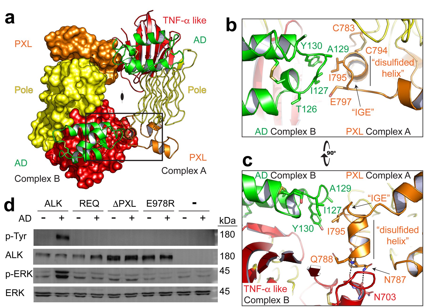
Fig. 3 |. Dimerization of ALK is directed by the PXL.

a, The ALK-ALKAL fusion complex forms a dimer (2:2, ALK:ALKAL). For clarity, the second receptor is shown in surface representation (left) with both bound ligands in green cartoon. b, An enlarged view of the boxed dimer interface from (a) to show details of the interaction. The PXL of one receptor (orange, Complex A) makes contacts with an epitope that combines ALKAL (green) and the TNF-α like region (red) of a second ligand-bound receptor (Complex B). c, An orthogonal view of the interface looking down the long axis of the Pole. d, NIH/3T3 cells stably expressing Halo-tagged ALK or ALK mutants were stimulated with a low concentration of purified ALKAL2-AD (0.5 nM for 10 min) to assess ligand responsiveness. REQ is a mutant that alters the three interface residues 795–797 (IGE, shown in b, c) to REQ. ΔPXL is a mutant that removes the entire “disulfided helix” 783–797 (shown in b,c). Data represent five independent measurements. For gel source data, see Supplementary Figure 1.
