Abstract
Despite good adherence to supervised endurance exercise training (EET), some individuals experience no or little improvement in peripheral insulin sensitivity. The genetic and molecular mechanisms underlying this phenomenon are currently not understood. By investigating genome-wide variants associated with baseline and exercise-induced changes (∆) in insulin sensitivity index (Si) in healthy volunteers, we have identified novel candidate genes whose mouse knockouts phenotypes were consistent with a causative effect on Si. An integrative analysis of functional genomic and transcriptomic profiles suggests genetic variants have an aggregate effect on baseline Si and ∆Si, focused around cholinergic signalling, including downstream calcium and chemokine signalling. The identification of calcium regulated MEF2A transcription factor as the most statistically significant candidate driving the transcriptional signature associated to ∆Si further strengthens the relevance of calcium signalling in EET mediated Si response.
Subject terms: Bioinformatics, Genetics, Physiology, Gene expression profiling
Introduction
Regular endurance exercise is a strong physiological stimulus that plays an important role in skeletal muscle homeostasis. It leads to a multitude of functional improvements when performed regularly (i.e. exercise training) and is considered a cornerstone in the prevention of type 2 diabetes1,2 by increasing tissue responsiveness to circulating insulin. Skeletal muscle contraction and peripheral insulin action are highly inter-twined3. In fact, up to 80% of the in vivo insulin-mediated glucose disposal in the postprandial state occurs in skeletal muscle4, making it quantitatively the most important tissue for systemic glucose disposal. However, we and others have demonstrated that healthy individuals are highly heterogeneous in their ability to improve peripheral insulin sensitivity (Si) in response to endurance exercise training (EET)5–7. Notably, despite good adherence to the EET program, a significant percentage of individuals (up to ~ 20%) show no changes in Si and some even demonstrate decreases in Si values5–7. Furthermore, we have previously shown that such phenomenon is likely to include a substantial genetic component8 and that healthy individuals with high and low Si responses to EET have different skeletal muscle gene expression patterns at baseline6.
A number of studies have been performed to understand the molecular basis of insulin resistance (IR), a pathological alteration in insulin sensitivity linked to many metabolic disorders, such as type 2 diabetes. Although the exact underlying cause of IR has not been fully elucidated, a number of major mechanisms, including oxidative stress, inflammation, insulin receptor mutations, endoplasmic reticulum stress, and mitochondrial dysfunction have been suggested9. Overall, however, the molecular mechanisms underlying variation in Si in a healthy population and the heterogeneous ability to improve Si through EET are currently not well understood.
Here we address this important question by computational analysis of genome-wide association study (GWAS) and skeletal muscle gene expression datasets derived from the HERITAGE Family Study. Our analysis identified several candidate genes linked to mechanisms of baseline Si, as well as training-induced changes in Si (ΔSi). Homozygous mouse knockouts of four of these candidates show alterations in glucose disposal and other relevant phenotypes, suggesting that our approach is likely to have identified genes causally linked to Si. Furthermore, analysis of both GWAS and skeletal muscle transcriptomics data shows that a molecular signature linked to calcium-regulated cholinergic signalling may be an important component of the observed variation in Si in a healthy population and predicts exercise-induced changes in Si both in HERITAGE and an independent clinical exercise study.
Methods
HERITAGE family study
The sample, study design, and EET protocol of the HERITAGE study have been described elsewhere10. Briefly, for the Caucasian sample of HERITAGE, 479 sedentary adults (233 males) from 99 nuclear families composed of parents (≤ 65 years old) and offspring (≥ 17 years old) were defined as completers (> 95% of all exercise session requirements) following exposure to a standardized and fully monitored progressive 20-week EET program (frequency of cycle ergometer sessions was three times per week). Participants were all sedentary, but healthy at baseline and not taking medications for hypertension, diabetes, or dyslipidemia. A detailed description of the study design and methodology (including a table with demographic data) can be found in the Supplementary Information section.
Intravenous glucose tolerance test (IVGTT) protocol
A frequently-sampled IVGTT was performed after an overnight fast (12 h), at baseline and post-intervention (24–36 h after the last exercise bout) following the protocol described in11. In premenopausal women, the test was scheduled to coincide with the follicular phase of the menstrual cycle. The Si index (mU/[L × min]), which measures the ability of an increment in plasma insulin to enhance the net disappearance of glucose from plasma was derived using the MINMOD Millennium software12. Changes in Si (∆Si) were calculated as post-training Si minus baseline Si.
GWAS genotype data processing
Single nucleotide polymorphism (SNP) genotyping (~ 325,000 SNPs, Illumina Human CNV370-Quad v3.0 BeadChips) on genomic DNA from lymphoblastoid cells was performed and subjected to extensive quality control as previously described13. SNPs excluded from association analyses were filtered according to the following criteria: (a) minor allele frequency < 5%, (b) violated Hardy–Weinberg equilibrium (p < 1 × 10⁻⁶), and (c) missing values in > 10% of individuals. SNPs are based on dbSNP build 151 with genomic coordinates for GRCh38 (hg38) assembly. To estimate linkage disequilibrium (LD), r2 correlation values between SNPs were calculated using default parameters in PLINK v1.9 (www.cog-genomics.org/plink/1.9/).
GWAS analysis
Baseline Si was adjusted for sex, age, log-transformed BMI, and weight-adjusted VO2max and ∆Si was adjusted for log-transformed baseline Si, sex, age, log-transformed BMI, and weight-adjusted VO2max. Associations between the normalized trait residuals and SNP genotypes were investigated using additive linear mixed effect (LME) models that accounted for within family correlations (function lme of the ‘nlme’ R package v3.114). Significance thresholds were calculated using the SimpleM method15 implemented in R programming language16. Conventional Bonferroni correction is overly conservative in genome-wide analyses due to high LD observed in genetic variants. The SimpleM method uses principal component analysis to calculate the effective number of independent tests, which resulted in 199,278 tests. A Bonferroni adjustment on this number results in a significance threshold of p < 2.51 × 10⁻⁷ (0.05/199,278). A suggestive significance threshold was set at p < 1 × 10⁻⁵. All statistical analyses were performed using R version 3.5.1. SNPs were mapped to genes based either on their position (located within a 20 kb window upstream and downstream of the gene) or if they have been identified as eQTL of a gene expressed in skeletal muscle tissue. Positional mapping was performed using MAGMA v1.07b and eQTL associations were retrieved from GTEx Portal release V817.
Candidate genes validation
We assessed the potential relevance of the candidate genes identified by the GWAS analysis by using a publicly available dataset from the International Mouse Phenotyping Consortium (IMPC) database18. We selected a panel of physiological measurements of relevance to Si. Details of the protocols are available from the database web site (https://www.mousephenotype.org/). Briefly, the ability to metabolize glucose has been assessed using three parameters derived from an intra-peritoneal glucose tolerance test (IPGTT). These were: (1) initial response to glucose challenge, (2) fasting blood glucose concentration and (3) the area of glucose response under the curve. Body composition was assessed by DEXA scan. Further characterization included a panel of blood measurements including insulin, cholesterol, glucose, glycerol, free fatty acids, and creatinine. In addition, respiratory exchange ratio was also available.
Functional GWAS
To test whether genes within specific biological pathways are enriched by genetic associations with lower p-values than expected by chance, we applied GLOSSI19 from cpvSNP R package (v 1.18.0)20. First, GWAS results were pruned to keep only independent SNPs (r2 > 0.8) resulting in 249,035 SNPs. After positionally mapping the remaining SNPs to genes (± 20 kb window), their p-values were used as input to compute the estimate of enrichment within a given biological pathway. Resulting p-values were corrected for multiple testing using Bonferroni correction. Gene set collections used KEGG pathways (c2.cp.kegg.v7.1.entrez) retrieved from MSigDB v7.121,22 and manually curated functional modules representing genes required for normal skeletal muscle activity23. Threshold for significantly enriched biological pathways was defined as padj < 0.05. In order to further investigate the most important pathways, we selected the most significant SNPs associations (alpha < 0.05) within the pathways identified by GLOSSI and remapped these on KEGG pathways using the web-based tool DAVID (version 6.8). Threshold for significant biological pathways was defined as padj < 0.05, provided in DAVID with Benjamini–Hochberg adjustment.
RNA extraction and global gene expression profiling
Vastus lateralis muscle biopsies were also obtained in a subsample of the SNP-genotyped participants (n = 41) before and after (~ 96 h after final exercise session) the intervention using a percutaneous needle. Each biopsy was immediately frozen in liquid nitrogen and stored at – 80 °C until RNA preparation. RNA extraction as well as reverse transcription were done as previously described24. Affymetrix U133 + 2 arrays were used to quantitate global mRNA expression levels. The raw microarray CEL files are deposited in the public Gene Expression Omnibus (GEO) database25 under accession number GSE117070.
Microarray data processing
Raw CEL files were Robust Multichip Average (RMA) normalized following removal of probes termed ‘absent’ in more than 80% of the samples by the MAS5 algorithm inside the ‘affy’ package (26,151 probesets discarded)26. Quality control plots of the polyA-control RNAs (spike-ins added right after RNA purification) highlighted a batch issue that was resolved by applying the COMBAT software27. The JetSet package was used to select a single ‘optimal’ probeset to represent each gene based on specificity, robustness against mRNA degradation, and MAS5 present call rate28. As the most representative probeset for each gene is selected, they have high splice isoform coverage.
Gene set enrichment analysis (GSEA)
The entire skeletal muscle transcriptome was ranked by individually regressing pre-training mRNA expression levels against baseline Si and ∆Si, using linear mixed effect models that accounted for within family correlations. Both outcome variables were adjusted by age, sex, log-transformed BMI, weight-adjusted VO2max, and type I fibre percentage (see29 for details on the fibre typing), with ∆Si also being adjusted for baseline Si. Based on this ranking (Student t-statistic), we performed a pre-ranked GSEA using the default parameters in clusterProfiler v3.14.3 R package30 to identify candidate biological pathways significantly enriched in genes that are associated with baseline Si and ∆Si (either top or bottom of the distribution). Gene set collections used were transcription factor targets and KEGG biological pathways (c3.tft.v7.1.entrez.gmt and c2.cp.kegg.v7.1.entrez, respectively) retrieved from MSigDB v7.121 and manually curated functional modules representing genes required for normal skeletal muscle activity23. Threshold for significant sets was defined as false discovery rate (FDR) < 0.05.
Transcriptomics-based model to predict ∆Si
To test if baseline expression of MEF2A target genes is predictive of exercise-induced ∆Si we applied a regression-based modelling approach allowing for pairwise interactions (function lm of the ‘stats’ R package16) between baseline expression of three genes. Only genes translating to proteins that interact with MEF2A were included and consisted of a total of 50 experimentally validated interactors with high confidence score (> 0.8) identified in the STRING database31 (Supplementary Table S1). All possible linear regression models based on all possible combinations of three-genes sets were examined (a total of 19,600 models). More precisely, we define:
where mRNA abundance is represented by θ and the noise model component by ε. Weight-adjusted VO2max and sex were included as covariates.
Ethics declaration
The study protocol was approved by the Institutional Review Boards at each of the five participating centers of the HERITAGE Family Study consortium (Indiana University, Laval University, University of Minnesota, Texas A&M University, and Washington University at St. Louis). Written informed consent was obtained from each study participant. The subjects who were under 18 years, one of the parents gave consent in addition to the participant. This was an easy procedure to follow as we were recruiting whole nuclear families. All research was performed in accordance with the Declaration of Helsinki.
Results
Overview of the analysis strategy
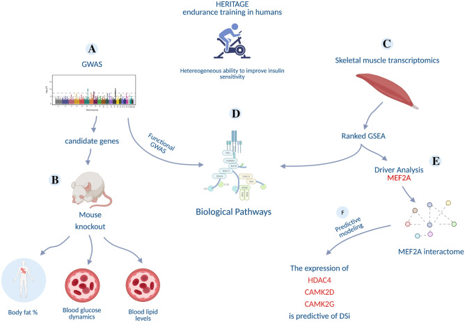
The overarching goal of this study is to investigate the genetic and molecular basis of the variation in Si and ∆Si following EET, across a healthy population. We address this by integrating a traditional GWAS approach with the analysis of skeletal muscle transcriptomics data within HERITAGE, one of the largest studies to evaluate the response of several physiological measurements to EET. The strategy, described in a schematic format in Fig. 1, involved:
The identification of genetic variants linked to Si and ∆Si (Fig. 1A) and the validation of the corresponding gene candidates in a mouse knock down experiment database (Fig. 1B).
The identification of transcriptional signatures linked to baseline Si and ∆Si (Fig. 1C).
The identification of transcription factors that may be driving the transcriptional signatures identified above (Fig. 1E).
Testing whether baseline expression of genes linked to TF drivers are good predictors of ∆Si (Fig. 1F).
Figure 1.
Overview of the study design, consisting of five interconnected steps: (A) identification of genetic variants linked to insulin sensitivity index (Si), (B) validation of the corresponding gene candidates in a mouse knock down experiment database, (C) identification of transcriptional signatures in skeletal muscles correlating with baseline Si and ∆Si, (D) integration of genetics and transcriptomics signatures, (E) identification of transcription factors likely to drive the transcriptional signatures linked to ∆Si which led to the identification of MEF2A transcription factor as main driver of the transcriptional profile, and (F) development of a statistical model that can predict ∆Si from the transcriptional state of MEF2A interacting genes.
In addition, by mapping gene candidates identified by GWAS and the transcriptional signatures we tested the hypothesis that genetic variation may be linked to downstream changes in gene expression (Fig. 1D).
GWAS analysis identifies putative loci linked to baseline and exercise-induced changes in Si
Investigation of genetic variants linked to inter-individual heterogeneity of ∆Si is based on the assumption that its underlying molecular mechanisms have a genetic component. Our analysis revealed that this assumption is indeed likely to be correct as 29% of the variance in ∆Si is accounted for by family membership (Supplementary Fig. S1). Moreover, from an ANOVA, there is 40% more variance between families than within families (p = 0.02), providing additional suggestive evidence that the changes in Si in response to exercise training are characterized by a significant heritable component.
Therefore, we set to identify specific genetic variants linked to Si and ∆Si by GWAS. As a first step we tested whether relevant physiological variables may be confounding factors that should be considered in the analysis. We discovered that variation in baseline Si was significantly linked to BMI, VO2max/kg, sex and age (Supplementary Fig. S2A). We also discovered that ΔSi was significantly linked to baseline Si and sex (Supplementary Fig. S2B), with 27% of the variance in ΔSi explained by baseline Si (Supplementary Fig. S2C). A similar amount of variance in post-training Si was also explained by baseline Si (Supplementary Fig. S2D). Based on this evidence, we performed a GWAS with the objective to identify SNPs linked to baseline Si and ∆Si, where both traits were adjusted for potential confounding variables.
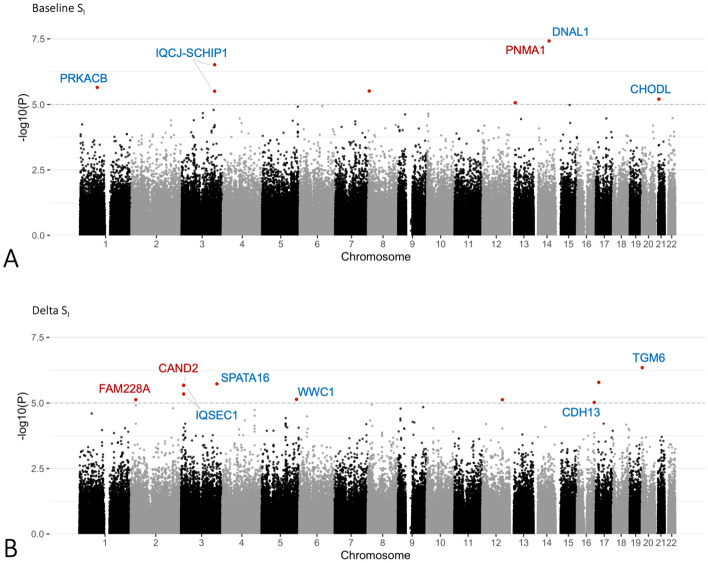
The GWAS analysis identified one SNP significantly associated to baseline Si (DNAL1 rs11622678, p = 3.79 × 10⁻⁸) plus seven SNPs with suggestive association (p < 10−5), and ten SNPs with suggestive association (p < 10−5) with ΔSi (Fig. 2). Positional and eQTL mapping revealed genes that are located within or near these SNPs (± 20 kb window) and/or have their expression correlated with them (Table 1).
Figure 2.
Manhattan plots showing genetic loci associated with baseline and ∆Si and their mapped genes. Genetic loci reaching at least suggestive association are represent by points highlighted in red. Genes mapped to these loci are annotated, where genes mapped by position are highlighted in blue, and genes mapped by eQTL are highlighted in red. Highlighted loci showing no gene annotation did not map to any genes according to the criteria we used. For additional details, see Table 1.
Table 1.
Genomic loci showing, at a minimum, suggestive association with ΔSi and baseline Si.
| SNP | Chr | Location (bp) | P | Gene symbol | Gene mapping | Gene location (bp) |
|---|---|---|---|---|---|---|
| Delta Si | ||||||
| rs4815227 | 20 | 2441096 | 4.50E−07 | TGM6 | Positional | 2380908–2432753 |
| rs12449918 | 17 | 15392862 | 1.63E−06 | – | – | – |
| rs6799845 | 3 | 172873569 | 1.86E−06 | SPATA16 | Positional | 172889357–173141268 |
| rs9211a | 3 | 12898047 | 2.10E−06 | IQSEC1 | Positional | 12897043–13282998 |
| – | – | – | LOC105376955 | Positional | 12882360–12885099 | |
| – | – | – | RP11-767C1.2 | Skeletal muscle eQTL | 12832219–12832728 | |
| – | – | – | CAND2 | Skeletal muscle eQTL | 12796472–12871916 | |
| rs14191a | 3 | 12898847 | 2.13E−06 | RP11-767C1.2 | Skeletal muscle eQTL | 12832219–12832728 |
| – | – | – | CAND2 | Skeletal muscle eQTL | 12796472–12871916 | |
| – | – | – | IQSEC1 | Positional | 12897043–13282998 | |
| – | – | – | LOC105376955 | Positional | 12882360–12885099 | |
| rs2600330 | 3 | 12926604 | 4.52E−06 | IQSEC1 | Positional | 12897043–13282998 |
| rs7722673 | 5 | 168417231 | 7.23E−06 | WWC1 | Positional | 168292060–168472303 |
| rs7595372 | 2 | 24409253 | 7.45E−06 | FAM228A | Skeletal muscle eQTL | 24175069–24200849 |
| rs969863 | 12 | 97202907 | 7.48E−06 | – | – | – |
| rs422455 | 16 | 83729855 | 9.51E−06 | CDH13 | Positional | 82626794–83796610 |
| Baseline Si | ||||||
| rs11622678* | 14 | 73655058 | 3.79E−08 | PNMA1 | Skeletal muscle eQTL | 73711783–73714372 |
| – | – | – | RP3-414A15.10 | Skeletal muscle eQTL | 73616700–73633941 | |
| – | – | – | DNAL1 | Positional | 73644875–73703728 | |
| – | – | – | RP3-414A15.2 | Skeletal muscle eQTL | 73530152–73530610 | |
| rs10936174 | 3 | 159198318 | 3.07E−07 | IQCJ | Positional | 159069252–159266307 |
| – | – | – | IQCJ-SCHIP1 | Positional | 159069252–159897366 | |
| rs7524898 | 1 | 84167162 | 2.24E−06 | PRKACB | Positional | 84077975–84238498 |
| rs10107799 | 8 | 6102222 | 3.07E−06 | – | – | – |
| rs7653174 | 3 | 159120808 | 3.13E−06 | IQCJ | Positional | 159069252–159266307 |
| – | – | – | IQCJ-SCHIP1 | Positional | 159069252–159897366 | |
| rs205666 | 21 | 18120899 | 6.27E−06 | CHODL | Positional | 17819595–18267373 |
| rs9511351b | 13 | 24575137 | 8.56E−06 | LOC101927375 | Positional | 24541663–24567321 |
| rs9553347b | 13 | 24557233 | 8.56E−06 | LOC105370295 | Positional | 24538602–24541913 |
| – | – | – | LOC101927375 | Positional | 24541663–24567321 | |
*Statistically significant association.
a,br2 > 0.8.
We next tested whether these candidate genes may be causally linked to insulin-regulated physiology. We used a publicly available dataset from the International Mouse Phenotyping Consortium database18. This database is the result of an international effort involving 19 assessment centres and reporting a wide range of physiology measurements from a collection of 6440 gene knockouts. Here we selected a panel of physiological measurements of relevance to Si from mouse knockouts in our putative gene lists (see methods section and Fig. 3 for a full list). Eight out of our twelve genes identified by the GWAS (67%) were represented in this database and had a full set of the selected measurements. These genes are PNMA1, DNAL1, IQCJ, CHODL for baseline Si and TGM6, SPATA16, CAND2, CDH13 for ΔSi). We found that DNAL1 and IQCJ (linked to baseline Si), and TGM6 and CAND2 (linked to ΔSi) had significant associations with one or more selected mouse phenotypes (Fig. 3).
Figure 3.
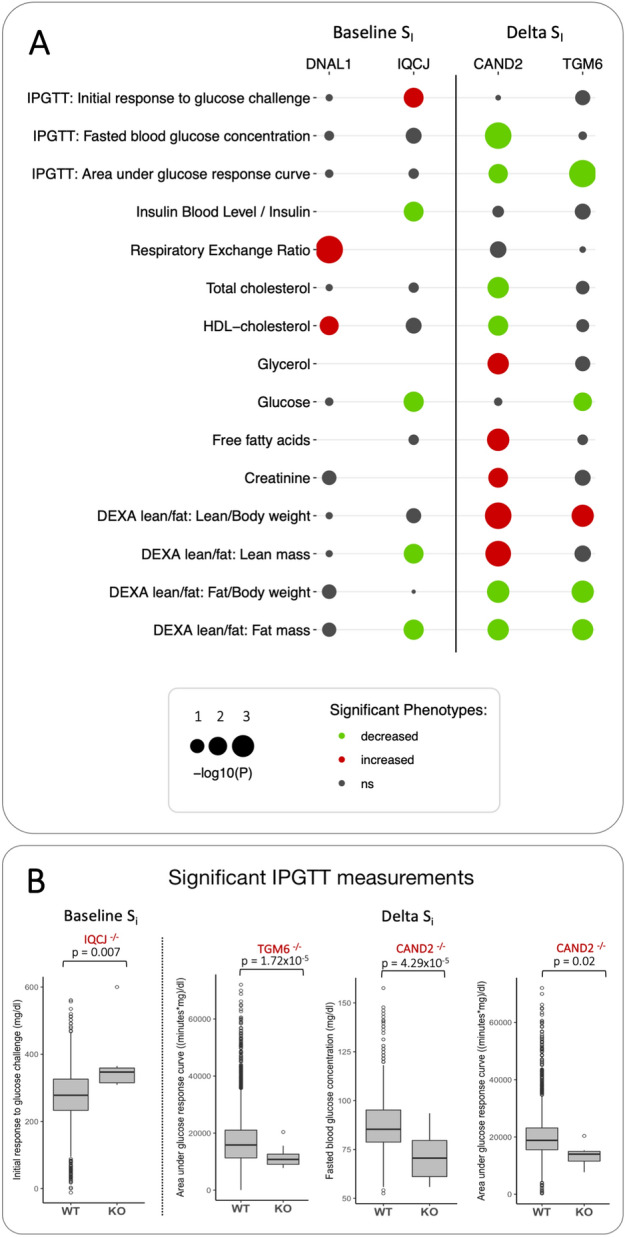
Validation of gene candidates from GWAS analysis in a mouse knock down experiment database. (A) Mouse knockout of DNAL1 and IQCJ (associated with baseline Si) and CAND2 and TGM6 (associated with ∆Si) lead to changes related to metabolism and skeletal muscle development (Padj < 0.05, ns = not significant). Details of each experiment can be found in the IMPC database18. (B) Boxplots detailing significant associations of IQCJ, CAND2 and TGM6 knockouts with measurements from IPGTT (intra-peritoneal glucose tolerance test) experiment in mice, which is analogous to intravenous glucose tolerance test (IVGTT) method used to measure Si in study participants.
The CAND2 knockout is characterized by lower glucose levels, improved glucose tolerance, lower levels of total and HDL cholesterol, increased levels of glycerol, free fatty acids and plasma creatinine, increased lean mass, and decreased fat mass. Interestingly, phenotypes associated with TGM6 knockout were found to be in accordance with those found associated with CAND2, leading also to lower levels of plasma glucose, improved glucose tolerance, lean mass increase, and fat mass decrease. It is worth noting that CDH13, CHODL, PNMA1, and SPATA16, although present in the database and tested for relevant phenotypes did not show significant associations with phenotypes relevant to Si.
Functional analysis of the genomic landscape associates the calcium signalling and cholinergic synapse pathways to Si
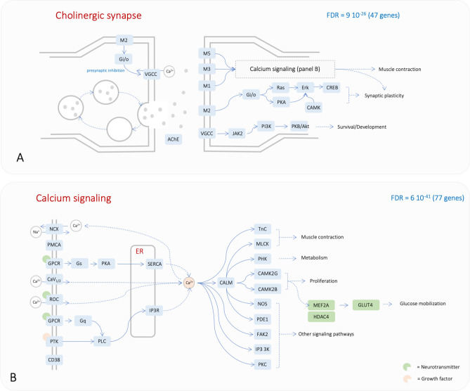
Complex traits are often characterised by a relatively small contribution of multiple genetic variants that all together contribute to the phenotype of interest. Traditional univariate GWAS analysis, which is often underpowered, may fail to identify these complex interactions. Functional GWAS analysis addresses this issue by testing whether genetic variants tend to cluster within given biological pathways. With this in mind, we analysed our GWAS results with GLOSSI, one such approach. After pruning GWAS results to select independent SNPs (R2 < 0.8) and positionally mapping those to genes (± 20 kb window), we identified significantly enriched biological pathways (KEGG pathways and curated skeletal muscle pathways22) for both baseline Si and ΔSi. This analysis initially identified 17 significant pathways linked to baseline Si and 8 pathways specifically linked to ΔSi (Supplementary Fig. S3). The 17 pathways linked to baseline Si included 470 genes linked to genetic variants with a lower nominal p value (p < 0.05). Interestingly, we saw that the four most significantly enriched pathways were sufficient to represent half of the 470 most significant genes. These pathways were calcium signalling (77 genes), axon guidance (63 genes), chemokine signalling (66 genes) and cholinergic synapse (47 genes) (Fig. 4, Supplementary Fig. S4 and Supplementary Table S2).
Figure 4.
Most significant pathways identified by functional GWAS. (A) cholinergic synapse pathway and (B) calcium signalling pathway. The two KEGG pathways are interconnected (B). The KEGG pathway calcium signalling has been edited to add three additional genes of relevance (green rectangles). Each node can represent multiple genes. Symbols in the two panels are the same as in the original KEGG pathway map (NCX: Na/Ca exchanger; PMCA: ATPase plasma membrane Ca2 + transporting; CaV12: calcium voltage-gated channel subunit alpha1 A and C; ROC: nicotinic acetylcholine receptor alpha-7; GPCR: cysteinyl leukotriene receptor 1; PTK: epidermal growth factor receptor; CD38: ADP-ribosyl cyclase 1; PLC: phosphatidylinositol phospholipase C; Gs: guanine nucleotide-binding protein G(s) subunit alpha; Gq: guanine nucleotide-binding protein subunit alpha-11; SERCA: P-type Ca2 + transporter type 2A; IP3R: inositol 1,4,5-triphosphate receptor type 1; TnC: troponin C; MLCK: myosin-light-chain kinase; PHK: phosphorylase kinase alpha/beta subunit; NOS: nitric-oxide synthase; PDE1: calcium/calmodulin-dependent 3',5'-cyclic nucleotide phosphodiesterase; FAK2: focal adhesion kinase 2; IP3 3 K: 1D-myo-inositol-triphosphate 3-kinase; M1: muscarinic acetylcholine receptor M1; M2: muscarinic acetylcholine receptor M2; M3: muscarinic acetylcholine receptor M3; M5: muscarinic acetylcholine receptor M5; VGCC: calcium voltage-gated channel subunit alpha1 C; AChE: acetylcholinesterase; Gi/o: G protein subunit beta 5; CAMK: CAMK2G and CAMK2B; CREB: cAMP responsive element binding protein 3; PKB/Akt: AKT serine/threonine kinase 3). Genes shown in this figure map to SNPs with p value < 0.05.
The same analysis performed with the top-most significant genes within the pathway enrichment set for ΔSi identified only 31 genes. While this limited number of genes preclude a systematic pathway enrichment analysis, 21 genes could be mapped within the adrenergic signalling in cardiomyocyte pathway. Importantly, five of these genes also mapped within the calcium signalling pathway (RYR2, SLC8A1, CACNA1C, CACNA1D and CACNA1S), providing a link with the analysis performed on the baseline Si. These included subunits of the ATPase NA+/K+ transporter (ATPA2/A4/B1/B3) and additional calcium voltage-gated channels. In addition, there were four subunits of the cytochrome C oxidase enzyme (COX4I2, COX6B1, COX7A1, COX7A2L).
Transcription factor driver analysis identifies the calcium dependent transcription factor MEF2A as the most significant driver of the ΔSi transcriptional signature
The results of the functional GWAS suggest a role of skeletal muscle in insulin dependent glucose uptake and the effects of exercise in remodelling this tissue. Therefore, we set to investigate the transcriptomic profile of skeletal muscles in a subset of the HERITAGE individuals. We wanted to identify baseline transcriptional signatures that correlate to Si and ΔSi as well as the transcription factors that may drive such signatures. More specifically, by using a GSEA based approach we searched for enrichment in transcription factor binding sites in the list of genes correlated to Si and ΔSi.
Only when including fibre type composition in the models we have identified gene sets (a total of 45) mapped to known transcription factor binding sites significantly enriched by genes whose skeletal muscle expression correlated to ΔSi (Supplementary Table S3). Fibre type composition has been previously linked to insulin-dependent glucose uptake in skeletal muscle32–34 and its addition to the ΔSi model also led to a larger number of significantly enriched biological pathways. These encompassed a variety of biological functions such as signalling, energy and amino acid metabolism, tissue homeostasis, protein degradation, immune system, and translation (Supplementary Fig. S5). Interestingly, the KEGG pathways chemokine signalling, neuroactive ligand receptor interaction, and the functional term calcium dynamics/homeostasis required for excitation–contraction coupling were reminiscent of the results for the functional GWAS analysis (Supplementary Table S2).
Remarkably, when we examined which transcription factor may be able to explain the expression of genes in the Si transcriptional signatures, we found that the top candidate gene was the calcium dependent transcription factor MEF2A (Fig. 5). The hypothesis that MEF2A drives a significant part of the transcriptional signature linked to ΔSi is supported by the observation that the global transcriptional signature associated to MEF2A knockdown in C2C12 can recapitulate the transcriptional signature correlated to ΔSi in the HERITAGE cohort (Fig. 1, Supplementary Information and Supplementary Table S4). This result is consistent with the findings from the functional GWAS that suggested a key role of calcium signalling in baseline Si. The linkage between genetic variation and the transcriptomics signatures in skeletal muscle associated with Si emerged through the linkage between calcium signalling and MEF2A.
Figure 5.
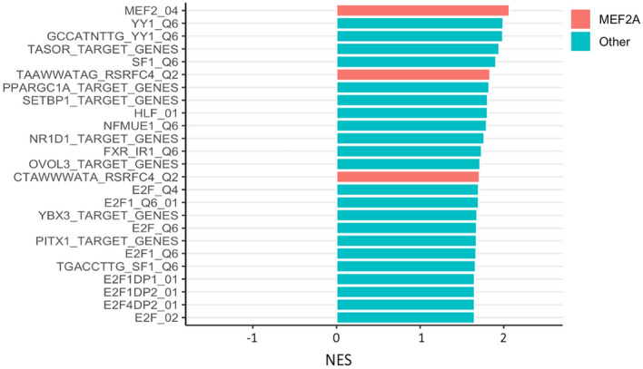
Top 25 transcription factor target gene sets significantly enriched in GSEA (Padj < 0.05) ranked by NES (normalised enrichment score). Gene sets with a positive NES are enriched by genes with skeletal muscle baseline expression positively correlated to ΔSi while genes with a negative NES are enriched by genes with skeletal muscle baseline expression negatively correlated to ΔSi. Sets corresponding to MEF2A target genes are highlighted in red. A full list of significant transcription factor target gene sets is available on Supplementary Table S3.
The development and validation of a baseline MEF2 transcriptional signature predictive of ΔSi
Ranked GSEA analysis revealed that genes with baseline expression levels showing higher (positive) or lower (inverse) correlation with ΔSi were found to be enriched by MEF2A targets (Supplementary Fig. S6 and Supplementary Table S5). We also show that the transcription factor MEF2A may be the main actor driving that transcriptional signature. We therefore set to develop a predictor of ΔSi based on the baseline expression of genes that are part of the MEF2A interactome. We focused on 50 experimentally validated interactors with a high confidence score (identified in the STRING database, see Supplementary Table S1 for an exhaustive list) and we performed a comprehensive analysis of all possible linear regression models based on gene expression levels of all possible combinations of three-genes sets (a total of 19,600 models). Remarkably, the most predictive statistical model (R2 = 48%, F-value = 6.4, p < 0.001; Table 2) included the direct activators of the MEF2 transcription factor (HDAC4, CAMK2D, and CAMK2G, Fig. 4B). In comparison, a linear regression model comprising only of sex and VO2max/kg as predictors showed poor predictive power (R2 = − 0.01%, F-value = 0.98 and p = 0.39), reinforcing the large contribution of these genes on predicting ΔSi. Consistent with the established hetero-multimeric nature of CaMKII, the model did show a highly significant interaction between the two isoforms CAMK2D and CAMK2G. Regression diagnostics confirmed conformity of the residuals to the assumptions of normality, linearity and homoscedasticity.
Table 2.
Result of the mRNA-based multivariate regression model for Si training response among the subset of White HERITAGE participants for which global gene expression data before and after the exercise program are available (n = 47).
| Variable | Regression coefficient | Standard error | t-value | p value |
|---|---|---|---|---|
| VO2max | − 0.03 | 0.004 | − 0.68 | 0.50 |
| Sex | − 2.15 | 0.83 | − 2.59 | 0.013 |
| CAMK2D mRNA | 40.73 | 27.98 | 1.46 | 0.15 |
| CAMK2G mRNA | 74.78 | 32.04 | 2.33 | 0.02 |
| HDAC4 mRNA | − 34.94 | 22.37 | − 1.56 | 0.13 |
| CAMK2D:CAMK2G | − 12.08 | 3.41 | − 3.54 | 0.001 |
| CAMK2D:HDAC4 | 6.94 | 1.63 | 4.27 | < 0.001 |
| CAMK2G:HDAC4 | − 0.40 | 2.90 | − 0.14 | 0.89 |
Importantly, the response variable (i.e. ∆Si) spanned a broad range (− 79 to + 120%) among the participants included in the current analysis. Sex and VO2max adjusted for body size were included as covariates in the model. Notably, the abundance of these transcripts was not responsive to the training intervention, but rather higher basal expression levels were associated with greater gains in Si. CAMK2D: 228555_at; CAMK2G: 212757_s_at; HDAC4: 228813_at.
To test the general applicability of the HERITAGE predictive gene signature, we took advantage of a previously published Affymetrix muscle gene expression dataset from a smaller independent mixed exercise training cohort35. Importantly, this cohort also spanned a broad range in terms of the training-induced change in the rate of peripheral glucose disappearance (Rd) (∆Rd ranging from − 20 to + 145%). Intriguingly, the baseline multi-gene RNA signature was able to explain 30% of the training-induced change in Rd among the healthy middle-aged male participants (n = 14) in the replication cohort (see Supplementary Fig. S7, a value close to the estimated ∆Si variance in the HERITAGE cohort when family membership has been accounted for).
Discussion
Here, we have shown that variation in insulin sensitivity across a normal healthy population and its modulation by EET is a complex trait where combined variation in genes linked to the KEGG pathways cholinergic signalling, calcium signalling, axon guidance and chemokine signalling is likely to be an important component. Despite such complexity, we have been able to identify genes that are causally linked to glucose disposal and other relevant phenotypic traits in mouse knockouts.
GWAS candidate genes associated to baseline and ∆Si
Our study is the first to investigate genome-wide associations with changes in Si in response to exercise training. Despite the SNPs we identified in the present study as significantly associated to baseline Si have not been previously associated to insulin sensitivity according to GWAS Catalogue database36, some of them have been previously linked to insulin resistance or related phenotypes. Importantly, mouse knockouts for four of these candidates (TGM6, CAND2, IQSEC1 and DNAL1) showed a relevant phenotype, suggesting a causal link with Si. Here we review the evidence in the literature that is consistent with our findings.
The function of TGM6 product, transglutaminase 6 (TG6), has not been studied extensively and is not yet well understood. However, transglutaminase 2 (TG2) has been implicated in glucose metabolism37,38 and glucose tolerance39. Transglutaminases catalyse serotonylation40, a process involved in the modulation of insulin secretion in pancreatic beta-cells41. The results of the TGM6 knockout experiment and the function of TG2 suggest a role for TG6 in Si and warrants further investigation.
There are several pieces of evidence that are consistent with a role of CAND2 in insulin sensitivity. CAND2 is mostly expressed in muscle tissues17,42. Beyond its role in myogenic differentiation43–45, CAND2 interacts with insulin receptor substrate 1 (IRS1) and is stimulated by insulin in type 2 diabetes patients, but not in non-diabetic controls46. CAND2 acts by modulating the assembly of ubiquitin–proteasome related complexes, such as E3 ligases47,48, which also have been implicated in insulin resistance and diabetes and are known to target key insulin signalling molecules49. Moreover, CAND2 has been shown to be upregulated during a 3 h hyperinsulinemic euglycemic clamp in vastus lateralis muscle of healthy subjects50. CAND2 has been previously mapped to SNPs associated with related phenotypes such as waist-hip ratio51 and waist circumference adjusted for BMI52,53.
SNP rs11622678 located in chromosome 14 reached a statistically significant association with baseline Si and was positionally mapped to DNAL1. Mutations in this gene cause primary ciliary dyskinesia as this gene affects movement of cilia and flagella54, and have been associated with respiratory diseases and lung function18,51. Interestingly, knockout of this gene led to increased respiratory exchange ratio (RER), with higher values indicating that carbohydrates are the main source of substrates being oxidized (Fig. 3).
Two SNPs (r2 = 0.49) have been positionally mapped to the fusion transcript IQCJ-SCHIP1 spanning two adjacent independent genes. Although the functions of IQCJ-SCHIP1 are still poorly understood, genetic variants tagging IQCJ have been associated to modulation of blood lipid levels in multiple independent studies55–58, while SCHIP1 has been implicated in axon guidance59–61 and was upregulated in differentiated myotubes compared to undifferentiated62 (see Supplementary Material for additional discussion on PNMA1, CHODL, SPATA16 and CDH13 which are either not present in the IMPC database or no relevant traits showed significant changes following knockout).
Furthermore, the observation that baseline Si negatively correlates with ∆Si (Supplementary Fig. S2C) is intriguing and suggestive of the existence of an upper limit for Si. This would result in a lower margin for improvement in individuals with a high Si value.
Is calcium mobilization triggered by muscle contraction potentially responsible for changes in Si?
In addition to the genes discussed above, we identified a consistent accumulation of SNPs correlated to baseline Si and ΔSi in calcium signalling and cholinergic synapse pathways (Supplementary Table S2). These findings suggest that cholinergic signalling via mobilization of calcium in skeletal muscle may mechanistically link muscle contraction to insulin sensitivity. A study investigating correlation between baseline gene expression and exercise-induced %ΔSi found that several significantly correlated genes in a validation cohort mapped to Ca2+ signalling, including CACNA1S and CAMK2D63.
There is considerable evidence that insulin signalling and muscle contraction are linked processes that activate multiple signalling cascades leading to glucose uptake64–67. Glucose uptake by skeletal muscle tissue is mediated by GLUT4, which upon stimulation by either insulin and/or contraction is translocated to the plasma membrane from vesicles. In insulin-mediated glucose uptake, insulin binding triggers a cascade of molecular reactions that lead to GLUT4 translocation, also triggering a transient Ca2+ influx in muscle cells. This process is suggested to enhance GLUT4 translocation and docking in the plasma membrane. Meanwhile, muscle contraction initiated by membrane depolarisation and increased concentrations in intracellular Ca2+ leads to activation of Ca2+ sensors such as Ca2+-calmodulin-dependent kinase II (CAMKII), which are key molecules in contraction-stimulated glucose transport. Activated CAMKIIs promote dissociation of HDAC4 from MEF2A transcription factor, leading to its activation and increased GLUT4 transcription68,69. Therefore, insulin- and contraction-mediated signalling pathways related to skeletal muscle glucose uptake are inter-twined, where GLUT4 increased expression and availability entrained by muscle contraction would also contribute to improved insulin-mediated glucose uptake.
It is therefore conceivable that variation in genes controlling muscle contraction (represented in the cholinergic synapse and calcium signalling pathways) could result in lower levels of intracellular Ca2+, leading to lower activation of the CAMKIIs and consequently reduced GLUT4 availability (Fig. 4B). Diminished localization of GLUT4 to the membrane, which is also Ca2+ dependent, could also contribute to glucose uptake impairment in skeletal muscle. Interestingly, none of the analyses shown here directly linked SCL2A4 (gene encoding for GLUT4) to either baseline or ΔSi. It is possible that our study, is not sufficiently well powered to capture SCL2A4 effect size, or that other mechanisms affecting GLUT4 regulation, such as post-translation modifications70, modulate insulin sensitivity. Additionally, none of the publicly available GWAS studies on insulin sensitivity (GWAS Catalog database36) reported associations with SLC2A4.
Genetic variation may not be the only mechanism controlling glucose mobilization and ultimately Si. A study investigating epigenetic patterns associated to type 2 diabetes has shown that first-degree relatives of patients with diabetes have differential DNA methylation patterns in genes related to insulin and Ca2+ signalling pathways compared to healthy individuals with no family history of the disease. Intriguingly, DNA methylation of genes involved in Ca2+ signalling pathways including MEF2A, which we also have identified in our approach, decreased after exercise71. The role of genetic and epigenetic variation in Ca2+ signalling in modulating inter-individual variability in insulin sensitivity warrants further investigation.
Other potential mechanisms linking genetic variation to Si
Our analyses suggest that genetic variation affecting other biological mechanisms could also be modulating Si. The chemokine signalling pathway has been identified in both GWAS and transcriptomics functional analyses, suggesting that variation in genes within this pathway could be affecting their expression and contributing to variation in exercised-induced Si response.
Several studies have suggested a role of chemokines and chemokine receptors in the development of insulin resistance, which is attributed in part to a state of low-grade inflammation due to elevated blood glucose and lipid levels induced by diet and excess adiposity72,73. This leads to induction of pro-inflammatory mediators such as chemokines that interfere with insulin signalling pathways. In the present study, we have identified gene expression profiles associated to inflammation-related pathways correlated with Si response, suggesting genetic variation affecting chemokine signalling could affect the inflammatory processes that naturally occur with exercise. Additionally, the emerging research field of ‘immunometabolism’74 has generated data indicating that a cross‐talk between immune- and metabolic-related molecules is essential to normal skeletal muscle physiology75.
Cell adhesion molecules pathway, which include key molecules involved in modulating ECM integrity, was also identified in both GWAS and transcriptomics functional analyses. There is some evidence linking insulin resistance to ECM remodelling75 with mechanisms attributed to physical impairment of insulin access to its receptor by increased ECM deposition, or to the roles of integrins in mediating insulin signalling76. Deletion of the muscle-specific integrin β1 (expressed by ITGB1) results in decreased insulin sensitivity, whereas the ECM of insulin resistant human muscle were reported to be associated to decreased abundance α-actinin 2 (expressed by ACTN2). Interestingly, in our functional investigation encompassing ∆Si associated genetic variants, two SNPs mapping to ACTN2 and ITGB1 genes are second and third top significant SNPs (p < 0.001), but several other top associated SNPs map to cell adhesion molecules, suggesting that mutations in ECM-related molecules could be influencing the Si-related traits at rest or in response to regular exercise.
Conclusions
The relatively large proportion of individuals who fail to improve metabolic fitness traits justify the importance of developing evidence-based personalized exercise prescription to maximize the health-promoting benefits of a physically active lifestyle. To develop such individualized recommendations for exercise, it is vital that the molecular basis driving phenotypic response variation be understood. Our multi-omics approach is a step in this direction as it provides evidence of a genetic component affecting calcium signalling that might be responsible for the large heterogeneity in ∆Si following a fully supervised EET program. The predictive RNA signature can potentially be used to stratify individuals before any intervention has taken place. Further studies are needed to test whether our signature could be predictive of response in different training protocols or whether ∆Si non-responders could benefit from different training regimes (e.g. high-intensity interval training or resistance exercise). This is important as skeletal muscle insulin resistance is one of the earliest hallmarks of the development of type 2 diabetes and other metabolic complications. Promisingly, muscle RNA abundance can now be more easily quantified due to the development of less invasive micro-needle biopsy sampling. Further, one-step multiplex real-time RT-PCR assays could offer a rapid, sensitive and cheap diagnostic option if a molecular predictor could be validated and replicated in multiple cohorts.
Supplementary Information
Acknowledgements
We thank Drs. Arthur S. Leon, D.C. Rao, James S. Skinner, Tuomo Rankinen, Jacques Gagnon, Treva Rice and the late Jack H. Wilmore for contributions to the planning, data collection, and conduct of the HERITAGE project, and for the data management of the IVGTT. Special thanks are also given to Richard S. Bergman from the Cedars-Sinai Medical Centre for his expertise in the analyses of the IVGTT data. This research was partially funded by National Heart, Lung, and Blood Institute Grants HL-45670, HL-47317, HL-47321, HL-47323, and HL-47327, all in support of the HERITAGE Family Study). L.Y. Takeshita has received funding from the Innovative Medicines Initiative 2 Joint Undertaking under the TransBioLine project with grant agreement No. 821283. This Joint Undertaking receives support from the European Union’s Horizon 2020 research and innovation programme and EFPIA. This communication reflects the author's view and neither IMI nor the European Union or EFPIA are responsible for any use that may be made of the information contained therein. P. K. Davidsen was supported by a PhD studentship funded by the Birmingham MRC-ARUK Centre for Musculoskeletal Ageing Research. C. Bouchard is partially funded by the John W. Barton Sr. Chair in Genetics and Nutrition. Z. S. Ghosh and C. Bouchard are partially supported by the National Institute of General Medical Sciences (NIGMS)-funded COBRE Grant 8-P30-GM-118430-01. S. Ghosh is supported in part by NIGMS Grant 2-U54-GM-104940, which funds the Louisiana Clinical and Translational Science Center and by the National Medical Research Council, Ministry of Health, Singapore (WBS R913200076263). M. A. Sarzynski is partially supported by NHLBI Grant R01HL146462 and NIGMS Grant P20GM103499, which funds the South Carolina IDeA Network of Biomedical Research Excellence.
Author contributions
M.K.C.H, P.S. and C.B. participated in the exercise training study design. L.Y.T., P.K.D. and F.F. formulated the data analysis plan. L.Y.T and P.K.D. performed data analysis. J.M.H, P.A. and F.F. contributed to overall data analysis. M.A.S., S.G. and C.B. contributed to the GWAS analysis. C.B. contributed to the quality control and modelling of the glucose and insulin data. L.Y.T., P.K.D. and F.F. drafted the manuscripts. F.F. produced Fig. 4 and L.Y.T. produced all other manuscript figures. S.J.W., J.M.R. and R.E.G. substantially contributed to manuscript revision proving expert knowledge on physiological aspects involving insulin sensitivity. All co-authors contributed to the critical review end editing of the manuscript. F.F. is the guarantor of this work and, as such, had full access to all the data in the study and takes responsibility for the integrity of the data and the accuracy of the data analysis.
Data availability
The datasets generated during and/or analysed during the current study are available from the corresponding author on reasonable request.
Competing interests
The authors declare no competing interests.
Footnotes
Publisher's note
Springer Nature remains neutral with regard to jurisdictional claims in published maps and institutional affiliations.
The authors contributed equally: Louise Y. Takeshita and Peter K. Davidsen.
Supplementary Information
The online version contains supplementary material available at 10.1038/s41598-021-98792-1.
References
- 1.Knowler WC, et al. Reduction in the incidence of type 2 diabetes with lifestyle intervention or metformin. N. Engl. J. Med. 2002;346:393–403. doi: 10.1056/NEJMoa012512. [DOI] [PMC free article] [PubMed] [Google Scholar]
- 2.Tuomilehto J, et al. Prevention of type 2 diabetes mellitus by changes in lifestyle among subjects with impaired glucose tolerance. N. Engl. J. Med. 2001;344:1343–1350. doi: 10.1056/NEJM200105033441801. [DOI] [PubMed] [Google Scholar]
- 3.Koval JA, et al. Effects of exercise and insulin on insulin signaling proteins in human skeletal muscle. Med. Sci. Sports Exerc. 1999;31:998–1004. doi: 10.1097/00005768-199907000-00012. [DOI] [PubMed] [Google Scholar]
- 4.Ferrannini E, et al. The disposal of an oral glucose load in patients with non-insulin-dependent diabetes. Metab. Clin. Exp. 1988;37:79–85. doi: 10.1016/0026-0495(88)90033-9. [DOI] [PubMed] [Google Scholar]
- 5.Boulé NG, et al. Effects of exercise training on glucose homeostasis: The HERITAGE family study. Diabetes Care. 2005;28:108–114. doi: 10.2337/diacare.28.1.108. [DOI] [PubMed] [Google Scholar]
- 6.Teran-Garcia M, Rankinen T, Koza RA, Rao DC, Bouchard C. Endurance training-induced changes In insulin sensitivity and gene expression. Am. J. Physiol. Endocrinol. Metab. 2005;288:E1168–1178. doi: 10.1152/ajpendo.00467.2004. [DOI] [PubMed] [Google Scholar]
- 7.Huffman KM, et al. Metabolite signatures of exercise training in human skeletal muscle relate to mitochondrial remodelling and cardiometabolic fitness. Diabetologia. 2014;57:2282–2295. doi: 10.1007/s00125-014-3343-4. [DOI] [PMC free article] [PubMed] [Google Scholar]
- 8.Bouchard C. Genomic predictors of trainability. Exp. Physiol. 2012;97:347–352. doi: 10.1113/expphysiol.2011.058735. [DOI] [PubMed] [Google Scholar]
- 9.Yaribeygi H, Farrokhi FR, Butler AE, Sahebkar A. Insulin resistance: Review of the underlying molecular mechanisms. J. Cell Physiol. 2019;234:8152–8161. doi: 10.1002/jcp.27603. [DOI] [PubMed] [Google Scholar]
- 10.Bouchard C, et al. Aims, design, and measurement protocol. Med. Sci. Sports Exerc. 1995;27:721–729. [PubMed] [Google Scholar]
- 11.Walton C, Godsland IF, Proudler AJ, Felton C, Wynn V. Evaluation of four mathematical models of glucose and insulin dynamics with analysis of effects of age and obesity. Am. J. Physiol. Endocrinol. Metab. 1992;262:E755–E762. doi: 10.1152/ajpendo.1992.262.5.E755. [DOI] [PubMed] [Google Scholar]
- 12.Boston RC, et al. MINMOD Millennium: A computer program to calculate glucose effectiveness and insulin sensitivity from the frequently sampled intravenous glucose tolerance test. Diabetes Technol. Ther. 2003;5:1003–1015. doi: 10.1089/152091503322641060. [DOI] [PubMed] [Google Scholar]
- 13.Bouchard C, et al. Genomic predictors of the maximal O2 uptake response to standardized exercise training programs. J. Appl. Physiol. 2011;1985(110):1160–1170. doi: 10.1152/japplphysiol.00973.2010. [DOI] [PMC free article] [PubMed] [Google Scholar]
- 14.Pinheiro, J., Bates, D., DebRoy, S., Sarkar, D., & R Core Team. nlme: Linear and Nonlinear Mixed Effects Models. (2020).
- 15.Gao X, Starmer J, Martin ER. A multiple testing correction method for genetic association studies using correlated single nucleotide polymorphisms. Genet. Epidemiol. 2008;32:361–369. doi: 10.1002/gepi.20310. [DOI] [PubMed] [Google Scholar]
- 16.R Core Team. R: A Language and Environment for Statistical Computing. (R Foundation for Statistical Computing, 2018).
- 17.GTEx Consortium et al. Genetic effects on gene expression across human tissues. Nature550, 204–213 (2017). [DOI] [PMC free article] [PubMed]
- 18.Dickinson ME, et al. High-throughput discovery of novel developmental phenotypes. Nature. 2016;537:508–514. doi: 10.1038/nature19356. [DOI] [PMC free article] [PubMed] [Google Scholar]
- 19.Chai H-S, et al. GLOSSI: A method to assess the association of genetic loci-sets with complex diseases. BMC Bioinform. 2009;10:102. doi: 10.1186/1471-2105-10-102. [DOI] [PMC free article] [PubMed] [Google Scholar]
- 20.McHugh, C., Larson, J. & Hackney, J. cpvSNP: Gene Set Analysis Methods for SNP Association p-Values that Lie in Genes in Given Gene Sets. (Bioconductor version: Release (3.11), 2020). 10.18129/B9.bioc.cpvSNP.
- 21.Subramanian A, et al. Gene set enrichment analysis: A knowledge-based approach for interpreting genome-wide expression profiles. Proc. Natl. Acad. Sci. U.S.A. 2005;102:15545–15550. doi: 10.1073/pnas.0506580102. [DOI] [PMC free article] [PubMed] [Google Scholar]
- 22.Kanehisa M, Furumichi M, Sato Y, Ishiguro-Watanabe M, Tanabe M. KEGG: Integrating viruses and cellular organisms. Nucleic Acids Res. 2021;49:D545–D551. doi: 10.1093/nar/gkaa970. [DOI] [PMC free article] [PubMed] [Google Scholar]
- 23.Mukund K, Subramaniam S. Co-expression network approach reveals functional similarities among diseases affecting human skeletal muscle. Front. Physiol. 2017;8:980. doi: 10.3389/fphys.2017.00980. [DOI] [PMC free article] [PubMed] [Google Scholar]
- 24.Phillips BE, et al. Molecular networks of human muscle adaptation to exercise and age. PLoS Genet. 2013;9:e1003389. doi: 10.1371/journal.pgen.1003389. [DOI] [PMC free article] [PubMed] [Google Scholar]
- 25.Edgar R. Gene expression omnibus: NCBI gene expression and hybridization array data repository. Nucleic Acids Res. 2002;30:207–210. doi: 10.1093/nar/30.1.207. [DOI] [PMC free article] [PubMed] [Google Scholar]
- 26.Gautier L, Cope L, Bolstad BM, Irizarry RA. affy—Analysis of Affymetrix GeneChip data at the probe level. Bioinformatics (Oxford, England) 2004;20:307–315. doi: 10.1093/bioinformatics/btg405. [DOI] [PubMed] [Google Scholar]
- 27.Johnson WE, Li C, Rabinovic A. Adjusting batch effects in microarray expression data using empirical Bayes methods. Biostatistics (Oxford, England) 2007;8:118–127. doi: 10.1093/biostatistics/kxj037. [DOI] [PubMed] [Google Scholar]
- 28.Li Q, Birkbak NJ, Gyorffy B, Szallasi Z, Eklund AC. Jetset: Selecting the optimal microarray probe set to represent a gene. BMC Bioinform. 2011;12:474. doi: 10.1186/1471-2105-12-474. [DOI] [PMC free article] [PubMed] [Google Scholar]
- 29.Rico-Sanz J, et al. Familial resemblance for muscle phenotypes in the HERITAGE Family Study. Med. Sci. Sports Exerc. 2003;35:1360–1366. doi: 10.1249/01.MSS.0000079031.22755.63. [DOI] [PubMed] [Google Scholar]
- 30.Yu G, Wang L-G, Han Y, He Q-Y. clusterProfiler: an R package for comparing biological themes among gene clusters. OMICS. 2012;16:284–287. doi: 10.1089/omi.2011.0118. [DOI] [PMC free article] [PubMed] [Google Scholar]
- 31.von Mering C, et al. STRING: known and predicted protein-protein associations, integrated and transferred across organisms. Nucleic Acids Res. 2005;33:D433–437. doi: 10.1093/nar/gki005. [DOI] [PMC free article] [PubMed] [Google Scholar]
- 32.Helge JW, Kriketos AD, Storlien LH. Insulin sensitivity, muscle fibre types, and membrane lipids. Adv. Exp. Med. Biol. 1998;441:129–138. doi: 10.1007/978-1-4899-1928-1_12. [DOI] [PubMed] [Google Scholar]
- 33.Fisher G, et al. Associations of human skeletal muscle fiber type and insulin sensitivity, blood lipids, and vascular hemodynamics in a cohort of premenopausal women. Eur. J. Appl. Physiol. 2017;117:1413–1422. doi: 10.1007/s00421-017-3634-9. [DOI] [PMC free article] [PubMed] [Google Scholar]
- 34.Pataky MW, et al. Skeletal muscle fiber type-selective effects of acute exercise on insulin-stimulated glucose uptake in insulin-resistant, high-fat-fed rats. Am. J. Physiol. Endocrinol. Metab. 2019;316:E695–E706. doi: 10.1152/ajpendo.00482.2018. [DOI] [PMC free article] [PubMed] [Google Scholar]
- 35.Meex RCR, et al. Restoration of muscle mitochondrial function and metabolic flexibility in type 2 diabetes by exercise training is paralleled by increased myocellular fat storage and improved insulin sensitivity. Diabetes. 2010;59:572–579. doi: 10.2337/db09-1322. [DOI] [PMC free article] [PubMed] [Google Scholar]
- 36.Buniello A, et al. The NHGRI-EBI GWAS Catalog of published genome-wide association studies, targeted arrays and summary statistics 2019. Nucleic Acids Res. 2019;47:D1005–D1012. doi: 10.1093/nar/gky1120. [DOI] [PMC free article] [PubMed] [Google Scholar]
- 37.Rossin F, et al. Transglutaminase 2 ablation leads to mitophagy impairment associated with a metabolic shift towards aerobic glycolysis. Cell Death Differ. 2015;22:408–418. doi: 10.1038/cdd.2014.106. [DOI] [PMC free article] [PubMed] [Google Scholar]
- 38.Kumar S, Donti TR, Agnihotri N, Mehta K. Transglutaminase 2 reprogramming of glucose metabolism in mammary epithelial cells via activation of inflammatory signaling pathways. Int. J. Cancer. 2014;134:2798–2807. doi: 10.1002/ijc.28623. [DOI] [PubMed] [Google Scholar]
- 39.Bernassola F, et al. Role of transglutaminase 2 in glucose tolerance: knockout mice studies and a putative mutation in a MODY patient. FASEB J. 2002;16:1371–1378. doi: 10.1096/fj.01-0689com. [DOI] [PubMed] [Google Scholar]
- 40.Bader M. Serotonylation: Serotonin signaling and epigenetics. Front. Mol. Neurosci. 2019;12:288. doi: 10.3389/fnmol.2019.00288. [DOI] [PMC free article] [PubMed] [Google Scholar]
- 41.Paulmann N, et al. Intracellular serotonin modulates insulin secretion from pancreatic beta-cells by protein serotonylation. PLoS Biol. 2009;7:e1000229. doi: 10.1371/journal.pbio.1000229. [DOI] [PMC free article] [PubMed] [Google Scholar]
- 42.Aoki T, et al. TIP120B: A novel TIP120-family protein that is expressed specifically in muscle tissues. Biochem. Biophys. Res. Commun. 1999;261:911–916. doi: 10.1006/bbrc.1999.1147. [DOI] [PubMed] [Google Scholar]
- 43.Aoki T, Okada N, Wakamatsu T, Tamura T. TBP-interacting protein 120B, which is induced in relation to myogenesis, binds to NOT3. Biochem. Biophys. Res. Commun. 2002;296:1097–1103. doi: 10.1016/s0006-291x(02)02031-4. [DOI] [PubMed] [Google Scholar]
- 44.Shiraishi S, et al. TBP-interacting protein 120B (TIP120B)/cullin-associated and neddylation-dissociated 2 (CAND2) inhibits SCF-dependent ubiquitination of myogenin and accelerates myogenic differentiation. J. Biol. Chem. 2007;282:9017–9028. doi: 10.1074/jbc.M611513200. [DOI] [PubMed] [Google Scholar]
- 45.Suzuki H, Suzuki A, Maekawa Y, Shiraishi S, Tamura T. Interplay between two myogenesis-related proteins: TBP-interacting protein 120B and MyoD. Gene. 2012;504:213–219. doi: 10.1016/j.gene.2012.05.022. [DOI] [PubMed] [Google Scholar]
- 46.Caruso M, et al. Increased interaction with insulin receptor substrate 1, a novel abnormality in insulin resistance and type 2 diabetes. Diabetes. 2014;63:1933–1947. doi: 10.2337/db13-1872. [DOI] [PMC free article] [PubMed] [Google Scholar]
- 47.You J, Wang M, Aoki T, Tamura T, Pickart CM. Proteolytic targeting of transcriptional regulator TIP120B by a HECT domain E3 ligase. J. Biol. Chem. 2003;278:23369–23375. doi: 10.1074/jbc.M212887200. [DOI] [PubMed] [Google Scholar]
- 48.Liu X, et al. Cand1-mediated adaptive exchange mechanism enables variation in F-box protein expression. Mol. Cell. 2018;69:773–786.e6. doi: 10.1016/j.molcel.2018.01.038. [DOI] [PMC free article] [PubMed] [Google Scholar]
- 49.Yang X-D, Xiang D-X, Yang Y-Y. Role of E3 ubiquitin ligases in insulin resistance. Diabetes Obes. Metab. 2016;18:747–754. doi: 10.1111/dom.12677. [DOI] [PubMed] [Google Scholar]
- 50.Rome S, et al. Microarray profiling of human skeletal muscle reveals that insulin regulates approximately 800 genes during a hyperinsulinemic clamp. J. Biol. Chem. 2003;278:18063–18068. doi: 10.1074/jbc.M300293200. [DOI] [PubMed] [Google Scholar]
- 51.Kichaev G, et al. Leveraging polygenic functional enrichment to improve GWAS power. Am. J. Hum. Genet. 2019;104:65–75. doi: 10.1016/j.ajhg.2018.11.008. [DOI] [PMC free article] [PubMed] [Google Scholar]
- 52.Tachmazidou I, et al. Whole-genome sequencing coupled to imputation discovers genetic signals for anthropometric traits. Am. J. Hum. Genet. 2017;100:865–884. doi: 10.1016/j.ajhg.2017.04.014. [DOI] [PMC free article] [PubMed] [Google Scholar]
- 53.Zhu Z, et al. Shared genetic and experimental links between obesity-related traits and asthma subtypes in UK Biobank. J. Allergy Clin. Immunol. 2020;145:537–549. doi: 10.1016/j.jaci.2019.09.035. [DOI] [PMC free article] [PubMed] [Google Scholar]
- 54.Mazor M, et al. Primary ciliary dyskinesia caused by homozygous mutation in DNAL1, encoding dynein light chain 1. Am. J. Hum. Genet. 2011;88:599–607. doi: 10.1016/j.ajhg.2011.03.018. [DOI] [PMC free article] [PubMed] [Google Scholar]
- 55.Bandesh K, et al. Genome-wide association study of blood lipids in Indians confirms universality of established variants. J. Hum. Genet. 2019;64:573–587. doi: 10.1038/s10038-019-0591-7. [DOI] [PubMed] [Google Scholar]
- 56.Vallée Marcotte B, et al. Plasma triglyceride levels may be modulated by gene expression of IQCJ, NXPH1, PHF17 and MYB in humans. Int. J. Mol. Sci. 2017;18:257. doi: 10.3390/ijms18020257. [DOI] [PMC free article] [PubMed] [Google Scholar]
- 57.Vallée Marcotte B, et al. Novel genetic loci associated with the plasma triglyceride response to an omega-3 fatty acid supplementation. J. Nutrigenet. Nutrigenom. 2016;9:1–11. doi: 10.1159/000446024. [DOI] [PubMed] [Google Scholar]
- 58.Rudkowska I, et al. Genome-wide association study of the plasma triglyceride response to an n-3 polyunsaturated fatty acid supplementation. J. Lipid Res. 2014;55:1245–1253. doi: 10.1194/jlr.M045898. [DOI] [PMC free article] [PubMed] [Google Scholar]
- 59.Kwaśnicka-Crawford DA, Carson AR, Scherer SW. IQCJ-SCHIP1, a novel fusion transcript encoding a calmodulin-binding IQ motif protein. Biochem. Biophys. Res. Commun. 2006;350:890–899. doi: 10.1016/j.bbrc.2006.09.136. [DOI] [PubMed] [Google Scholar]
- 60.Klingler E, et al. The cytoskeleton-associated protein SCHIP1 is involved in axon guidance, and is required for piriform cortex and anterior commissure development. Development. 2015;142:2026–2036. doi: 10.1242/dev.119248. [DOI] [PubMed] [Google Scholar]
- 61.Martin P-M, et al. Schwannomin-interacting protein 1 isoform IQCJ-SCHIP1 is a multipartner ankyrin- and spectrin-binding protein involved in the organization of nodes of Ranvier. J. Biol. Chem. 2017;292:2441–2456. doi: 10.1074/jbc.M116.758029. [DOI] [PMC free article] [PubMed] [Google Scholar]
- 62.Tsumagari K, et al. Gene expression during normal and FSHD myogenesis. BMC Med. Genom. 2011;4:67. doi: 10.1186/1755-8794-4-67. [DOI] [PMC free article] [PubMed] [Google Scholar]
- 63.Barberio MD, et al. PDPR gene expression correlates with exercise-training insulin sensitivity changes. Med. Sci. Sports Exerc. 2016;48:2387–2397. doi: 10.1249/MSS.0000000000001041. [DOI] [PMC free article] [PubMed] [Google Scholar]
- 64.Zierath JR, Krook A, Wallberg-Henriksson H. Insulin action and insulin resistance in human skeletal muscle. Diabetologia. 2000;43:821–835. doi: 10.1007/s001250051457. [DOI] [PubMed] [Google Scholar]
- 65.Lanner JT, Bruton JD, Katz A, Westerblad H. Ca2+ and insulin-mediated glucose uptake. Curr. Opin. Pharmacol. 2008;8:339–345. doi: 10.1016/j.coph.2008.01.006. [DOI] [PubMed] [Google Scholar]
- 66.Kjøbsted R, et al. Enhanced muscle insulin sensitivity after contraction/exercise is mediated by AMPK. Diabetes. 2017;66:598–612. doi: 10.2337/db16-0530. [DOI] [PubMed] [Google Scholar]
- 67.Sylow L, Kleinert M, Richter EA, Jensen TE. Exercise-stimulated glucose uptake—regulation and implications for glycaemic control. Nat. Rev. Endocrinol. 2017;13:133–148. doi: 10.1038/nrendo.2016.162. [DOI] [PubMed] [Google Scholar]
- 68.Ojuka EO, Goyaram V, Smith JAH. The role of CaMKII in regulating GLUT4 expression in skeletal muscle. Am. J. Physiol. Endocrinol. Metab. 2012;303:E322–E331. doi: 10.1152/ajpendo.00091.2012. [DOI] [PubMed] [Google Scholar]
- 69.Niu Y, et al. Exercise-induced GLUT4 transcription via inactivation of HDAC4/5 in mouse skeletal muscle in an AMPKα2-dependent manner. Biochimica et Biophysica Acta (BBA) Mol. Basis Dis. 2017;1863:2372–2381. doi: 10.1016/j.bbadis.2017.07.001. [DOI] [PubMed] [Google Scholar]
- 70.Sadler JBA, Bryant NJ, Gould GW, Welburn CR. Posttranslational modifications of GLUT4 affect its subcellular localization and translocation. Int. J. Mol. Sci. 2013;14:9963–9978. doi: 10.3390/ijms14059963. [DOI] [PMC free article] [PubMed] [Google Scholar]
- 71.Nitert MD, et al. Impact of an exercise intervention on DNA methylation in skeletal muscle from first-degree relatives of patients with type 2 diabetes. Diabetes. 2012;61:3322–3332. doi: 10.2337/db11-1653. [DOI] [PMC free article] [PubMed] [Google Scholar]
- 72.Yao L, Herlea-Pana O, Heuser-Baker J, Chen Y, Barlic-Dicen J. Roles of the chemokine system in development of obesity, insulin resistance, and cardiovascular disease. J. Immunol. Res. 2014;2014:e181450. doi: 10.1155/2014/181450. [DOI] [PMC free article] [PubMed] [Google Scholar]
- 73.Xu L, Kitade H, Ni Y, Ota T. Roles of chemokines and chemokine receptors in obesity-associated insulin resistance and nonalcoholic fatty liver disease. Biomolecules. 2015;5:1563–1579. doi: 10.3390/biom5031563. [DOI] [PMC free article] [PubMed] [Google Scholar]
- 74.Hotamisligil GS. Foundations of immunometabolism and implications for metabolic health and disease. Immunity. 2017;47:406–420. doi: 10.1016/j.immuni.2017.08.009. [DOI] [PMC free article] [PubMed] [Google Scholar]
- 75.Mukund K, Subramaniam S. Skeletal muscle: A review of molecular structure and function, in health and disease. WIREs Syst. Biol. Med. 2020;12:e1462. doi: 10.1002/wsbm.1462. [DOI] [PMC free article] [PubMed] [Google Scholar]
- 76.Williams AS, Kang L, Wasserman DH. The extracellular matrix and insulin resistance. Trends Endocrinol. Metab. 2015;26:357–366. doi: 10.1016/j.tem.2015.05.006. [DOI] [PMC free article] [PubMed] [Google Scholar]
Associated Data
This section collects any data citations, data availability statements, or supplementary materials included in this article.
Supplementary Materials
Data Availability Statement
The datasets generated during and/or analysed during the current study are available from the corresponding author on reasonable request.


