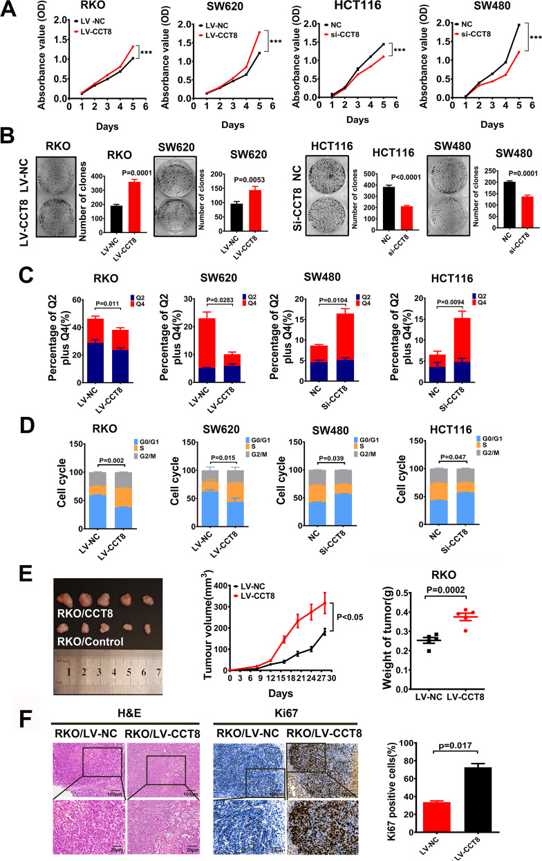
Fig. 3. CCT8 promoted the proliferation of CRC cells in vivo and in vitro.
A The CCK8 assay was performed to study the effects of CCT8 on CRC cell proliferation in loss- and gain-of-function analysis. B Representative figures and data of colony formation assay in indicated cells. Bars of the right panel represented the number of formed cells. C Bars represent apoptosis in vitro after treatment of fluorouracil for 12 h. D Bars represent the cell cycle analysis in CCT8 overexpressed and silenced CRC cell lines. E Resected xenograft tumors were injected with RKO/LV-CCT8 and RK0/LV-NC. The tumor growth curve over time and the final tumor weight in the scatter plot graph. F Representative figures of HE staining of subcutaneous tumor were shown. Proliferative ability was measured by the Ki-67 proliferative labeling index. Bars of the right panel represent the Ki-67 index of subcutaneous tumor.

