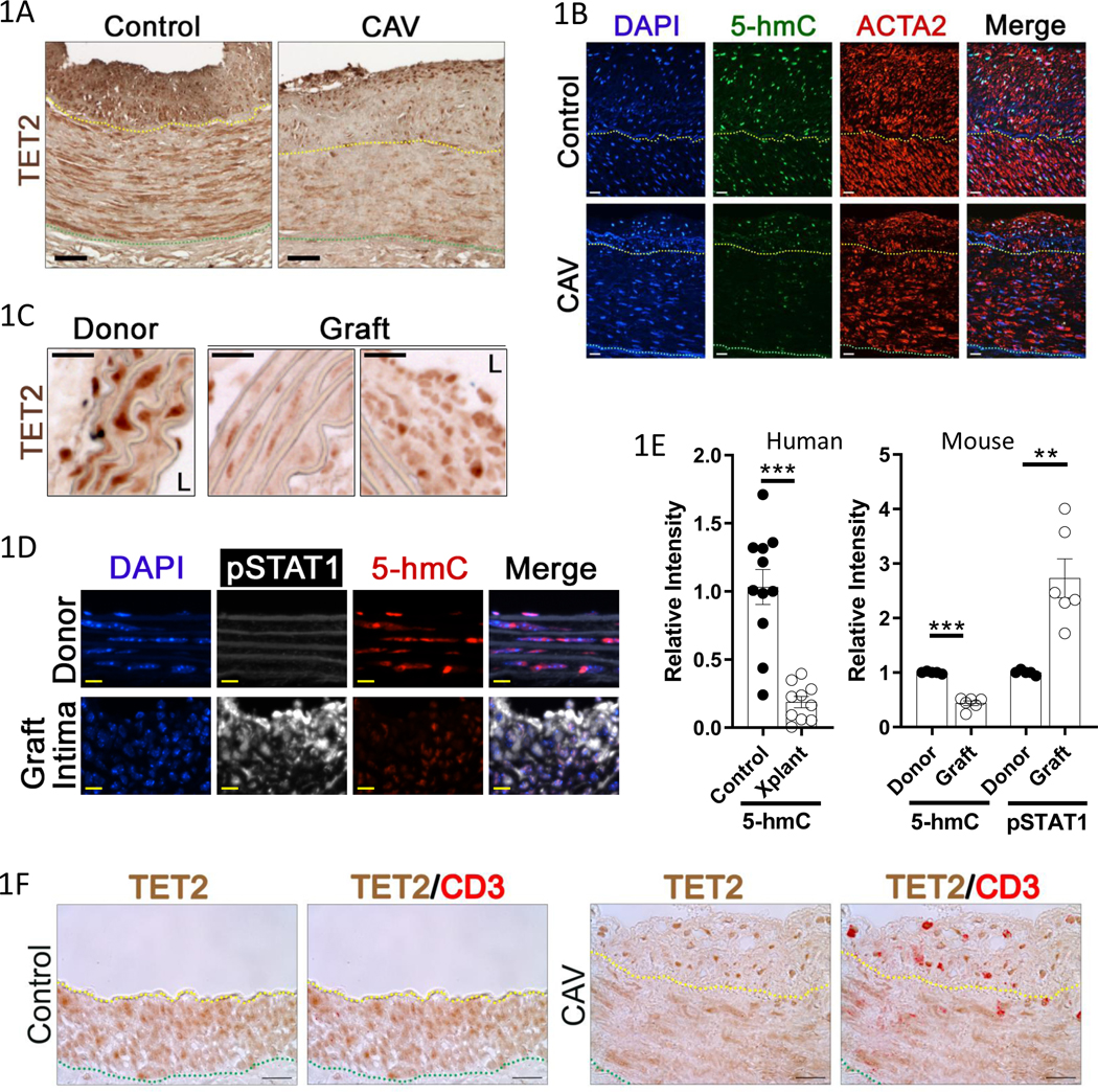
Figure 1: TET2 expression and 5-hmC are repressed in allograft vasculopathy; coincident with canonical IFNγ signaling.
Formalin-fixed artery tissue was obtained from similarly aged, deceased patients who had received heart transplants (CAV, N=7), kidney transplants (N=3), or who died from non-cardiovascular diseases and had mild atherosclerotic intimal thickening (Control coronaries N=10, control kidney N=1)). Sections of coronary arteries were immunostained for A) TET2 or B) 5-hmC and smooth muscle alpha actin (ACTA2), with DAPI nuclear counterstain. Representative images shown. Cryosections of 28day sex-mismatched murine aortic interposition grafts were similarly immunostained for C) TET2 (L denotes lumen), or D) 5-hmC and phospho-STAT1 (pSTAT1), with DAPI. Representative images shown. N=3 E) Quantitation of Left) relative 5-hmC signal intensity in human samples (Control N=11, Transplant (Xplant) N=10), Right) relative 5-hmC and pSTAT1 signal intensity at 28days after aortic transplant in mouse. (Donor N=5, Graft N=6). Scale bars: A) 50μM, B) 50μm, C) 25μm, D) 10μm. F) Normal, and transplant, human coronary artery sections were immunostained for TET2 and CD3. Representative images shown (Control N=11, CAV (and kidney transplant GA) N= 10). Yellow dotted line indicates internal elastic lamina (IEL), green dotted line indicates external elastic lamina (EEL). Scale bar: 20μm. E) Left: Welch’s t test, Right: one sample t test. **P<0.01, ***P<0.001.

