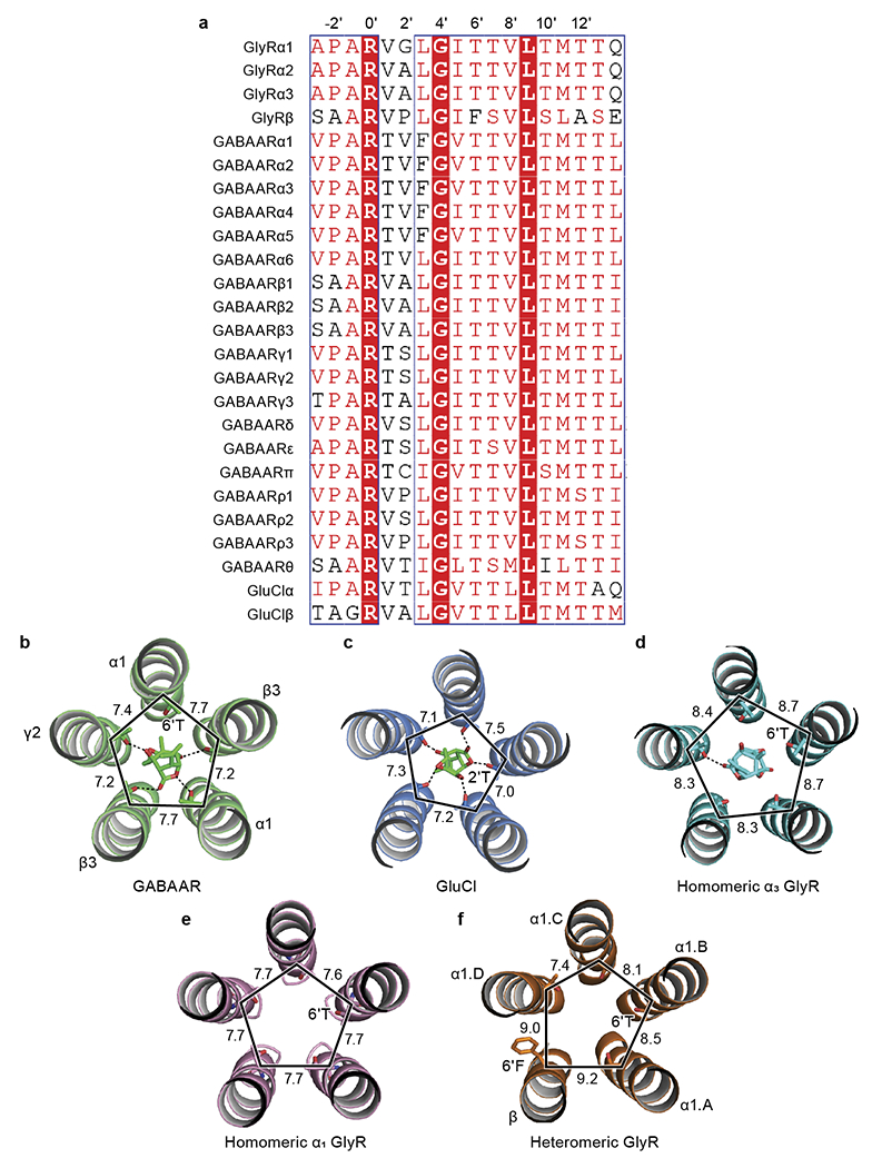
Extended Data Fig. 6. Analysis associated with TMD.

a, Sequence alignment of M2 helices among GABAAR, GlyR and GluCl. Higher prime numbers approach ECD, lower prime numbers approach intracellular domain. The −2’ position is the first amino acid of M2 helix. Sequence alignment was performed by PROMALS3D. b-d, Isolated M2 helices bound with picrotoxin from GABAAR (b; PDB ID: 6HUG), GluCl (c; PDB ID: 3RI5) and homomeric GlyR (d; PDB ID: 6UD3). The important amino acids 6’T or 2’T interacting with picrotoxin are labeled. The M2 helices and picrotoxin are shown in cartoon and stick representation, respectively. e-f, Isolated M2 helices from native homomeric GlyR (e) and heteromeric GlyR (f), respectively. The 6’T and 6’F are shown in stick representation. The M2 helices are shown in cartoon representation. All distances are denoted in Å.
