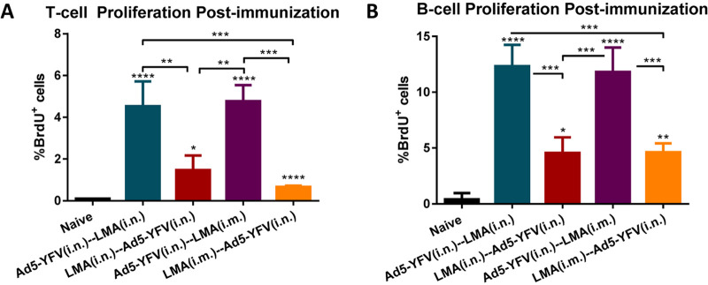
FIG 6.
T- and B-cell proliferation in response to heterologous prime-boost vaccination during long-term study. Spleens were collected 21 days after the last vaccination dose from a cohort (n = 5 per group) of immunized and naive control mice as described for Fig. 5. The isolated splenocytes were stimulated with rF1-V (100 μg/ml) for 72 h and 37°C, and then BrdU was added at a final concentration of 10 μM during the last 18 h of incubation with rF1-V to be incorporated into newly synthesized DNA of the splenocytes. Subsequently, the BrdU-labeled splenocytes were surface stained for T- and B-cell markers followed by BrdU and 7-AAD staining. The splenocytes were then subjected to flow cytometry, and the percentage of BrdU-positive cells in CD3 (A)- and CD19 (B)-positive populations were calculated using FACSDiva software. Statistical significance was determined using one-way ANOVA with Tukey’s post hoc test. Asterisks above columns represented comparison to the control group, while asterisks with comparison bars denoted significance between the indicated groups. *, P < 0.05; **, P < 0.01; ***, P < 0.001; ****, P < 0.0001. Two biological replicates were performed and data plotted. In vitro studies had 3 replicates.

