Abstract
Objectives:
To evaluate maxillary and mandibular alveolar and basal bone widths using cone-beam computed tomography (CBCT) and to verify the correlation between CBCT images and posteroanterior (PA) cephalograms.
Materials and Methods:
The CBCT scans and PA cephalograms were obtained from 20 men (age range = 24.0–29.1 years; mean age = 27.2 years; SD = 2.8 years) and 20 women (age range = 20.3–28.1 years; mean age = 26.4 years; SD = 3.2 years) with normal occlusion. On CBCT images, maxillary and mandibular bone widths were measured at three posterior sites and five bone levels. The differences between maxillary and mandibular bone widths were calculated and compared with conventional transverse width of PA cephalograms.
Results:
Statistically significant differences in maxillary and mandibular bone widths were detected at different levels and sites. Bone widths were significantly increased from the alveolar crest toward the basal bone in the maxillary molar and mandibular second premolar and molar areas. A statistically significant correlation was only found between CBCT images and PA cephalograms for maxillomandibular width at the first molar area.
Conclusion:
The results of this study suggested that three-dimensional assessment of maxillomandibular width is mandatory for the transverse analysis.
Keywords: Maxillomandibular width; CBCT, Transverse analysis
INTRODUCTION
Transverse maxillary dimension is one of the critical aspects of a functional and stable occlusion.1 Undiagnosed transverse discrepancy leads to adverse periodontal response, unstable dental camouflage, and less than optimal dentofacial esthetics.2 Betts et al.3 developed a cephalometric analysis for posteroanterior (PA) cephalograms, which calculates the transverse maxillomandibular width differential; transverse discrepancies up to 5 mm might be treated with camouflage but beyond that a surgical approach to expansion should be considered.
The most common assessment of skeletal transverse discrepancy compares the widths between the right and left jugale points and between the right and left antegonial points.4 The jugale point to the antegonial point ratio might not be as reliable an indicator of transverse relationships as has been commonly believed.5 Superimposition of anatomic structures on the view reduces the clarity of the landmarks and increases identification errors.6,7 Head orientation, such as any rotation or tipping of the head when taking the cephalogram, affects the horizontal relationships of the landmarks, making it difficult to evaluate symmetry and measure horizontal distances.8 With these problems, the practitioner is often challenged to make a proper diagnosis using PA cephalograms.
To overcome these limitations, computed tomography (CT) for the assessment of the transverse dimensions of the maxilla was introduced by Timms et al.9 in the 1980s. However, the use of conventional CT scans in orthodontics has been limited because of cost and radiation concerns.10 Cone-beam CT (CBCT) was developed for imaging hard tissues of the maxillofacial region; it results in minimum distortion, costs less, and has lower radiation exposure than conventional CT. The high resolution of CBCT images is due to the isotropic voxel (equal in all three dimensions), which produces submillimeter resolutions ranging from 0.4 mm to as low as 0.125 mm.10 Several investigations have shown the high accuracy of CBCT images for quantitative and qualitative analysis.11–14 However, few studies have assessed maxillary and mandibular skeletal and dental transverse relationships based on CBCT.
The aim of this study was to evaluate maxillary and mandibular alveolar and basal bone widths at the first premolar (P1), second premolar (P2), and first molar (M1) using CBCT images and to verify the correlation with PA cephalograms.
MATERIALS AND METHODS
Twenty men (age range = 24.0–29.1 years; mean age = 27.2 years; SD = 2.8 years) and 20 women (age range = 20.3–28.1 years; mean age = 26.4 years; SD = 3.2 years) with normal occlusions (Angle's Class I canine and molar relation) were enrolled in this study. Subjects with skeletal dysplasia, asymmetric dental arch, moderate to severe crowding, missing tooth, and history of prosthetic or orthodontic treatment were excluded. All subjects provided informed consent to participate in this study, and this study was approved by the Institutional Review Board for Medical Science at the Chonnam National University Hospital, Gwangju, Korea (I-2008-12-156).
The CBCT scans were obtained by CB MercuRay (Hitachi Medical Co, Tokyo, Japan) under the following conditions: 120 kV, 15 mA, voxel size 0.3 mm (P mode), and field of view 150 mm. The CBCT scan data were processed and reformatted by the OnDemand3D Application program version 1.0, (CyberMed Inc, Seoul, Korea).
Conventional PA cephalograms were obtained using cephalometric x-ray equipment (OrthoCeph OC100, Instrumentarium Imaging Co, Tuusula, Finland). A photostimulable phosphor plate was used as the detector and was positioned 150 mm from the midsagittal plane. The source-midsagittal plane distance was 1500 mm. The plate was scanned at 650 dpi (Kodak DirectView CR975 system, Carestream Health, Rochester, NY).
Measurement of Maxillary and Mandibular Bone Width on the CBCT Image
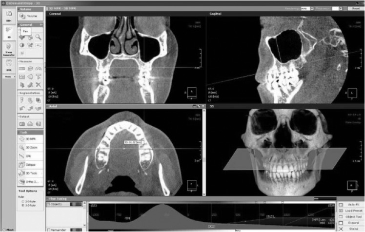
On CBCT images, maxillary and mandibular bone widths were measured at three posterior sites (P1, P2, and M1) and at five bone levels (alveolar crest, furcation, root apex, and at 2 mm and 5 mm below the root apex from M1); the differences between maxillary and mandibular bone widths were also calculated. To obtain standardized axial and coronal slices, the following references were used. Reference planes were used to ensure that the two-dimensional coronal slices were consistently oriented: (1) the axial plane was defined as the functional occlusal plane15; (2) the coronal plane was perpendicular to the axial plane, passing through the buccal groove of the maxillary right first molar; and (3) the sagittal plane was perpendicular to both the axial and coronal planes, passing through the midpoint between the medial rims of the orbits (Figure 1). The vertical levels of the measurements were established at the alveolar crest, furcation, root apex, and 2 mm and 5 mm below the root apex from M1. This was accomplished by moving the horizontal reference line based on the millimeter ruler. By using the axial slice, the final measurements were made through the buccal portion of the cortical bone where the horizontal reference line contacted the endosteal surface.
Figure 1.
Measurement of maxillary bone width on CBCT images using orientations of three orthogonal views.
Measurement of Maxillary and Mandibular Width on PA Cephalogram
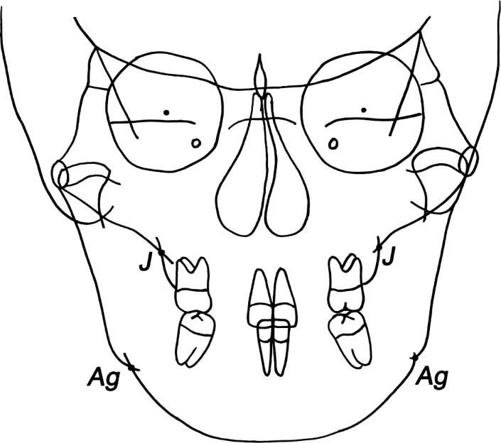
The PA cephalograms were imported into V-ceph software (version 6.0, CyberMed Inc, Seoul, Korea), and two linear measurements commonly used in conventional transverse analysis were chosen. The distances between the right and left jugale points and antegonial points that represent maxillary width and mandibular width, respectively, were measured (Figure 2). The differences between maxillary and mandibular bone widths were calculated.
Figure 2.
Measurement of maxillary and mandibular width on PA cephalogram. J indicates jugale; Ag, antegonion.
Statistical Analysis
To assess the reliability of measurements, all measurements were repeated after 2 weeks by the same investigator, and the mean of two measurements was used in the statistical analysis. The systematic intraexaminer error between two measurements was determined using a paired t-test. Also, the magnitude of the measurement error was assessed by calculating the intraclass correlation coefficient (ICC). Repeated measures analysis of variance with the general linear model was used to identify differences at different levels and sites, and multiple comparisons were performed with the Bonferroni test. Mauchly's sphericity test was used to test the equality of the variances of the differences between the levels of the repeated measures factor. Pearson correlation analysis was used to assess the relationship between measurements from CBCT images and conventional transverse widths of PA cephalograms. All analyses were performed with version 18.0 of the PASW software package (SPSS Inc, Chicago, Ill).
RESULTS
The systematic intraexaminer error was evaluated at P < .05 and was found to be statistically insignificant. The ICC measurement indicated excellent reliability with a mean ICC of 0.874 (ICC = 0.79–0.94). Before assessing maxillary and mandibular alveolar and basal bone widths, sex difference in bone width was assessed. There was no statistically significant difference between male and female subjects according to the t-test (P > .05). Based on the results of those tests, data were combined for men and women.
Means and standard deviations for maxillary and mandibular alveolar and basal bone widths are shown in Table 1. Statistically significant differences in maxillary and mandibular bone widths were detected at five different bone levels and at three sites.
Table 1.
Data of Maxillary and Mandibular Widths on CBCT Images and Comparison of the Five Areas (unit: mm)
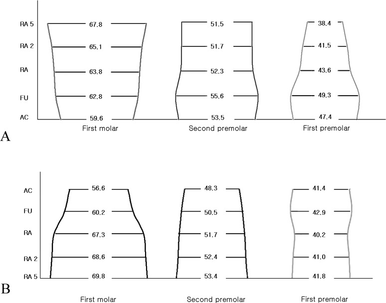
In the maxilla, the mean alveolar and basal bone widths at P1 and P2 were significantly increased until the furcation level but then decreased toward 5 mm below the root apex. At M1, the mean maxillary alveolar and basal bone widths were significantly increased from the alveolar crest level toward 5 mm below the root apex level.
In the mandible, the alveolar and basal bone widths at P2 and M1 were significantly increased from the alveolar crest level toward 5 mm below the root apex level. However, at P1 the alveolar and basal bone widths were exhibited consistently according to the five levels in contrast to those in the maxilla and mandible.
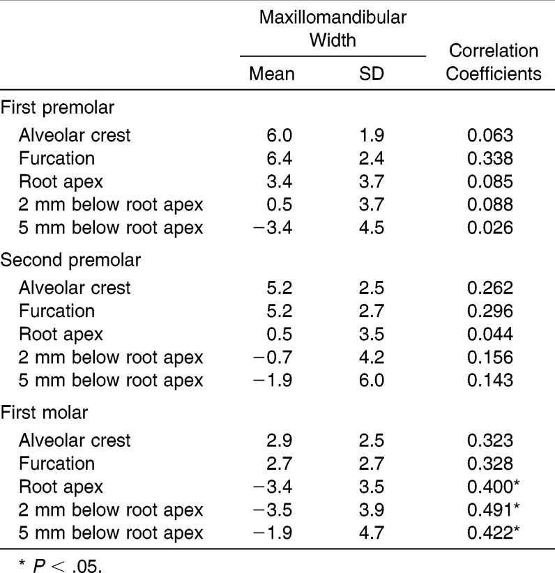
Associations of maxillomandibular width between CBCT and PA cephalograms are shown in Table 2. The value of maxillomandibular width on PA cephalograms was 24.6 mm on average (SD = 4.8 mm). There was a statistically significant correlation between CBCT images and PA cephalograms for the M1 area only. Maxillomandibular width of M1 at three levels (root apex and 2 mm and 5 mm below root apex) showed a statistically significant correlation with those on PA cephalograms. However, maxillomandibular widths of P1 and P2 did not show any correlation with PA cephalograms.
Table 2.
Maxillomandibular Widths on CBCT Images and Correlation with PA Cephalograms (unit: mm)
DISCUSSION
It is generally acknowledged that maxillofacial deformities and malocclusions require diagnosis in all three planes of space.16,17 Podesser et al.18 suggested a method of quantification of factors in the transverse dimension of the maxilla based on CT scanning. Primozic et al.19 performed three-dimensional evaluation of crossbite correction using laser-scanned study casts. This study measured palatal surface area and palatal volume.
Recently, with the introduction of CBCT imaging in orthodontics, a diagnosis in three planes can be easily obtained with minimal radiation. In addition, the spatial limitations of two-dimensional imaging, especially in PA cephalograms, can be overcome. In particular, overlaps of molars on PA films that complicate identification can be overcome by the ability to use CBCT to view coronal slices at different thicknesses; consequently, determination of molar and posterior alveolar inclination is facilitated.20
In this study, our objectives were to evaluate maxillary and mandibular alveolar and basal bone widths and to verify the correlation between CBCT images and PA cephalograms. As the literature contains few studies with a detailed examination of maxillomandibular width using CBCT images, we had to create our own evaluation protocol. Differences in bone width were found according to the five levels of each tooth (Figure 3). At the M1 area, the alveolar and basal bone widths significantly increased from the alveolar crest toward 5 mm below the root apex in both the maxilla and mandible. At the P1 and P2 areas, however, variations existed depending on the five levels. These patterns might be explained by different anatomic characteristics in the maxillary and mandibular posterior areas. Such differences in bone width between five levels and their neighboring sites should be considered, especially when evaluating the effect of expansion treatment.
Figure 3.
Diagrams of three-dimensional maxillary (A) and mandibular (B) bone widths according to the 5 levels of each tooth on CBCT. Data are presented as mean: AC indicates, alveolar crest; FU, furcation; RA, root apex; RA 2, 2 mm below root apex; RA 5, 5 mm below root apex.
We found no significant correlation of maxillomandibular width between CBCT images and PA cephalograms except at the M1 area. These findings revealed that assessment of skeletal transverse discrepancy using PA cephalograms with jugale and antegonial points might have limitations or cause possible misunderstanding.
Miner et al.21 investigated transverse dimension relationships using CBCT scans of patients with and without crossbite. They analyzed the width of the jaws and the inclination of the first molars. We did not perform dental measurements, such as inclination of the molars. We sought to verify a correlation of transverse discrepancy between CBCT images and PA cephalograms. With regard to dental compensation in patients with transverse discrepancy, further studies involving such dental measurements as inclination of molars are required.
CONCLUSIONS
Statistically significant differences in maxillary and mandibular bone widths were detected at different levels and sites on CBCT images.
There was a statistically significant correlation of the maxillomandibular width at the first molar area between CBCT images and PA cephalograms.
ACKNOWLEDGMENT
This study was supported by a grant (CRI13035-1) from the Chonnam National University Hospital Biomedical Research Institute.
REFERENCES
- 1.Vanarsdall RL, White RP., Jr Three-dimensional analysis for skeletal problems. Int J Adult Orthodon Orthognath Surg. 1994;9:159. [PubMed] [Google Scholar]
- 2.Vanarsdall RL., Jr Transverse dimension and long-term stability. Semin Orthod. 1999;5:171–180. doi: 10.1016/s1073-8746(99)80008-5. [DOI] [PubMed] [Google Scholar]
- 3.Betts NJ, Vanarsdall RL, Barber HD, Higgins-Barber K, Fonesca RJ. Diagnosis and treatment of transverse maxillary deficiency. Int J Adult Orthodon Orthognath Surg. 1995;10:75–96. [PubMed] [Google Scholar]
- 4.Ricketts RM. Perspectives in the clinical application of cephalometrics, the first fifty years. Angle Orthod. 1981;51:115–150. doi: 10.1043/0003-3219(1981)051<0115:PITCAO>2.0.CO;2. [DOI] [PubMed] [Google Scholar]
- 5.Ghafari J, Cater PE, Shofer FS. Effect of film-object distance on posteroanterior cephalometric measurements: suggestions for standardized cephalometric methods. Am J Orthod Dentofacial Orthop. 1995;108:30–37. doi: 10.1016/s0889-5406(95)70063-3. [DOI] [PubMed] [Google Scholar]
- 6.Thurow R. Cephalometric methods in research and private practice. Angle Orthod. 1951;21:104–116. doi: 10.1043/0003-3219(1951)021<0104:CMIRAP>2.0.CO;2. [DOI] [PubMed] [Google Scholar]
- 7.Major P, Johnson D, Hesse K, Glover K. Landmark identification error in posterior anterior cephalometrics. Angle Orthod. 1994;64:447–454. doi: 10.1043/0003-3219(1994)064<0447:LIEIPA>2.0.CO;2. [DOI] [PubMed] [Google Scholar]
- 8.Major P, Johnson D, Hesse K, Glover K. Effect of head orientation on posterior anterior cephalometric landmark identification. Angle Orthod. 1996;66:51–60. doi: 10.1043/0003-3219(1996)066<0051:EOHOOP>2.3.CO;2. [DOI] [PubMed] [Google Scholar]
- 9.Timms DJ, Preston CB, Daly PF. A computed tomographic assessment of maxillary movement induced by rapid expansion—a pilot study. Eur J Orthod. 1982;4:123–127. doi: 10.1093/ejo/4.2.123. [DOI] [PubMed] [Google Scholar]
- 10.Scarfe WC, Farman AG, Sukovic P. Clinical applications of cone-beam computed tomography in dental practice. J Can Dent Assoc. 2006;72:75–80. [PubMed] [Google Scholar]
- 11.Lagravere MO, Carey J, Toogood RW, Major PW. Three-dimensional accuracy of measurements made with software on cone-beam computed tomography images. Am J Orthod Dentofacial Orthop. 2008;134:112–116. doi: 10.1016/j.ajodo.2006.08.024. [DOI] [PubMed] [Google Scholar]
- 12.Ballrick JW, Palomo JM, Ruch E, Amberman BD, Hans MG. Image distortion and spatial resolution of a commercially available cone-beam computed tomography machine. Am J Orthod Dentofacial Orthop. 2008;134:573–582. doi: 10.1016/j.ajodo.2007.11.025. [DOI] [PubMed] [Google Scholar]
- 13.Timock AM, Cook V, McDonald T, et al. Accuracy and reliability of buccal bone height and thickness measurements from cone-beam computed tomography imaging. Am J Orthod Dentofacial Orthop. 2011;140:734–744. doi: 10.1016/j.ajodo.2011.06.021. [DOI] [PubMed] [Google Scholar]
- 14.Patcas R, Müller L, Ullrich O, Peltomäki T. Accuracy of cone-beam computed tomography at different resolutions assessed on the bony covering of the mandibular anterior teeth. Am J Orthod Dentofacial Orthop. 2012;141:41–50. doi: 10.1016/j.ajodo.2011.06.034. [DOI] [PubMed] [Google Scholar]
- 15.Jacobson A. Update on the “Wits” appraisal. Angle Orthod. 1988;58:205–219. doi: 10.1043/0003-3219(1988)058<0205:UOTWA>2.0.CO;2. [DOI] [PubMed] [Google Scholar]
- 16.Broadbent BS. A new x-ray technique and its application to orthodontia. Angle Orthod. 1931;1:45–66. [Google Scholar]
- 17.Thurow RC. Fifty years of cephalometric radiography. Angle Orthod. 1981;51:39–40. [PubMed] [Google Scholar]
- 18.Podesser B, Williams S, Bantleon HP, Imhof H. Quantitation of transverse maxillary dimensions using computed tomography: a methodological and reproducibility study. Eur J Orthod. 2004;26:209–215. doi: 10.1093/ejo/26.2.209. [DOI] [PubMed] [Google Scholar]
- 19.Primozic J, Richmond S, Kau CH, Zhurov A, Ovsenik M. Three-dimensional evaluation of early crossbite correction: a longitudinal study. Eur J Orthod. 2013;35:7–13. doi: 10.1093/ejo/cjq198. [DOI] [PubMed] [Google Scholar]
- 20.Maki K, Inou N, Takanishi A, Miller AJ. Computer-assisted simulations in orthodontic diagnosis and the application of a new cone beam x-ray computed tomography. Orthod Craniofac Res. 2003;6(suppl 1):95–101. doi: 10.1034/j.1600-0544.2003.241.x. [DOI] [PubMed] [Google Scholar]
- 21.Miner RM, Al Qabandi S, Rigali PH, Will LA. Cone-beam computed tomography transverse analysis. Part I: Normative data. Am J Orthod Dentofacial Orthop. 2012;142:300–307. doi: 10.1016/j.ajodo.2012.04.014. [DOI] [PubMed] [Google Scholar]


