Abstract
Objective To evaluate the role of serum alkaline phosphatase (ALP) and ultrasonography (USG) in monitoring the progress of treatment in diaphyseal non-unions.
Methods This prospective observational cohort study included adult patients with diaphyseal fractures of major long bones previously treated with internal fixation and eventually resulting in non-union. Following the definitive treatment for non-union, the patients were followed-up periodically for six months, and serial monitoring of the levels of ALP and USG were performed along with radiographs (X-rays) to ascertain the status of the union.
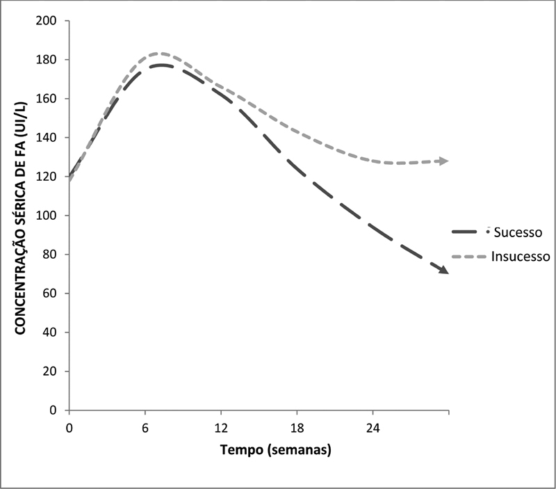
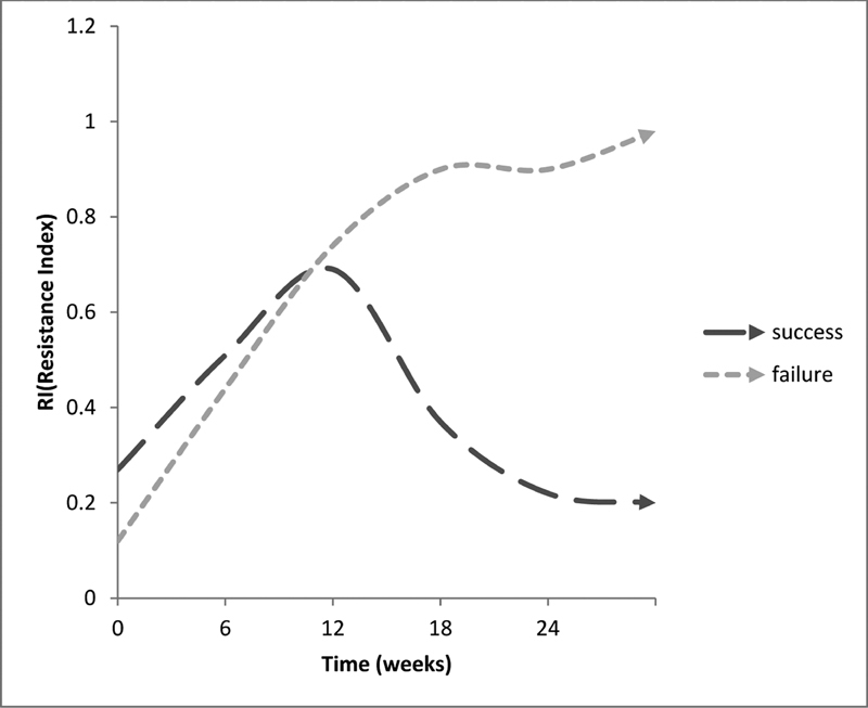
Results After an initial rise at seven weeks, ALP levels declined to normal values in fractures which united, whereas they remained high in cases of persistent non-union. Similarly, after an elevation of the vascular resistive index (RI) at around 12 weeks in all the patients, it decreased in cases progressing to union, while it remained persistently high even at 24 weeks in fractures failing to unite. Cases of persistent non-union continued to show hypoechogenic callus at 24 weeks instead of converting into hyperechogenic callus, as observed in cases which progressed to union.
Conclusion Significant changes suggestive of union appeared simultaneously on the X-rays, USG and ALP levels during the follow-up. However, a serial examination of the ALP levels and USG during the follow-up gave a hint of the direction of progress in the healing process of fracture non-union. Their role in monitoring the outcome of non-union is more complimentary than supplementary to the X-rays.
Keywords: bony callus, ultrasonography, alkaline phosphatase
Introduction
Fracture non-union is an outcome difficult to predict, and even though it is an established complication, in the clinical practice, both clinicians and patients deem it a poor outcome. 1 There is variability in the clinical assessment of the status of union in fractures and a lack of consensus on the the reliability of various methods to assess the progress of the union. Clinical appreciation combined with X-rays is used for the assessment of both acute fractures and non-unions, even though other methods to monitor and document bone healing, like computed tomography (CT), magnetic resonance imaging (MRI), bone scintigraphy etc., have been used sparingly in select cases. However, in addition to the concern regarding availability, these advanced investigations represent an added financial burden and/or pose a risk of radiation exposure to the patient. The objective of the present study was to evaluate the role of alternative investigative methods: serum alkaline phosphatase (ALP) and ultrasonography (USG), and compare them with the conventional radiograph (X-ray) in monitoring the progress of treatment in diaphyseal non-unions. In addition to being inexpensive and easily available, USG and the measurementof the levels of ALP were selected to represent physical and biochemical markers of union respectively.
Methodology
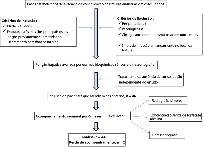
This prospective observational study was performed at a state-run tertiary care hospital attached to a postgraduate teaching institute from April 2018 to September 2019, with due permission from the Institutional Ethics Committee. Diaphyseal fractures of major long bones that is, the femur, tibia (with or without the fibula), humerus and the radius and ulna (individually / combined), previously treated with any type of internal fixation in adults (> 18years), and eventually resulting in non-union, were included in the study. Only those patients with normal liver function confirmed by USG and serum biochemical tests were included in the study. The treatment of non-union remained independent of the study. All patients were followed up for six months at six-week intervals. At each follow-up visit, all patients were assessed clinically and by plain radiographs for signs of union. In addition to serial ALP measurements, at each follow-up, the fractures were evaluated by USG for echogenecity and measurement of the vascular resistive index (RI) of the callus. The measurement of the ALP levels was performed spectrophotometrically using the p-nitrophenylphosphate (PNPP) kinetic International Federation of Clinical Chemistry (IFCC) method with a laboratory reference range of 28 IU/L to 111 IU/L. The USG was performed using a Philips (Amsterdam, The Netherlands) iU22 camera equipped with a linear probe with a resolution of 12 MHz to 5 MHz, MSK Superficial program and 3D Power Color Doppler. Whereas the authors evaluated fracture healing using plain radiographs, the USG examination of the fracture was performed by an independent radiologist without the knowledge of the X-ray status. Fig. 1 shows a brief overview of the methodology.
Fig. 1.
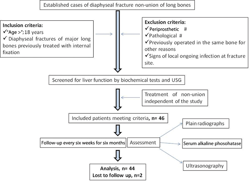
Flowchart showing the basic methodology applied to the present study.
Observations and Results
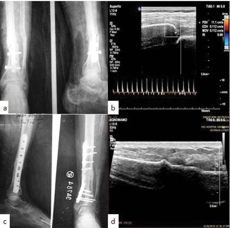
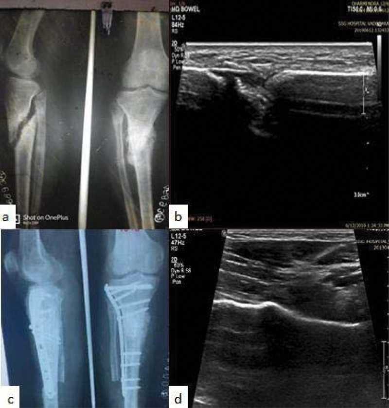
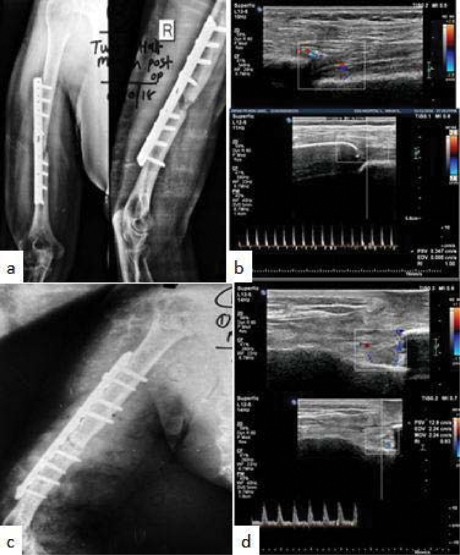
The sample for the final follow-up and analysis was composed of 44 patients (36 male, 8 female). These included 20 cases of upper-limb and 24 cases of lower-limb diaphyseal non-unions. Morphologically, there were 24 cases of atrophic, 6 cases of hypertrophic and 14 cases of comminuted non-unions. The average age of the patients was 37 (range: 18 to 75) years. In total, 28 patients underwent cancellous/tricortical/fibular bone grafting, 6 were stabilized by additional fixation, 4 patients were subjected to implant extraction followed by functional bracing, 4 patients were treated by late dynamisation of statically interlocked nail, and the remaining 2 were treated by an Illizarov ring fixator. Table 1 shows a summary of the observations with reference to the outcome of the study. Illustrative cases are shown in Figs. 2 , 3 and 4 .
Table 1. Summary of the observations regarding the outcomes of the study.
| Outcome→ | Union | Non-union | Total/Gross |
|---|---|---|---|
| Parameters ↓ | |||
| Number of cases | 30 | 14 | 44 |
| Type of non-union | |||
| Atrophic | 18 | 6 | 24 |
| Hypertrophic | 4 | 2 | 6 |
| Comminuted | 8 | 6 | 14 |
| Average level of serum akaline phosphatase (28–111 IU/L) | |||
| At admission | 121 | 118 | 120 |
| At the final follow-up | 94 | 128 | 105 |
| Peak value during the study | 179 | 181 | 180 |
| Average resistive index (ultrasonography) | |||
| At admission | 0.28 | 0.12 | 0.22 |
| At the final follow-up | 0.22 | 0.9 | 0.44 |
| Peak value during the study | 0.79 | 1.04 | 0.87 |
| Follow-up (weeks) | |||
| Peak level of serum akaline phosphatase | 7.2 | 6.9 | 7.0 |
| Peak value of the resistive index | 14.4 | 21.4 | 16.6 |
| Hyperechoic callus on ultrasonography | 20 | Not observed | 20 |
| Callus on X-ray (union time) | 18.8 | Not observed | 18.8 |
Fig. 2.
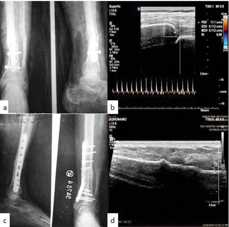
Illustrative Case1. ( A ) X-ray showing non-union of the distal tibia; ( B ) ultrasonography (USG) showing fracture gap with high resistive index; ( C ) fracture union after bone grafting at 24 weeks; and ( D ) USG at 24 weeks showing hyperechoic signals and absent vascular resistance.
Fig. 3.
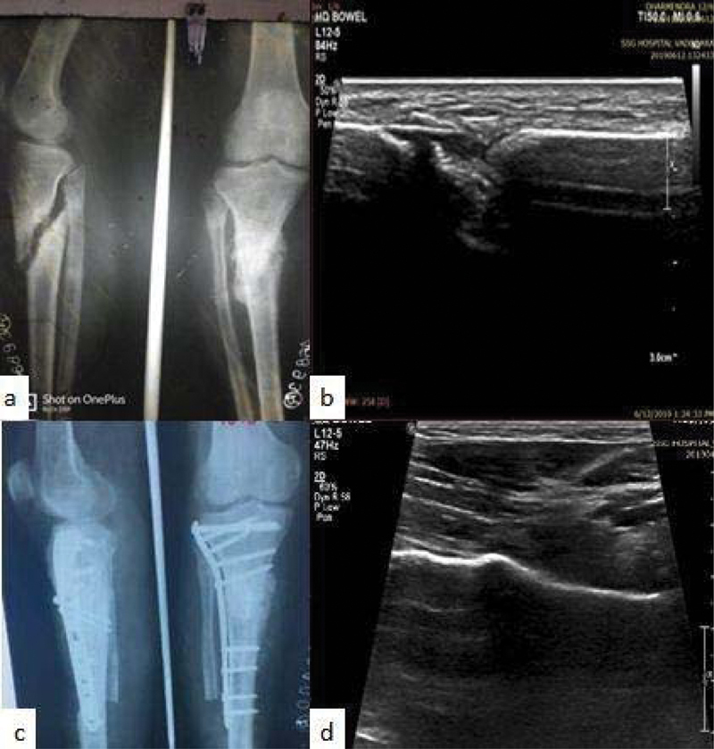
Illustrative Case2. ( A ) X-ray showing non-union of the proximal tibia; ( B ) USG showing heterogenous hypoechoic signals of non-union; ( C ) fracture union after bone grafting at 18 weeks; and ( D ) USG at 18 weeks showing hyperechoic signals.
Fig. 4.
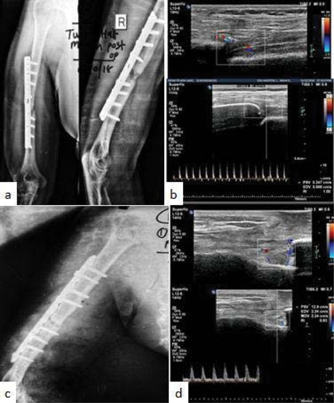
Illustrative Case3. ( A ) X-ray showing non-union shaft of the humerus; ( B ) USG showing fracture gap with high resistive index; ( C ) persistent fracture non-union after 24 weeks of the revision surgery; and ( D ) USG at 24 weeks showing fracture gap, hypoechoic signals and high vascular resistance.
X-ray
We considered a fracture radiologically united only when the X-rays showed bridging bone across the fracture in both orthogonal views. A total of 30 patients showed signs of union, and 14 patients showed persistent non-union on the X-rays at the final follow-up ( Figs. 2d , 3d and 4d ). During the follow-up, calluses were observed as early as 12 weeks and as late as 24 weeks. The average union time as assessed by the X-rays was of 18.8 (range: 12 to 24) weeks. Bone grafting ( n = 22) showed more promising results in achieving union.
Serum Alkaline Phosphatase
It has been our observation that patients with established non-union who present within six months of the primary surgery usually have ALP levels > 110 IU/L. The average ALP level as measured when the patients were recruited to the study was of 120 (range: 42 to 202) IU/L. The average peak level reached during the study was of 180 (range: 136 to 216) IU/L. The levels of ALP initially increased in all cases and reached a peak at around 7 weeks. In fractures which progressed toward union, the ALP levels dropped to normal by the end of the study period, whereas, in cases which failed to unite, the ALP levels continued to remain high following the initial spike until the end of the study period ( Fig. 5 ). The average ALP level at the end of the study period in united and persistent non-union cases were of 94 IU/L and 128 IU/L respectively.
Fig. 5.
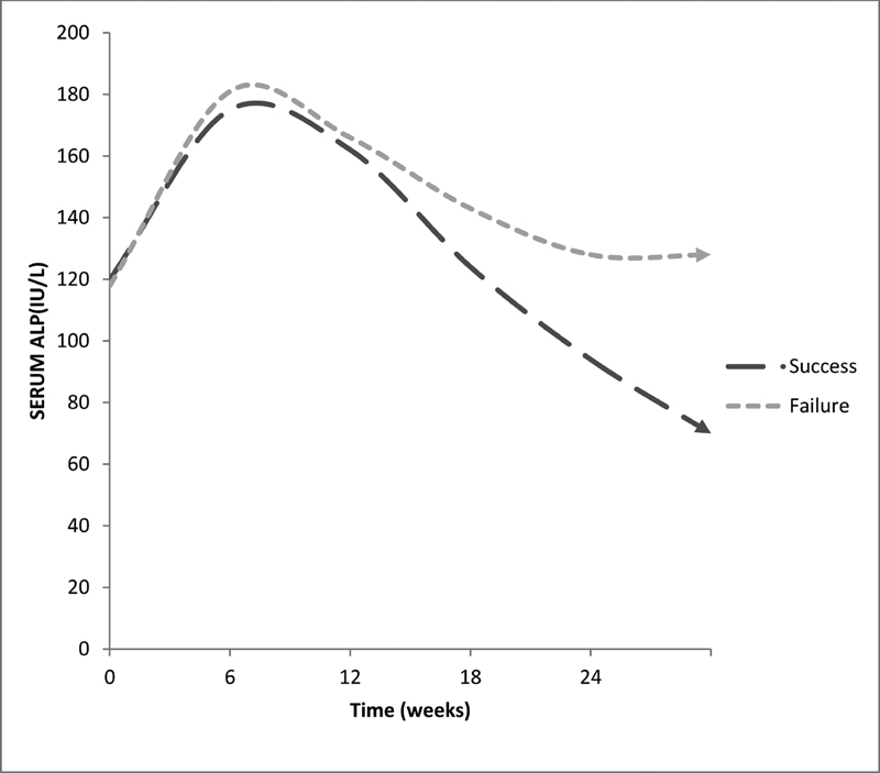
Trend of average serum alkaline phosphatase levels in cases of union and non-union.
Ultrasonography
The assessment of union in fractures by USG was performed qualitatively, by the echogenicity of the callus, and quantitatively, by measuring the vascular RI. 2
In the sample, there were established cases of non-union. Hence, a hypoechoic structure (fibrous tissue) was observed since the first day of recruitment. However, in contrast to the homogenously hypoechoic signals of a newly-formed hematoma converting to soft callus, in our cases, gray-scale USG at recruitment showed heterogenous-hypoechogenicity due to the fibrosclerosis that had ensued within the non-union ( Figs. 2b , 3b and 4b ). The average time taken for hyperechoic callus suggestive of mineralization to appear in uniting fractures was of 20 (range: 18 to 24) weeks. In cases which failed to unite, the USG examination continued to show hypoechogenicity at 24 weeks ( Fig. 4d ).
The average of RI score as measured when the patients were recruited to the study was of 0.22, with 30 patients showing non-recordable levels (due to fibrosclerosis). Irrespective of the outcome, a steady elevation in RI scores was observed in all cases until around 12 to 18 weeks. The RI scores decreased following their elevation in patients progressing to union. In fractures failing to unite, they persistently remained high even at 24 weeks ( Fig. 6 ). The average RI scores at the final follow-up in united and persistent non-union cases were of 0.22 and 0.90 respectively ( Fig. 2d , 3d and 4d ).
Fig. 6.
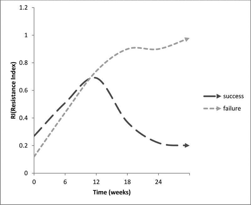
Trend of average vascular resistive index in cases of union and non-union.
A gross analysis of the findings of the study ( Table 2 ) was attempted using the frequency with which all parameters that is, the X-ray, ALP levels, hyperechoic callus and RI scores, showed positive signs of union at different intervals during the follow up. Bridging callus on X-ray, ALP levels within the laboratory reference range, hyperechoic signals on gray-scale USG and a lower RI score as compared with the previous reading on the Doppler USG were considered signs of progress of the union. In the present analysis, the cases of each parameter were mutually inclusive for that follow-up time. The earliest time when the ALP levels and the USG predicted a possible outcome of the treatment (12 to 18 weeks) coincided with the early changes observed on the X-rays.
Table 2. Analysis of the findings of the present study.
| Investigation | Number of patients | |||
|---|---|---|---|---|
| At 6 weeks | At 12 weeks | At 18 weeks | At 24 weeks | |
| X-ray (bridging callus) | 0 | 4 | 22 | 30 |
| Level of serum akaline phosphatase (< 111 IU/L) | 0 | 0 | 8 | 30 |
| Ultrasonography (hyperechoic signal+ decreasing resistive index) | 0 | 0 | 20 | 30 |
Discussion
The non-union of diaphyseal fractures to long bones, which otherwise have a lower incidence, represent an uphill task to the treating surgeon. Although much information is available in the literature regarding non-union, to the best of our knowledge, we could not find any perfectly-matching study that described the monitoring of the progress of the treatment in cases of long-bone non-union. A detailed evaluation of studies regarding the value of the laboratory and imaging studies in cases of long-bone non-union in animals was performed by Zimmermann et al. 3 However, most of these studies cannot be properly reproduced in the routine clinical practice. Clinical improvement, such as painless weight bearing, use of the affected extremity, and lack of swelling and tenderness have always comforted both patient and surgeon as a reflection of a fair progress. Supportive radiological evidences such as that of X-rays and CT scans have reinforced such clinical impressions. In the qualitative assessment, the line, margins and gaps of the fracture, the external callus, the callus-to-cortex ratio and the bridging callus are examined. Advanced diagnostic methods, like dual-energy X-ray absorptiometry (DEXA) scans, multidirectional CT and laboratory tests, like the measurment of the levels of done-specific alkaline phosphatase, matrix metalloproteases and specific growth factors 4 etc., have booked a place in theoretical texts, but have yet to find place in the routine clinical practice.
Several studies 5 6 7 8 have been published supporting the evidence that the ALP levels can be used as an adjunct biomarker to the clinical and radiological evidences of healing. In clinical trials, 9 10 ALP levels showed a significantly higher baseline and a tendency towards a slow increase during the first two months of healing in acute fractures, and this might help to predict delayed fracture healing. This increase in ALP activity observed is assumed to happen due to the rising concentration of the bone isoenzyme of ALP during the healing process, 11 and it coincides with the beginning of mineralization. 12 In line with the studies related to biological markers in acute fractures, ALP levels temporarily increased after the intervention for up to seven weeks in all patients, followed by normalization in those showing signs of union in the present study. Based on this, we believe that, as observed in acute fractures, serial ALP measurements have significance in monitoring the treatment of fracture non-unions as well.
The evaluation of the callus by USG has been well described in the literature, which reports that the callus can be observed much sooner on the USG than on plain X-rays. 13 14 15 16 Significant changes in the mineral composition of the callus during fracture healing changes the speed of the reflected wave (echo) picked up by the transducers. Based on this echo pattern, the callus is labeled either as hypoechoic or hyperechoic. 17 Early (soft) calluses in fresh fractures are seen as homogenous-hypoechoic which, with mineralization, mature to homogenous-hyperechoic (hard) calluses over time. The presence of a hyperechoic ultrasonographic signal from the fracture site was found to have 100% of correlation with the presence of a hard callus on the biopsy. 14 Fractures which fail to progressively unite show persistence of the hypoechgenic pattern. Fibrous tissue is seen as a heterogenous hypoechoic strutucture in cases of non-union. 12 Chachan et al. 18 compared the efficacy of plain radiography versus USG in monitoring fracture healing, and found the sensitivity and specificity of USG to diagnose fracture union to be of 100% and 80% respectively. In the present study, over the course of up to 16 weeks, all patients showed hypoechoic calluses, which converted to hyperechoic calluses in the patients showing signs of union, and remained hypoechoic in those with persistent non-union. Fibrosclerotic tissue within the non-union site was a significant limiting factor in the qualitative evaluation of the callus by USG in the present study. In our sample consisting exclusively of cases of non-union, the absence of hematoma-induced periosteal reaction and mixed echogenic images due to a standstill in the initial healing process might have been the factors that led to the underestimation of the onset of healing as diagnosed by USG.
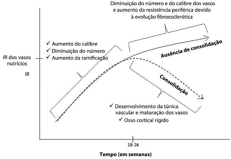
The process of fracture healing can be followed with color Doppler imaging and spectral analysis. 19 The USG is able to follow the formation of new vessels at the fracture site and to assess flow characteristics in them during the development of the fracture callus. In addition to following the formation of new vessels at the fracture site, Doppler USG can measure the RI, which reflects the resistance to blood flow caused by a microvascular bed distal to the site of measurement. In patients with normal callus development, the caliber of the afferent vessels progressively increases, and their number decreases. The RI progressively increases, up to values similar to those of nutrient vessels (0.36 to 0.90). Within the second month of the fracture, a muscular tunic develops in the vessel wall, converting to a mature vessel with the progressing mineralization of the cortex, thus causing the RI to progressively decrease and finally disappear. 11 On the other hand, patients with non-union and delayed healing have persistent higher resistance related to a poor formation of neovasculature and increasing peripheral resistance in the residual vessels because of the fibrosclerotic involution of bone healing 11 ( Fig. 7 ). In consistence with the aforesaid description, in the present study, the RI steadily increased until around 12 weeks in all patients; subsequently, at 24 weeks, patients who progressed to union showed low RIs, and those who progressed toward non-union had persistently high RIs.
Fig. 7.
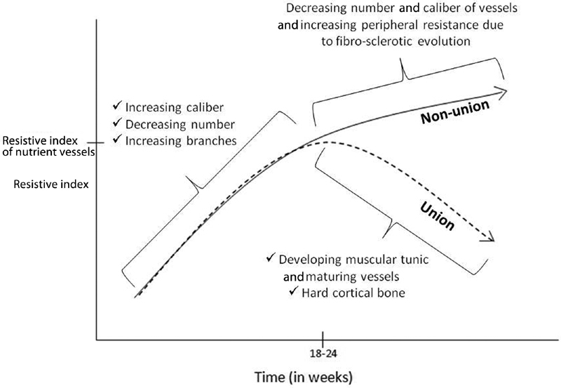
Vascular changes and their effect on the resistive index in cases of union and non-union.
A gross analysis of the findings of the present study showed that changes in ALP levels, USG and X-rays occurred simultaneously during the follow-up in cases of fracture non-union. None of them proved superior in terms of early prediction of the outcome. Nevertheless, a serial examination of ALP levels and USG during the follow-up gave a hint of the direction of progress in the healing process of the fracture. There is still a great need for scientific research before biological markers like the ALP can be used to make major treatment decisions. Although USG has been established to be a reliable technique for the early diagnosis of healing in acute fractures, its role in outcome evaluation in cases of non-union is yet to be determined. Whereas spot X-rays provide qualitative information regarding the changes during fracture healing, isolated ALP and USG findings have no defined laboratory limits for the period of follow-up after fixation, which can be used to separate individual cases into favorable and unfavorable categories. Furthermore, the USG requires training and technical expertise to demonstrate the required results. This could possibly be a limitation to the widespread use of USG for such cases. Although the equipment is easy to procure, not all institutions have on staff professionals trained to evaluate the healing process. Moreover, a study on the cost-effectiveness of USG could further validate the role of USG in monitoring fracture healing. The conventional plain radiograph will surely remain a popular method of assessment because of its long time-tested history, practicality, and lower costs. 1 18
Conclusion
The limited sample size of the present study ruled out any statistical conclusion. Significant changes suggestive of union were simultaneously observed on X-rays, USG and ALP levels during the follow-up. The earliest period when ALP levels and USG predicted a possible outcome coincided with the early changes observed on the X-rays. However, the serial evaluation of ALP levels and USG definitely gave a hint regarding the direction of the progress of the treatment. At present, their role in monitoring the outcome of non-union is more complimentary than supplementary to the X-rays.
Funding Statement
Suporte Financeiro Não houve suporte financeiro de fontes públicas, comerciais, ou sem fins lucrativos.
Financial Support There was no financial support from public, commercial, or non-profit sources.
Conflito de Interesses Os autores declaram não haver conflito de interesses.
Estudo desenvolvido no Departamento de Ortopedia, Medical College Baroda e SSG Hospital, Vadodara, Gujarat, Índia.
Study developed at the Department of Orthopedics, Medical College Baroda SSG Hospital, Vadodara, Gujarat, India.
Referências
- 1.Nandra R, Grover L, Porter K. Fracture non-union epidemiology and treatment. Trauma. 2016;18(01):3–11. [Google Scholar]
- 2.Wawrzyk M, Sokal J, Andrzejewska E, Przewratil P. The role of ultrasound imaging of callus formation in the treatment of long bone fractures in children. Pol J Radiol. 2015;80:473–478. doi: 10.12659/PJR.894548. [DOI] [PMC free article] [PubMed] [Google Scholar]
- 3.Zimmermann G, Müller U, Wentzensen A.The value of laboratory and imaging studies in the evaluation of long-bone non-unions[published correction appears in Injury. 2007;38(10):1224]Injury 20073802S33–S37. [DOI] [PubMed] [Google Scholar]
- 4.Taniguchi T, Matsumoto T, Shindo H. Changes of serum levels of osteocalcin, alkaline phosphatase, IGF-I and IGF-binding protein-3 during fracture healing. Injury. 2003;34(07):477–479. doi: 10.1016/s0020-1383(02)00380-7. [DOI] [PubMed] [Google Scholar]
- 5.Ajai S, Sabir A, Mahdi A A, Srivastava R N. Evaluation of serum alkaline phosphatase as a biomarker of healing process progression of simple diaphyseal fractures in adult patients. Int Res J Biol Sci. 2013;2:40–43. [Google Scholar]
- 6.Nilsson B E, Westlin N E. The plasma concentration of alkaline phosphatase, phosphorus and calcium following femoral neck fracture. Acta Orthop Scand. 1972;43(06):504–510. doi: 10.3109/17453677208991272. [DOI] [PubMed] [Google Scholar]
- 7.Muljačić A, Poljak-Guberina R, Turčić J, Živković O, Guberina M, Klaić B. The changes of bone-specific alkaline phosphatase (BALP) associated with callus formation and rate of bone healing. Croat Chem Acta. 2010;83(03):315–321. [Google Scholar]
- 8.Das S, Ghosh S, Pal K, Chaudhuri A, Datta S. Changes in biochemical markers in blood and urine in case of malunion and non-union after fracture of long bones. Saudi J Sports Med. 2015;15(03):269–275. [Google Scholar]
- 9.Emami A, Larsson A, Petrén-Mallmin M, Larsson S. Serum bone markers after intramedullary fixed tibial fractures. Clin Orthop Relat Res. 1999;(368):220–229. [PubMed] [Google Scholar]
- 10.Oni O O, Mahabir J P, Iqbal S J, Gregg P J. Serum osteocalcin and total alkaline phosphatase levels as prognostic indicators in tibial shaft fractures. Injury. 1989;20(01):37–38. doi: 10.1016/0020-1383(89)90042-9. [DOI] [PubMed] [Google Scholar]
- 11.Bowles S A, Kurdy N, Davis A M, France M W, Marsh D R.Serum osteocalcin, total and bone-specific alkaline phosphatase following isolated tibial shaft fracture Ann Clin Biochem 199633(Pt 3):196–200. [DOI] [PubMed] [Google Scholar]
- 12.Kachewar S G, Kulkarni D S. Utility of diagnostic ultrasound in evaluating fracture healing. J Clin Diagn Res. 2014;8(03):179–180. doi: 10.7860/JCDR/2014/4474.4159. [DOI] [PMC free article] [PubMed] [Google Scholar]
- 13.Craig J G, Jacobson J A, Moed B R. Ultrasound of fracture and bone healing. Radiol Clin North Am. 1999;37(04):737–751. doi: 10.1016/s0033-8389(05)70126-3. [DOI] [PubMed] [Google Scholar]
- 14.Moed B R, Kim E C, van Holsbeeck M. Ultrasound for the early diagnosis of tibial fracture healing after static interlocked nailing without reaming: histologic correlation using a canine model. J Orthop Trauma. 1998;12(03):200–205. doi: 10.1097/00005131-199803000-00012. [DOI] [PubMed] [Google Scholar]
- 15.Allen G M, Wilson D J. Ultrasound and the diagnosis of orthopaedic disorders. J Bone Joint Surg Br. 1999;81(06):944–951. doi: 10.1302/0301-620x.81b6.10456. [DOI] [PubMed] [Google Scholar]
- 16.Moed B R, Subramanian S, van Holsbeeck M. Ultrasound for the early diagnosis of tibial fracture healing after static interlocked nailing without reaming: clinical results. J Orthop Trauma. 1998;12(03):206–213. doi: 10.1097/00005131-199803000-00013. [DOI] [PubMed] [Google Scholar]
- 17.Cunningham J L, Kenwright J, Kershaw C J. Biomechanical measurement of fracture healing. J Med Eng Technol. 1990;14(03):92–101. doi: 10.3109/03091909009015420. [DOI] [PubMed] [Google Scholar]
- 18.Chachan S, Tudu B, Sahu B. Ultrasound monitoring of fracture healing: is this the end of radiography in fracture follow-ups? J Orthop Trauma. 2015;29(03):e133–e138. doi: 10.1097/BOT.0000000000000207. [DOI] [PubMed] [Google Scholar]
- 19.Sun M H, Leung K S, Zheng Y P. Three-dimensional high frequency power Doppler ultrasonography for the assessment of microvasculature during fracture healing in a rat model. J Orthop Res. 2012;30(01):137–143. doi: 10.1002/jor.21490. [DOI] [PubMed] [Google Scholar]


