Introduction
The importance of the pericardial space has received increasing attention as interventional techniques such as epicardial mapping and ablation have overcome the limitations of intracardiac approaches (1). Although several photographs and illustrations have attempted to show the anatomy of the human pericardial space (2), three-dimensional understanding of its complex anatomy in relation to surrounding mediastinal structures and the entry sites of cardiac nerves has been challenging. The main difficulty in understanding is that the space is a ‘potential’ space, which is immediately distorted once the pericardium is opened to define its anatomy. Furthermore, visualizing three-dimensional orientations of pericardial reflections has rarely been achieved due to its complexity and fragility. To overcome these limitations, we performed dissections of human hearts with intact pericardium and successfully visualized three-dimensional morphology of the pericardial space using specialized photography (2), air/liquid/agar inflation technique, cross-sectional imaging, and stereoscopic display (3).
Figure 1: Phrenic Nerve, Anterior Pericardial Reflection, and Pulmonary Hila.
The pericardial space is filled with 400 mL of 70% ethanol. The heart is viewed from the anterior (left upper panel), right lateral (left lower panel), and left lateral (right lower panel) directions, showing anterior pericardial reflection, bilateral pulmonary hila and phrenic nerves, and the sterno-pericardial ligament. Both phrenic nerves are identically located anterior to both pulmonary hila. As the cardiac apex is rotated leftward, the right and left phrenic nerves are commonly related to the sinus venarum portion of the right atrium and basal-mid left ventricular lateral wall, respectively. However, this spatial relationship is variable due to the wide variation in the horizontal rotation of individual hearts, as the heart, except the hila of the heart, is free from the pericardium on which the phrenic nerves are fixed. Right upper panel is a line diagram from the Dr. Wallace A. McAlpine collection (2), reproduced with permission of the UCLA Cardiac Arrhythmia Center.
Figure 2: Vestigial Fold and Orifices of the Pericardial Recesses and Sinuses.
From a left supero-lateral opening of the pericardium, the vestigial fold arising from the ridge between the left atrial appendage and left pulmonary veins toward the left pulmonary artery is observed (left upper panel). The vestigial fold is the common location of the ligament/vein of Marshall as well as the persistent left superior caval vein, if any. A tube is placed in the left superior pulmonary vein. Lower panels show the right lateral (left panel) and left lateral (right panel) aspects of the entrance/exit of the pericardial recesses and sinuses viewed after removing all but the basal portion of the pericardium. Yellow dotted lines indicate the cut edge of the pericardium. In contrast to the transverse sinus, the left (right upper and lower panels) and right (left lower panel) pulmonic recesses are blind-ended without communication with the transverse sinus. The left pulmonic recess is located posterior to the vestigial fold, and the transverse sinus is located anterior to the fold. The left atrial appendage floats within the pericardial space and marks the left entrance/exit of the transverse sinus.
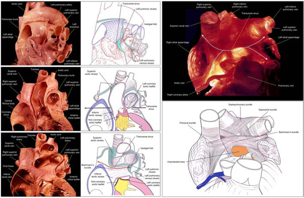
Figure 3: Vestigial Fold and Transverse Sinus as Described by Wallace A. McAlpine.
Photographs and corresponding traced line diagrams are reproduced from the Dr. Wallace A. McAlpine collection (2) with permission of the UCLA Cardiac Arrhythmia Center. Left upper panels display the left lateral view (refer to Figure 2), and left middle and lower panels demonstrate the anterior view. Left lower panel images highlight the progressive dissection of the great arteries identified in the left middle panel images. The spatial relationship between the vestigial fold sandwiched by the anteriorly located transverse sinus and the posteriorly located left pulmonic recess is shown. The transverse sinus has superior and inferior extension right posterior to the aortic root, referred to as the inferior and superior aortic recesses, respectively. The inferior aortic recess reaches close to the nadir of the non-coronary aortic sinus. The superior aortic recess reaches close to the aortic arch anterior to the superior caval vein. Thus, the superior aortic recess is referred to as the aorto-caval recess. Right panels show the transverse sinus located between the proximal great arteries and both atria. The left and right atrial appendages are located at bilateral entrances/exits of the sinus. As the apices of both atrial appendages are free within the pericardial space, the transverse sinus may be entered via either from anterior and posterior side of both appendages (white dotted double-headed arrows). The middle inferior part of the transverse sinus beneath the Bachmann’s bundle is sandwiched between the aortic root and the left atrial anterior wall. Note the extremely thin left atrial wall in this region (transilluminated) referred to as the unprotected area.
Figure 4: Three-dimensional Reconstruction of the Contrast Material Injected into the Pericardial Space.
Upper panels show multiplanar reconstruction images of computed tomographic datasets before and after contrast media injection into the pericardial space of the heart ex vivo, submerged in a container. The air-fluid level (yellow arrows) elevates after 400 mL of contrast media is injected into the pericardial space. Lower panels are the three-dimensional reconstruction images of the pericardial space demonstrated in stereoscopic display aligned for the cross-eyed method, viewed from the supero-posterior direction. When applying stereopsis with the cross-eyed method, the left and right eye images were interchanged such that the left eye viewed the image on the right and vice versa (3). Yellow circles are placed at the bottom of each panel for cross-fusion of the bilateral panels during stereoscopic viewing using the cross-eyed method. By referring to this three-dimensional image, careful dissection is carried out without destruction of the pericardium as shown in Figures 5–8. In this case, left atrial roof that is not covered by the pericardium is inaccessible via pericardial approach and corresponds to the posterior hilum. Note the wide variation in this shape, as evident between cases in Figure 7 and 13.
Figure 5: Location and Alignment of the Pericardial Reflections.
Upper panel shows pericardium-preserved heart viewed down from the supero-posterior direction. The pericardial space is fully inflated with air. Both red and sky-blue lines denote the locations of pericardial reflections exclusively surrounding two orifices of the heart, referred to as the superior and posterior hila of the heart, respectively. Although the recesses demarking the posterior hilum appear complex, they are symmetric except for the reflection surrounding the inferior caval vein. Similar to the left pulmonic recess, the right pulmonic recess is also blind-ended and referred to as the retro-caval recess. Lower panels show photograph (left panel) and corresponding traced line diagram (right panel) from the Dr. Wallace A. McAlpine collection reproduced with permission of the UCLA Cardiac Arrhythmia Center. Lower panels are useful to understand spatial relationship between the transverse sinus and the left pulmonic recess divided by the vestigial fold. Clinically, guiding a catheter through the transverse sinus is challenging as the transverse sinus is not a simple conduit but takes a tightly curved course. Note how the superior caval vein and vestigial fold separate both pulmonic recesses from the transverse sinus (Figure 2). As both pulmonic recesses are blind-ended with paper-thin pericardium in the plane of the transverse sinus, manipulation of catheters within these recesses can cause perforation at the posterior mediastinum toward the posterior hilum.
Figure 6: Air Inflation Technique to Analyze Intact Anatomy of the Pericardial Space.
Pericardium-preserved heart before (left upper panel) and after (left lower and right upper panels) 400 mL of air inflation. This technique allows visualization of the exact location of pericardial reflections. Left and right images in each panel except right lower panel are viewed from the anterior and supero-posterior directions, respectively. Right upper panel images show “lantern” images to enhance the pericardial space. Without inflation, the dimensions of the sinuses and recesses are difficult to discern. Right lower panels show the fluoroscopic left anterior oblique views during pericardial access via subxiphoid puncture. Entire epicardial surface that are not related to the superior and posterior hila of the heart can be readily approached during epicardial access. Note a guidewire (arrowheads) takes the inferior course then reach the epicardial region near the left atrial appendage (left). A guidewire further proceeds anterior to both arterial trunks (right) and surrounds both ventricular bases to reach the epicardial region at the basal inferior right ventricle.
Figure 7: Pericardial Space Replaced with Blue-Colored Agar Showing the Hilum of the Heart.
Pericardial space is replaced with agar colored with methylene blue to improve visibility and to maintain its three-dimensional anatomy. In our experience, colored agar is the most useful material to maintain the architecture and visualize the complexity of pericardial space. Red and blue strings mark pericardial reflections surrounding superior (arterial) hilum and posterior (venous) hilum, respectively (Supplementary Videos 1 and 2). The superior hilum encircles ascending aorta and pulmonary trunk, and the posterior hilum encircles pulmonary and caval veins. Both hila do not communicate with each other as they are separated by the transverse sinus. The two hila are the exclusive entries/exits for the extracardiac nerves and vessels. Some of these nerves can be observed macroscopically (red and sky-blue arrowheads). The roof of the left atrium and the antrum of the pulmonary veins are the exact location of the posterior hilum directly facing to the mediastinum and extra-pericardial right pulmonary artery. Perforation of the left atrium corresponding to the posterior hilum, therefore, does not result in cardiac tamponade but rather a mediastinal hematoma. In this heart, if the ganglionated plexuses are located within this posterior hilum, they cannot be ablated via an epicardial approach without traversing part of the reflection surrounding it. In similar fashion, epicardial ablation of atrial fibrillation cannot be applied within the atrial roof surrounded by the posterior hilum. Although the posterior hilum of this heart had a length of 18.5 mm in the antero-superior to postero-inferior direction, the shape of the posterior hilum is variable. In some cases, two adjacent reflections between the transverse sinus and oblique sinus can fuse (Figures 14, 15). Tubes are placed in the pulmonary veins to maintain their shape. Right lower panels are transthoracic echocardiographic images (parasternal long axis view) during systole (left) and diastole (right) showing pericardial effusion in the transverse and oblique sinuses. Refer to Figure 15.
Figure 8: Stereoscopic Display of the Heart with Intact Pericardium.
Stereoscopic display aligned for cross-eyed method. Yellow circles are placed at the bottom of each panel for fusion of the bilateral panels during stereoscopic viewing using the cross-eyed method. Upper panels are viewed from the anterior direction. Middle panels are viewed from the posterior direction. Lower magnified panels are viewed from the supero-posterior direction to focus on the posterior hilum of the heart. Red and blue strings mark pericardial reflections surrounding superior (arterial) hilum and posterior (venous) hilum, respectively. Tubes are placed in the pulmonary veins to maintain their shape.
Figure 9: Transection of the Transverse Sinus.
Pericardium-preserved heart with methylene blue colored-agar replacing the pericardial space is transected at the level of the transverse sinus and four pulmonary veins. Upper panel indicates the transection plane relative to the heart. Lower panels show the transected surface of the inferior part viewed from the superior direction (left) and the transected surface of the superior part viewed from the inferior direction (right). Both surfaces are aligned in opened-up fashion along the right side of the heart.
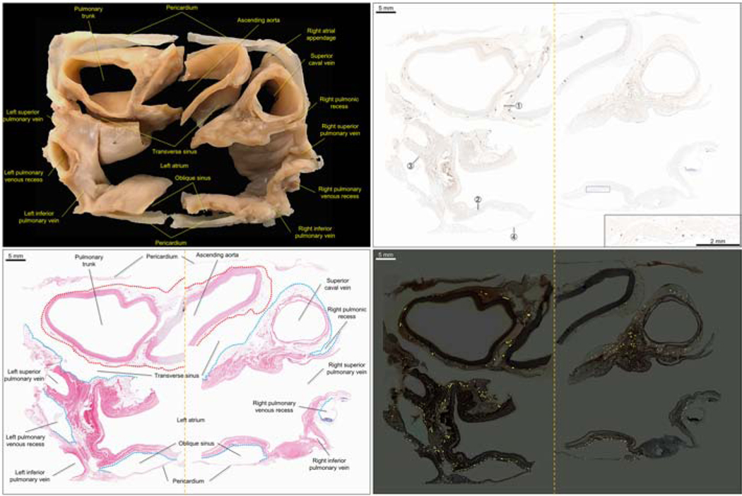
Figure 10: Slab-Cut Hilar Section for the Histological Analysis.
Additional transection is performed approximately 1 cm beneath the initial transection surface to obtain a tissue slab (left upper panel) for histology. Left lower panel shows the transected surface of the second inferior cut viewed from the superior direction. Red and sky-blue dotted lines indicate the superior hilum and the posterior hilum of the heart, respectively. Orange dashed line indicates additional bisecting line. Yellow arrowhead denotes the pericardial reflection between the right inferior pulmonary vein and inferior caval vein, that prevents communication between oblique sinus from the right postero-inferior pericardial space (Figures 1, 14, 15). White dashed ellipse encircles the epicardial adipose tissue containing the left coronary artery, above the left ventricular summit. Right panels are comparable computed tomographic sections reconstructed from a case with pericardial effusion. These images are viewed from the supero-posterior direction. Note that in the left panels, cardiac chambers and intrapericardial arterial trunks collapse due to injection of the colored agar into the pericardial space.
Figure 11: Processed Tissue and Hematoxylin-Eosin and S100 Staining of the Hilum of the Heart.
Left upper panel shows the slab-cut sample after tissue processing to be fit into two tissue cassettes. Left lower panel shows corresponding histological image stained with hematoxylin-eosin. Red and sky-blue dotted lines denote the superior hilum and the posterior hilum, respectively. Note the transverse sinus separating both hila as well as four pulmonary veins are successfully demonstrated in this single section. Right upper panel shows S100 staining (brown) of the hilum of the heart to show the course and density of the cardiac innervation via both hila of the heart. Inset at the right lower corner is a magnification image of the epicardial aspect of the left atrial roof wall facing the oblique sinus. Right lower panel is a digital enhancement of the nerves stained with S100 that can be traced with lower magnification. When compared with left panels, relationship between nerve distribution and both cardiac hila can be appreciated. Circled numbers indicate magnified image location in Figure 12. Note the epicardium-predominant innervation pattern that can surely be accessed via an epicardial approach.
Figure 12: Representative Histological Magnified Images to Show Cardiac Nerves.
Circled numbers correspond to those in Figure 11. Left and right columns show hematoxylin-eosin and S100 staining, respectively. Panels ① show a large myelinated nerve fiber surrounded by perineurium located between the ascending aorta and pulmonary trunk (superior hilum). Panels ② demonstrate magnified left atrial roof musculature. Although it is difficult to detect nerve fibers with hematoxylin-eosin staining, S100 reveals many nerve fibers running between the myocardial cells. Panels ③ show a ganglion found at the epicardium adjacent to the left superior pulmonary vein and left pulmonary venous recess. Panels ④ shows the nerves found at the pericardium posterior to the oblique sinus, which may explain clinical findings such as chest pain during pericarditis.
Figure 13: Progressive Dissection with a Paired Sketch to Show Vagal Innervation.
Left upper panels shows the pericardium-preserved heart with the trachea and the esophagus. The right vagus nerve distributes the posterior vagal trunk. The second physiological narrowing of the esophagus can be noted. The esophagus as well as esophageal branches are removed in the left lower panels, showing bronchial branches/plexus posterior to the trachea and bronchi. The trachea and bronchus as well as bronchial branches/plexus are removed in the right upper panels showing cardiac branches/plexus. Pericardial space is inflated with air. To further clarify the relationship between innervation and both hila of the heart, pulmonary arteries are resected, and the pericardial space is replaced with methylene blue-colored agar (right lower panels). Red and blue asterisks show the nerves communicating with the superior and posterior hila, respectively. In this heart, two adjacent reflections between the transverse sinus and oblique sinus fuse with each other, rendering the posterior hilum quite narrow. Given this anatomy, almost the entire left atrial roof is accessible via an epicardial approach, as long as the catheter can be delivered to each recess and sinus. Perforation of the left atrial roof, the region surrounded by the four pulmonary veins, azygos vein, and branch of the right pulmonary artery would result in cardiac tamponade, in contrast to mediastinal hematoma in the heart shown in Figure 7. Tubes are placed in the pulmonary veins to maintain their shape.
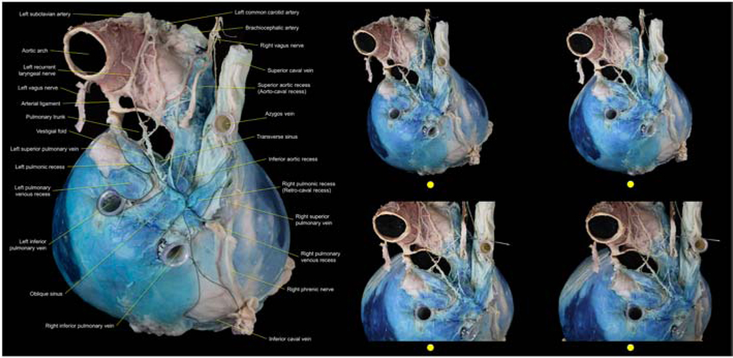
Figure 14: Gross Anatomy Demonstrating Vagal Innervation at the Hilum of the Heart with Stereoscopic Display.
Pericardial space is replaced with agar colored with methylene blue to improve visibility and to maintain its three-dimensional anatomy (left panel). Red and black dotted line denote pericardial reflections surrounding superior (arterial) and posterior (venous) hila, respectively. Compare with Figure 7. Two adjacent reflections between the transverse sinus and oblique sinus are fusing. Vestigial fold and the fused plane between the transverse and the oblique sinuses receive cardiac branches derived mainly from the left recurrent laryngeal nerve (Supplementary Videos 3 and 4). This blue-colored agar inflation beautifully shows alignment of the pericardial reflections between the right inferior pulmonary vein and inferior caval vein. This apposition prevents communication between right and left postero-inferior pericardial spaces posterior to the inferior caval vein (Figures 1, 10, 15). Right panels are stereoscopic display aligned for cross-eyed method. Yellow circles are placed at the bottom of each panel for cross-fusion of the bilateral panels during stereoscopic viewing using the cross-eyed method. Upper panels are viewed from the supero-posterior direction. Lower panels are magnified mages.
Figure 15: Cardiac computed tomographic images reconstructed from a case of hemorrhagic pericardial effusion and epicardial hematoma.
Superior panels show serial axial images. Inferior panels are multiplanar reconstruction images corresponding to apical two-chamber (left), three-chamber (second right), and four-chamber (right) images during transthoracic echocardiography. Second left panel is the plane between the two- and three-chamber images, at the center of the left atrial posterior wall. This case demonstrates a thin persistent left superior caval vein. With profound understanding of detailed anatomy of the pericardial space learned using the dissected hearts (Figures 1–14), not only the recesses and sinuses, but also the vestigial fold and fused reflections between the transverse and oblique sinuses (white arrowheads) can be precisely appreciated in these clinical images. Note how the oblique and transverse sinuses can be observed in a single three-chamber image, as often in the parasternal long axis view during transthoracic echocardiography (Figure 7). Similar to the anatomy demonstrated in Figure 14, the pericardial space surrounds a substantial portion of the left atrial roof and posterior wall, indicating a narrow posterior hilum in this case. The pericardial reflections (yellow arrowhead) prevent communication between right and left postero-inferior pericardial spaces posterior to the inferior caval vein (Figures 1, 10, 14). Note that the variable shape of the transverse sinus in each panel implies potential difficulty in passing a catheter through this sinus. Risk of esophageal injury via an epicardial, as opposed to an endocardial, approach is self-evident.
Supplementary Material
Red and blue strings mark pericardial reflections surrounding superior (arterial) hilum and posterior (venous) hilum, respectively. See Figure 7.
Red and blue strings mark pericardial reflections surrounding superior (arterial) hilum and posterior (venous) hilum, respectively. See Figure 7.
The pericardial space has been replaced with methylene blue-colored agar. See Figure 14.
The pericardial space has been replaced with methylene blue-colored agar. See Figure 14.
Acknowledgments:
We wish to thank individuals who have donated their bodies and tissues for the advancement of education and research. We are also grateful to Travis G. Siems and Alex Rodriguez with the UCLA Donated Body Program and Edwin Ng with the UCLA Surgical Sciences Laboratory for their support in the dissections. We are thankful to Anthony Smithson and Arvin Roque-Verdeflor with the Translational Research Imaging Center at UCLA for their support in computed tomographic data acquisition, and Yunfeng Li, Joanne Olivas, Ko Kiehle, Voicu Ciobanu, Joseph L. Bautista, and Clara Magyar with the Translational Pathology Core Laboratory at UCLA for their assistance in preparation of slides of histological images. We also appreciate Masataka Suzuki, and Yu Izawa, for their support for collection of clinical images.
Funding: This work was made possible by support from NIH grants OT2OD023848 to KS
Footnotes
Conflict of interest: None
Publisher's Disclaimer: This is a PDF file of an unedited manuscript that has been accepted for publication. As a service to our customers we are providing this early version of the manuscript. The manuscript will undergo copyediting, typesetting, and review of the resulting proof before it is published in its final form. Please note that during the production process errors may be discovered which could affect the content, and all legal disclaimers that apply to the journal pertain.
References
- 1.Bradfield JS, Shivkumar K. Epicardial Interventions in Electrophysiology: Transformation to an Established Approach. Card Electrophysiol Clin. 2020;12(3):xv. Epub 2020/08/11. doi: 10.1016/j.ccep.2020.06.003. PubMed PMID: 32771197. [DOI] [PubMed] [Google Scholar]
- 2.McAlpine WA. Heart and Coronary Arteries: An Anatomical Atlas for Clinical Diagnosis, Radiological Investigation, and Surgical Treatment. New York: Springer-Verlag; 1975. [Google Scholar]
- 3.Mori S, Izawa Y, Nishii T. Simple Stereoscopic Display of 3-Dimensional Living Heart Anatomy Relevant to Electrophysiological Practice. J Am Coll Cardiol EP. 2020;6:1473–1477. [DOI] [PubMed] [Google Scholar]
Associated Data
This section collects any data citations, data availability statements, or supplementary materials included in this article.
Supplementary Materials
Red and blue strings mark pericardial reflections surrounding superior (arterial) hilum and posterior (venous) hilum, respectively. See Figure 7.
Red and blue strings mark pericardial reflections surrounding superior (arterial) hilum and posterior (venous) hilum, respectively. See Figure 7.
The pericardial space has been replaced with methylene blue-colored agar. See Figure 14.
The pericardial space has been replaced with methylene blue-colored agar. See Figure 14.


