Abstract
Drosophila’s white gene encodes an ATP-binding cassette G-subfamily (ABCG) half-transporter. White is closely related to mammalian ABCG family members that function in cholesterol efflux. Mutants of white have several behavioral phenotypes that are independent of visual defects. This study characterizes a novel defect of white mutants in the acquisition of olfactory memory using the aversive olfactory conditioning paradigm. The w1118 mutants learned slower than wildtype controls, yet with additional training, they reached wildtype levels of performance. The w1118 learning phenotype is also found in the wapricot and wcoral alleles, is dominant, and is rescued by genomic white and mini-white transgenes. Reducing dietary cholesterol strongly impaired olfactory learning for wildtype controls, while w1118 mutants were resistant to this deficit. The w1118 mutants displayed higher levels of cholesterol and cholesterol esters than wildtype under this low-cholesterol diet. Increasing levels of serotonin, dopamine, or both in the white mutants significantly improved w1118 learning. However, serotonin levels were not lower in the heads of the w1118 mutants than in wildtype controls. There were also no significant differences found in synapse numbers within the w1118 brain. We propose that the w1118 learning defect may be due to inefficient biogenic amine signaling brought about by altered cholesterol homeostasis.
Keywords: olfactory learning, white, Drosophila, cholesterol, serotonin, dopamine, ABCG transporter, freeze tolerance
1. Introduction
Drosophila’s white gene, necessary for normal eye pigmentation, has one of the longest and most impactful histories of any gene [1,2,3]. Loss-of-function white alleles are also commonly found in Drosophila experimental genotypes [4,5]. Despite the usefulness of these easily observable genetic markers, the presence of white mutant alleles can pose a confounding problem in many experiments because they have pleiotropic behavioral and neural phenotypes, including aspects of learning and memory. Some behaviors are due to poor visual acuity [6,7,8,9], while others appear independent of visual perception [10,11,12,13,14,15,16]. The molecular mechanisms by which the mutant white alleles produce these non-visual phenotypes remain mostly uncertain.
One possible explanation for the pleiotropy is the molecular role of white as a broadly selective ATP-binding cassette (ABC) transporter of the G-subfamily [17,18,19]. Many eukaryotic ABC transporters are efflux proteins that transport a broad array of molecules out of the cell and include the breast cancer resistance protein (BCRP, ABCG2) [20,21]. Additionally, some ABC transporters, like white in the pigment granules of the Drosophila eye and Malpighian tubules, are involved in transport from the cytoplasm into intracellular compartments [22]. Many ABC transporters are largely involved in the movement of hydrophobic compounds and are associated with metabolism, secretion, and homeostasis [23]. Structurally, most ABC transporters have a minimum of four domains—two nucleotide-binding domains (NBD) and two transmembrane domains (TMD) [20]. Functionally, the TMDs bind substrates and are allosterically coupled to the NBD so that substrate binding enables ATP binding and subsequent ATP hydrolysis to facilitate substrate transport [24,25]. The ABC G-subfamily (ABCG), which includes the white protein, is distinctive because it consists entirely of half transporters that form obligate homo- or heterodimers for function [26,27].
Many ABCG family members have roles in sterol homeostasis [27]. Mammalian ABCG1 and ABCG4 are co-expressed in the brain, most likely as homodimers, and are involved with intracellular sterol transport and regulation of cholesterol homeostasis [27,28,29,30,31]. The ABCG5 and ABCG8 transporters form obligate heterodimers, crucial for the excretion of cholesterols [32,33]. The Drosophila ATP transporter expressed in trachea (Atet) is located in the fly’s prothoracic gland and functions in exocytotic vesicles as an ecdysone pump [34]. Additionally, the Drosophila ABCG gene CG9663 is transcriptionally upregulated by Drosophila hormone receptor 96 (DHR96), a nuclear receptor that responds to changes in cholesterol abundance, suggesting a possible role in cholesterol homeostasis [35]. To our knowledge, the white gene has not been previously examined for a role in cholesterol homeostasis.
The white gene has roles in several learning paradigms. Loss-of-function w1118 mutants have severe defects in novelty habituation during exploration, which appear to be primarily due to visual defects [9]. The w1118 mutants have also exhibited attenuated operant place memory when tested in an operant heat box assay [12]. While the w1118 mutants displayed normal levels of short-term olfactory memory in the standard long program paradigm, w1118 short-term memory was higher than wildtype when the electric shock voltage, used as the unconditioned stimulus, was reduced [11]. This enhanced learning found in w1118 is likely due to an increased electric shock sensitivity found in this mutant [11].
Some behavioral differences are hypothesized to result from white’s putative role in serotonin (5-HT) biosynthesis through its participation in tryptophan and guanine transport [12,14]. Consistent with this hypothesis, the defect in operant place memory is phenocopied by inhibition of 5-HT synthesis but not by the inhibition of dopamine biosynthesis [12]. Some measurements from whole fly heads have found a lower level of 5-HT, dopamine, and histamine in w1118 compared to wildtype Canton-S or Oregon-R flies [12,36]. However, the 5-HT levels in wildtype heads from these studies were much higher than previously measured by others [12,36,37,38,39,40]. Moreover, a later study found no difference in 5-HT levels between the dissected brains of w1118 and wildtype Canton-S, suggesting that white may not have a limiting effect in the synthesis of biogenic amines within the central nervous system [40].
In this paper, we demonstrate a role for the white gene in olfactory associative learning, and we explore possible mechanisms related to cholesterol homeostasis. We present evidence that suggests w1118 mutants are defective in regulating levels of cholesterol and cholesterol esters. Furthermore, increasing either dopamine or 5-HT levels in the w1118 mutants partially rescues the learning defect. Yet, we found that 5-HT levels in the w1118 heads were somewhat higher than those of our wildtype control flies. We propose a hypothesis whereby defects in cholesterol homeostasis in the w1118 mutants lead to less effective signaling through 5-HT and dopamine receptors and, consequently, account for less efficient acquisition of learning.
2. Results
2.1. Mutants of white Have Poor Learning Acquisition
In the Drosophila “long program” olfactory learning paradigm, 12 electric shocks are delivered during a one-minute presentation of the odorant-paired conditioning stimulus (CS+) [41,42]. Each electric shock represents a single training trial, and an acquisition curve can be generated by varying the number of shocks paired with the CS+ odorant. The 12 shocks presented in the long program saturate the amount of learning in wildtype lines [41,43]. Acquisition curves can be used to identify mutants that have defects in their rate of learning, while maintaining wildtype memory formation or recall.
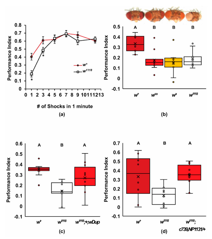
The w1118 mutants used in this study were previously shown to have normal levels of learning in the long program [11,41]. We found that the w1118 loss-of-function mutants had defects in acquiring olfactory associative memory and that these defects were overcome by additional training trials (Figure 1a). These mutants had significantly lower performance indices at one and three training trials, but by five training trials had caught up to the performance of the wildtype Canton-S controls (two-way ANOVA, Adj. R2 = 0.475, F6,121 = 20.157, p < 0.0001, n = 10–13; Tukey post hoc w+ versus w1118, p < 0.05, n = 64). We did not find any significant differences between naïve responses of w1118 and wildtype flies and the olfactory or electric shock controls (Supplementary Table S1). These data indicate a slower rate of learning phenotype for the w1118 mutants.
Figure 1.
Mutants of white have a defect in acquisition that can be rescued by white+ and mini-white transgenes. (a) Olfactory learning acquisition curves are shown for w1118 mutants and wildtype Canton-S (w+). The w1118 mutant has lower associative STM with one and three shock pairings to odor (two-way ANOVA, Adj. R2 = 0.475, F6,121 = 20.157, p < 0.0001, n = 10–13; Tukey post hoc w+ versus w1118, p < 0.05, n = 64); (b) one-trial olfactory learning for wildtype and the wcoral, wapricot, and w1118 mutants are shown. Representative photos of the heads are provided above the graph to demonstrate the level of white activity remaining in eye pigment cells for each mutant. All mutants were equally poor in associative STM and significantly different from wildtype (ANOVA, Adj. R2 = 0.321, F3,32 = 6.524, p < 0.001, n = 9); (c) poor one-trial learning for w1118 was rescued by a genomic duplication (w1118; Dp(1;3)DC050- wDup; ANOVA, Adj. R2 = 0.356, F2,27 = 9.022, p < 0.001; Tukey post hoc, p < 0.05; n = 10); (d) poor one-trial learning for w1118 was rescued by two mini-white marked GAL4 drivers (ANOVA, Adj. R2 = 0.254, F2,25 = 5.590, p < 0.01; Tukey post hoc, p < 0.05; n = 8–10). In panels b, c, and d, groups that do not share letters above the box plots are significantly different from each other according to the Tukey post hoc test (* p < 0.05).
To verify that the learning defect in w1118 was due to a deficiency in white gene activity, we examined independent alleles and transgenic rescue constructs using the one-trial learning protocol (Figure 1b–d). The hypomorphic whiteapricot (wa) and whitecoral (wco) mutants also had significantly lower performance indices after one-trial learning, comparable to the amorphic w1118 mutants (Figure 1b; ANOVA, Adj. R2 = 0.321, F3,32 = 6.524, p < 0.001, n = 9). The decrease in performance for all three w mutants was about 50%, regardless of the strength of the eye phenotype, suggesting that olfactory learning is very sensitive to reduced levels of white activity. The w1118 one-trial learning phenotype was rescued by including a genomic duplication of the w locus (Figure 1c; ANOVA, Adj. R2 = 0.356, F2,27 = 9.022, p < 0.001; Tukey post hoc, p < 0.05; n = 10). This duplication was a 93,901 base pair genomic fragment of the X chromosome, containing the entire white gene near its center, and also included the CG12498, CG32795, and the IncRNA:CR43494 genes; this duplication was docked on the left arm of chromosome 3 [44]. We further found that learning was restored in w1118 flies by the addition of two copies of mini-white contained within two separate GAL4 driver lines, c739 and NP1131 (Figure 1d, ANOVA, Adj. R2 = 0.254, F2,25 = 5.590, p < 0.01; Tukey post hoc, p < 0.05; n = 8–10). The mini-white construct used in these GAL4 drivers contains an hsp70 promoter that drives relatively high levels of mini-white gene expression [45]. These allele and rescue data indicate that relatively high levels of white activity are required for wildtype rates of olfactory learning.
2.2. Screen for Additional ABCG Learning Mutants
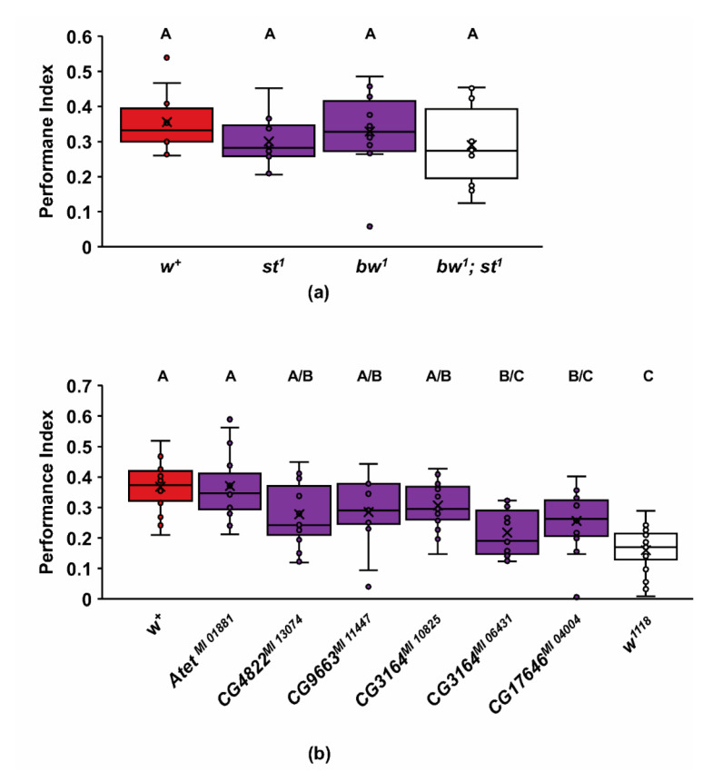
White can homodimerize or heterodimerize with other ABCG half-transporters for normal functions. For example, white is known to heterodimerize with both brown and scarlet ABCG transporters to produce the drosopterin and ommochrome screening pigments, respectively, within the eye’s pigment cells [46,47]. However, mutants of scarlet1 (st1), brown1 (bw1), and the bw1; st1 double mutant all show levels of learning comparable to wildtype Canton-S flies (Figure 2a; ANOVA, Adj R2 = −0.015, F3,36 = 0.809, p = 0.497; Tukey post hoc, p < 0.05; n = 10). The bw1 and st1 are both loss of function mutations, and the bw1; st1 double mutants are white-eyed, lacking all ommochromes and drosopterins [48,49]. This double mutant’s failure to mimic the w1118 olfactory learning phenotype suggests that white’s function in the eye is, not surprisingly, independent of its role in learning. Moreover, the transport functions of the white–brown and white–scarlet transporters are not required for olfactory learning.
Figure 2.
Assessment of one-trial learning in Drosophila ABCG mutants. (a) Outcrossed mutants of ABCG transporters scarlet (st1), brown (bw1), and bw1; st1 were not statistically different from wildtype (w+) (ANOVA, Adj R2 = −0.015, F3,36 = 0.809, p = 0.497; Tukey post hoc, p < 0.05; n = 10); (b) the AtetMI 01881, CG4822MI 13074, CG9663MI 11447, CG3164MI 10825, CG3164MI 06431, and CG17646MI 04004 homozygous mutants were compared to both w+ flies and w1118 mutants. All ABCGs except AtetMI 01881 had lower acquisition performance than w+. CG3164MI 06431 and CG17646MI 04004 were both significantly different from w+ and not different from w1118 after Tukey post hoc analysis. (ANOVA, Adj. R2 = 0.309, F7,114 = 8.717, p < 0.0001; Tukey post hoc, p < 0.05; n = 15). Groups that do not share letters above the box plots are significantly different from each other according to the Tukey post hoc test (p < 0.05).
In search of other potential heterodimerization partners, we examined five additional ABCGs that are expressed in Drosophila adult heads [50] and are highly homologous to white for defects in one-trial olfactory learning (Figure 2b) [18]. To accomplish this, we examined homozygotes for Mi{MIC} element insertions within these genes, all predicted to severely disrupt activity [51]. Although several of the ABCG mutants we tested appeared to trend lower than wildtype Canton-S, only the CG17646MI 04004 and CG3164MI 06431 mutants were significantly different from wildtype, while not different from w1118 (Figure 2b; ANOVA, Adj. R2 = 0.309, F7,114 = 8.717, p < 0.0001; Tukey post hoc, p < 0.05; n = 15). All ABCGs except w1118 had lower naïve odor avoidance for MCH and/or 3-OCT (Supplementary Table S1). In contrast, all the ABCG mutants had normal shock avoidance (Supplementary Table S1). The AtetMI 01881 mutant showed robust learning levels despite low odor avoidance. This result suggests that the odor detection threshold required for maximal olfactory learning may be lower than the threshold for naïve avoidance. Nevertheless, we cannot rule out at this time that the learning defects seen in the CG3164MI 06431 and CG17646MI 04004 mutants are possibly due to defects in olfactory sensitivity.
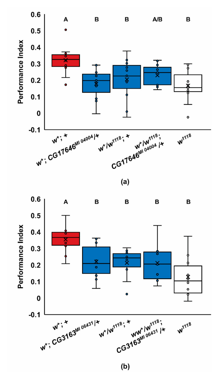
We further examined the learning phenotypes of the CG17646MI 04004 and CG3164MI 06431 mutants by asking if the poor learning phenotypes of these ABCG mutants and w1118 were dominant and if there were phenotypic interactions with w1118 in learning (Figure 3). Since white is on the X chromosome, we used only females in these experiments. There were significant effects of genotype in this experiment (Figure 3a; ANOVA, Adj. R2 = 0.205, F4,50 = 4.485, p < 0.004; Tukey post hoc, p < 0.05; n = 11; Figure 3b; ANOVA, Adj. R2 = 0.299, F4,45 = 6.227, p < 0.001; Tukey post hoc, p < 0.05; n = 10). Interestingly, the w1118 learning phenotype was completely dominant for one-trial learning (Figure 3a,b; p < 0.05). This dominance, together with the wco and wa phenotypes, indicates that learning is highly sensitive to white activity and that the wildtype phenotype requires high levels of white expression. In addition, since the w+/w1118 heterozygotes were generated from wildtype female virgins, this indicates that the learning phenotype is not maternally inherited. Similar to w1118, both the CG3164MI 06431 and CG17646MI 04004 mutant alleles also had dominant learning phenotypes (Figure 3a,b). The CG17646MI 04004/+ heterozygotes had mostly wildtype odor avoidance, suggesting the mutant avoidance phenotype is recessive, or perhaps semi-dominant (Supplementary Table S1). In contrast, the CG3164MI 06431/+ heterozygotes’ odor avoidance defects were fully dominant (Supplementary Table S1). We failed to find any additive genetic interactions between w1118 and the CG17646MI 04004 or CG3164MI 06431 alleles in the respective transheterozygotes, consistent with these genes affecting the same learning processes (Figure 3); however, given the low level of learning in these genotypes, floor effects may have limited our ability to detect additive interactions. Together these data suggest the possibility that white may heterodimerize with either CG17646MI 04004 or CG3164MI 06431, or both, to support learning.
Figure 3.
Reduced one-trial learning for Drosophila ABCG mutants is not additive with w1118. (a) Crosses were made to produce CG17646MI 04004 heterozygotes, w1118 heterozygotes, or transheterozygotes of w1118 and CG17646MI 04004. The w1118 and CG17646MI 04004 alleles have dominant learning phenotypes and are not additive for reduced acquisition (ANOVA, Adj. R2 = 0.205, F4,50 = 4.485, p < 0.004; Tukey post hoc, p < 0.05; n = 11); (b) crosses were made to produce CG3164MI 064314 heterozygotes, w1118 heterozygotes, or trans-heterozygotes of w1118 and CG3164MI 06431. The w1118 and CG3164MI 06431 alleles have dominant learning phenotypes and are not additive for reduced acquisition (ANOVA, Adj. R2 = 0.299, F4,45 = 6.227, p < 0.001; Tukey post hoc, p < 0.05; n = 10). In panels a and b, groups that do not share letters above the box plots are significantly different from each other according to the Tukey post hoc test (p < 0.05).
2.3. A Role for white in Cholesterol Homeostasis
A potential mechanism for white’s role in learning, and possibly also for either CG17646 or CG3164, is suggested by the roles of the mammalian ABCG1, ABCG4, ABCG5, and ABCG8 transporters in cellular cholesterol efflux [32,33,52]. Unlike mammals that are capable of de novo cholesterol synthesis, Drosophila melanogaster is a cholesterol auxotroph [53]. If white participates in the efflux of sterols, reducing the levels of white should affect cholesterol abundance and availability within the nervous system. Even subtle changes in cholesterol levels could affect presynaptic vesicle release and recycling, and postsynaptic receptor trafficking and localization in the plasma membrane in a way that reduces the rate of olfactory learning [54,55,56].
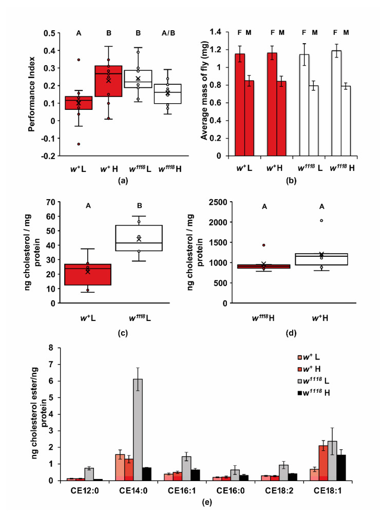
To test this possibility, we first asked if olfactory learning is sensitive to levels of dietary cholesterol (Figure 4a). Wildtype and w1118 flies were grown from embryos on a minimal diet of sucrose, yeast, and agar, with or without 0.1 mg/mL of cholesterol. Wildtype flies raised on a low-cholesterol diet had significantly lower performance in one-trial learning than the flies raised on a high-cholesterol diet, indicating a wildtype need of dietary cholesterol for olfactory learning (two-way ANOVA, Adj. R2 = 0.183, F3,44 = 4.508, p < 0.008; Tukey post hoc, p < 0.05; n = 12). The w1118 mutant flies performed significantly better than wildtype flies on the low-cholesterol diet, in contrast to their performance on normal food (Tukey post hoc, p < 0.05). Hence, the w1118 mutation leads to a resistance to the effect of low dietary cholesterol on learning. Increasing dietary cholesterol in w1118 flies did not lead to further increases in learning in w1118 flies as it did in wildtype flies (Figure 4a; p = 0.249). It is interesting that the slow learning phenotype of w1118 mutants was not present under these sterol restrictive diets. These results suggest that cholesterol homeostasis is altered in w1118 mutants in a manner that changes the optimal level of dietary cholesterol for learning and perhaps other cholesterol-dependent phenotypes.
Figure 4.
The w1118 mutants have differential responses to dietary cholesterol in one-trial learning and in cholesterol accumulation in their corpses. Canton-S (w+) and w1118 flies were reared on a low- or high-cholesterol (0.1 mg/mL added) diet. (a) Dietary cholesterol had a significant effect on one-trial learning (two-way ANOVA, Adj. R2 = 0.183, F3,44 = 4.508, p < 0.008; Tukey post hoc, p < 0.05; n = 12). Lower dietary cholesterol led to impaired learning in wildtype flies (p < 0.05), but not in w1118 mutants (p = 0.249); (b) fly mass was unaffected by genotype or dietary cholesterol for both males and females (two-way ANOVA, Adj. R2 = −0.138, F3,19 = 0.111, p = 0.952, n = 5–6); (c) the cholesterol levels in the w1118 mutants were significantly higher than wildtype flies grown when both were raised on the low-cholesterol diet (t(10) = 3.292, p < 0.01, n = 6); (d) The cholesterol levels in w1118 mutants were not significantly higher than wildtype flies grown when both were raised on the high-cholesterol diet (t(10) = 1.185, p = 0.263, n = 6); (e) w1118 mutants raised on the low-cholesterol diet have greatly increased cholesterol esters compared to wildtype controls and w1118 mutants raised on the high-cholesterol diet (n = 3).
To test if differences in cholesterol homeostasis could account for the w1118 learning phenotype, we compared cholesterol and cholesterol ester levels in w1118 mutants and wildtype control flies fed either a low- or high-cholesterol diet (Figure 4). The dietary cholesterol changes in these experiments did not affect the mass of either wildtype or w1118 mutants (Figure 4b; two-way ANOVA, Adj. R2 = −0.138, F3,19 = 0.111, p = 0.952, n = 5–6). To analyze the uptake and metabolism of dietary cholesterol under each condition, the flies were raised on the same diets used in the previous learning experiment, and then levels of cholesterol and cholesterol esters from frozen whole flies were measured by LC-MS. On the low-cholesterol diet, the w1118 mutants had approximately twice the level of cholesterol compared to the wildtype control (Figure 4c; t(10) = 3.292, p < 0.01, n = 6). On the high-cholesterol diet, the levels of cholesterol extracted from the corpses of both genotypes were approximately 50-fold higher and with greater variability as compared to the low-cholesterol diet; the differences in cholesterol levels between the w1118 and wildtype flies on this high-cholesterol diet were, however, not significant (Figure 4d; t(10) = 1.185, p = 0.263, n = 6).
Since cellular cholesterol is esterified for storage and transport [57], we also measured the levels of cholesterol esters in the corpses of w1118 and control flies on low- and high-cholesterol diets. There were increases in several individual cholesterol ester levels of w1118 flies on the low-cholesterol diet compared to all other groups (Figure 4e; n = 3). While the low sample number makes significance testing less reliable, the effect of a low-cholesterol diet on elevation of cholesterol esters for w1118 flies was consistent and most noticeable for CE 14:0 (cholesterol myristate) in Figure 4e. The effect of diet on cholesterol ester levels of wildtype flies was not appreciatively different except for CE18:1 (Figure 4e). There were also no significant differences in total cholesterol esters between the w1118 mutants grown on the high-cholesterol diet and either wildtype group (Figure S1; Tukey, p < 0.05). However, there was a strong increase in the total cholesterol esters of w1118 flies fed a low-cholesterol diet (Figure S1; Tukey, p < 0.05). These differences in total cholesterol and cholesterol ester levels and response to dietary cholesterol levels in w1118 mutants are consistent with a role for white in regulating cholesterol homeostasis.
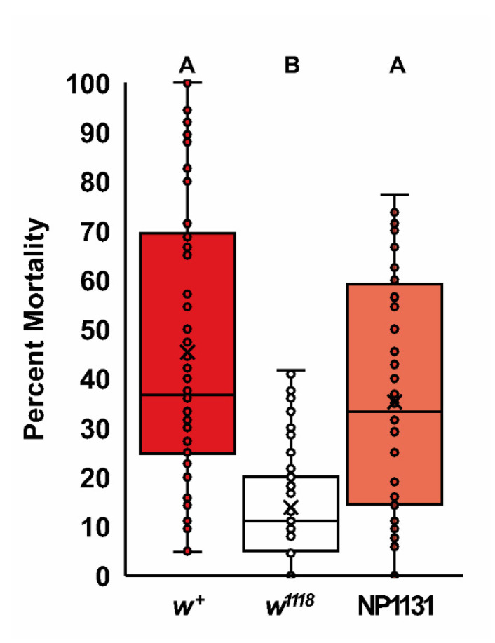
Cellular injury from subfreezing temperatures is primarily due to membrane damage [58]. The susceptibility to this membrane damage can be partially ameliorated by increasing membrane fluidity through changes in membrane lipid composition, including through higher cholesterol levels [59,60]. Cholesterol levels affect freeze tolerance in Drosophila, where flies with higher levels of cholesterol can better survive subfreezing temperatures [59]. Since w1118 mutants have altered cholesterol homeostasis, we asked if they also have an altered freeze tolerance. In this experiment, we examined the ability of control flies, w1118 mutants, and w1118 mutants containing a mini-white transgene (NP1131) to survive a two-hour incubation at −5 °C (Figure 5). There was a significant effect of genotype in this experiment (Kruskal–Wallis, k = 73.404, p < 0.0001, n = 64–68). The w1118 mutants displayed greatly reduced mortality after freezing compared to wildtype flies (Dunn’s procedure, p < 0.0001), and this w1118 freeze-tolerance phenotype was reversed by the presence of a mini-white transgene (Dunn’s procedure, p < 0.0001), suggesting the increased tolerance is due to a reduction in white activity. This change in freeze tolerance is consistent with w1118 mutants having increased membrane fluidity through higher levels of cholesterol and/or other lipids.
Figure 5.
The w1118 mutants have increased freeze tolerance. Wildtype Canton-S (w+), w1118, and w1118; NP1131(mini-white) genotypes were subjected to 120 min at −5 °C, followed by a 24 h recovery at 25 °C. The median percentage of mortality after this recover period is shown as the center line, with the mean indicated by the x. There were significant differences in mortality between genotypes (Kruskal–Wallis, k = 73.404, p < 0.0001, n = 64–68). The w1118 flies survived the subfreezing temperature significantly better than w+ and w1118; NP1131 (Dunn’s procedure, p < 0.0001; n = 64–68). Groups that do not share letters above the box plots are significantly different from each other according to the Dunn’s post hoc test (p < 0.05).
2.4. Biogenic Amine Signaling and the w1118 Learning Phenotype
Another possible mechanism for the role of white in learning is through differences in biogenic amine signaling. Serotonin (5-HT) and dopamine levels were previously reported to be reduced in w1118, bw1, and st1 mutants [12,36]. However, a subsequent study has failed to find a difference between w1118 and the wildtype in biogenic amine levels using a different detection method [40]. Considering both 5-HT and dopamine function in olfactory learning and memory, reducing either the levels of these amines or reducing the efficacy of their signaling pathways could impact the synaptic processes involved in forming olfactory associative memories [61,62,63].
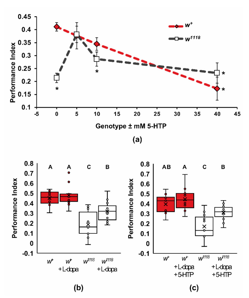
To test the hypothesis that reduced biogenic amine signaling in w1118 mutants causes the learning defect, we examined the impact of artificially raising the levels of 5-HT and dopamine through dietary supplementation (Figure 6). Wildtype and w1118 flies were fed 0, 5, 10, and 40 mM 5-HTP, the metabolic precursor to 5-HT (Figure 6a). These concentrations of 5-HTP, when fed to adult Drosophila, raise the levels of 5-HT extracted from heads [64]. In wildtype flies, increasing the levels of 5-HT led to a dose-dependent decrease in one-trial olfactory learning, with significantly lower performance found at 40 mM 5-HTP (two-way ANOVA, Adj. R2 = 0.348, F7,140 = 12.208, p < 0.0001, Dunnett post hoc, * p < 0.01). The w1118 mutants had a more complex response to increasing levels of 5-HT. A slight increase in 5-HT levels, induced by 5 mM 5-HTP feeding, resulted in a rescue of the learning phenotype (Figure 6a; Dunnett post hoc, p < 0.01). At 10 mM and 40 mM 5-HTP, the performance of w1118 was significantly lower than the untreated wildtype control (Dunnett post hoc, p < 0.01) and not significantly different from each other or untreated w1118 (Tukey, p < 0.05), supporting the previous findings by Yarali, et al. [40], who found no difference in either dopamine or 5-HT levels in w1118 mutants. These data suggest that 5-HT signaling in the w1118 mutants is insufficient to support wildtype rates of learning.
Figure 6.
5-HTP and L-dopa partially rescue wildtype acquisition for w1118 mutants. (a) Canton-S (w+) and w1118 flies were fed 5-HTP or vehicle and then examined for one-trial learning. Vehicle-fed flies for all three experiments were averaged for 0 mM, n = 37. Compared to the optimal acquisition of wildtype controls, 0mM, 10 mM, and 40 mM 5-HTP-fed w1118, and 40 mM 5-HTP-fed wildtype flies showed reduced learning (two-way ANOVA, Adj. R2 = 0.348, F7,140 = 12.208, p < 0.0001, Dunnett post hoc for w+ 0 mM 5-HTP as the control group for comparison, * p < 0.01). We found that 5 mM of 5-HTP improved w1118 acquisition compared to untreated w1118, while 10 mM and 40 mM 5-HTP-fed w1118 flies retained low acquisition (Tukey post hoc, p < 0.05); (b) Canton-S (w+) and w1118 flies were fed L-dopa or vehicle and examined for one-trial learning. L-dopa partially rescued wildtype acquisition for w1118 mutants. (two-way ANOVA, Adj. R2 = 0.522, F3,39 = 16.308, p < 0.0001; Tukey post hoc, p < 0.05, n = 10–11); (c) feeding both 5-HT and L-dopa partially rescued the w1118 one-trial learning phenotype (two-way ANOVA, Adj. R2 = 0.467, F3,40 = 13.544, p < 0.0001; Tukey post hoc, p < 0.05, n = 11). In panels b and c, groups that do not share letters above the box plots are significantly different from each other according to the Tukey post hoc, test (p < 0.05).
We also asked if raising dopamine levels in the w1118 mutants could impact learning. Feeding flies 2 mg/mL of the dopamine precursor L-dopa increases dopamine to levels that are sufficient for different behavioral responses [8,65]. This treatment resulted in a partial but significant increase in learning in the w1118 mutants, but it did not affect wildtype learning (Figure 6b; two-way ANOVA, Adj. R2 = 0.522, F3,39 = 16.308, p < 0.0001; Tukey post hoc, p < 0.05, n = 10–11). These data suggest that dopamine signaling in w1118 is also insufficient to achieve wildtype rates of learning. We further asked if there is a significant interaction between increasing 5-HT and L-dopa in the w1118 mutants (Figure 6c). Feeding wildtype flies both 5mM 5-HTP and 2 mg/mL L-dopa did not significantly affect learning (two-way ANOVA, Adj. R2 = 0.467, F3,40 = 13.544, p < 0.0001; Tukey post hoc, p < 0.05, n = 11); however, the w1118 mutants achieved a partial rescue of learning, like that of L-dopa alone (p < 0.05).
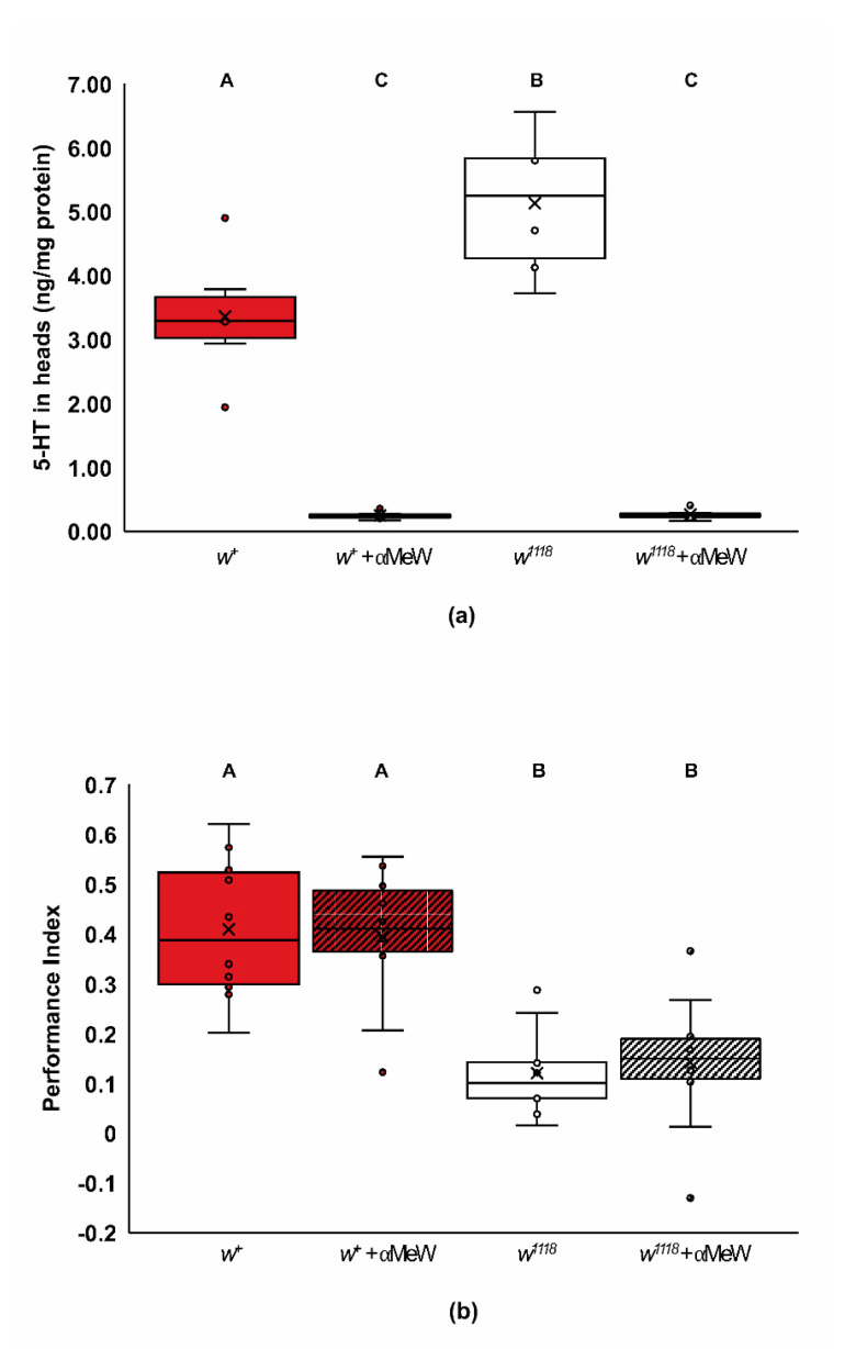
Our data suggest that w1118 mutants have complex defects in 5-HT signaling that impair their rate of learning (Figure 6a). One possibility for this defect in 5-HT signaling is that the w1118 mutants are defective in 5-HT synthesis. This hypothesis predicts lower levels of 5-HT in the heads of w1118, and that reducing levels of 5-HT will mimic the lower rate of learning found in w1118 mutants. We quantified 5-HT levels of wildtype and w1118 mutant flies fed either vehicle or 20 mM α-methyl-tryptophan (αMeW) (Figure 7a). The αMeW compound has been used successfully to inhibit 5-HT synthesis and signaling in Drosophila melanogaster [12,64,66]. Vehicle-fed w1118 heads had statistically significant higher levels of 5-HT than vehicle-fed wildtype heads (Figure 7a; two-way ANOVA, Adj. R2 = 0.891, F3,20 = 63.549, p < 0.0001; Tukey post hoc for genotype, p < 0.01; n = 6), and, as predicted, treatment with αMeW lowered 5-HT levels in both genotypes (Figure 7a; Tukey post hoc for treatment, p < 0.0001). Surprisingly, wildtype flies treated with 20 mM αMeW, which had reduced 5-HT levels (Figure 7a), did not show reduced performance after one training trial (Figure 7b; two-way ANOVA, Adj. R2 = 0.519, F3,36 = 15.05, p < 0.0001; Tukey post hoc, p < 0.05; n = 10). These data suggest that the learning phenotype found after reducing white activity is not due to a generalized reduction in 5-HT levels. Instead, the altered performance of w1118 flies given different doses of 5-HTP and L-dopa is likely due to a lack of biogenic amine signaling efficacy.
Figure 7.
w1118 mutants have increased levels of 5-HT within their heads. (a) 5-HT levels normalized against extracted protein concentrations are shown. Wildtype Canton-S (w+) and w1118 mutant flies were treated with vehicle or 20 mM α-methyl-tryptophan (αMeW) for 40 h. In vehicle-fed flies, 5-HT was significantly higher in the w1118 mutants compared to wildtype flies (two-way ANOVA, Adj. R2 = 0.891, F3,20 = 63.549, p < 0.0001; Tukey post hoc for genotype, p < 0.01; n = 6). Additionally, αMeW significantly lowered the amount of 5-HT in the heads of both wildtype and w1118 mutants (Tukey post hoc for treatment, p < 0.0001); (b) wildtype Canton-S (w+) and w1118 mutant flies were fed vehicle or 20 mM αMeW and trained with a single trial. Acquisition in vehicle-fed wildtype and w1118 mutants were significantly different, however there was no effect of αMeW treatment on one-trial learning (two-way ANOVA, Adj. R2 = 0.519, F3,36 = 15.05, p < 0.0001; Tukey post hoc, p < 0.05; n = 10). In panels a and b, groups that do not share letters above the box plots are significantly different from each other according to the Tukey post hoc test (p < 0.05).
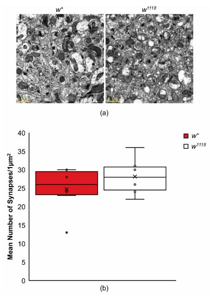
One possible explanation for the reduced rate of learning and efficacy of 5-HT and dopamine signaling in w1118 mutants is a reduction in synapse numbers. Cholesterol has been shown to modify synapse numbers in patched mutants and wildtype flies fed a high-cholesterol diet have more synaptic connections per unit area [67]. We examined the number of synapses in w1118 and wildtype controls in an area that included the mushroom body calyxes (Figure 8). In these experiments, there were no significant differences in the number of synaptic connections found between w1118 and wildtype flies (Figure 8; t10 = 1.04, p = 0.32). These data suggest that the reduction in the efficacy of biogenic amine signaling is not due to a reduced number of synaptic connections in w1118 mutants.
Figure 8.
The average number of synapses are not significantly different between wildtype and w1118 mutants. (a) Representative micrographs of wildtype Canton-S (w+) and w1118 mutants in an area adjacent to the mushroom body calyces are shown. Scale bar = 1 mm. (b) The average number of central synapses were quantified in wildtype and w1118 mutants; w1118 mutants were not significantly different from wildtype (t10 = 1.04, p = 0.32, n = 6).
3. Discussion
The white gene of Drosophila has a prodigious history, leading to several fundamental discoveries in genetics [68,69]. Recently, mutants of white have been found to have several phenotypes involved in complex behaviors [9,10,11,12,13,14,16,70]. Several of these behaviors are believed to be due to changes in 5-HT signaling in these mutants [12,14]. In this study, we have demonstrated that white mutants have a slower rate of learning for olfactory associative short-term memories (Figure 1a). The white gene is both necessary and sufficient for the wildtype acquisition of memory (Figure 1b–d). Known binding partners of the white half transporter, scarlet and brown, have normal acquisition of these associative memories, suggesting that for its role in learning, white might be homodimeric, or it partners with other Drosophila ABCG transporters such as CG17646 and CG3164 (Figure 2). Several ABCG transporters, including CG17646, are involved in lipid transport and perhaps in cholesterol homeostasis [18,52,71].
This study suggests that dietary cholesterol plays a role in olfactory learning for the w+ wildtype, which is possibly mimicked by w1118 mutant flies (Figure 4a). High-cholesterol diets improve learning compared to low-cholesterol diets for wildtype flies, similar to low dietary cholesterol in the w1118 mutants. Higher dietary cholesterol might impair learning in these mutants, though our results were not statistically significant. These differential responses to cholesterol support a role for white in cholesterol homeostasis. Consistent with this role, w1118 flies have altered levels of cholesterol and cholesterol esters (Figure 4c–e), and have increased freeze tolerance (Figure 5)—a phenotype associated with increased cellular cholesterol levels [59]. However, we do not believe this role in homeostasis necessitates a direct cholesterol transport function for white.
The precise roles of 5-HT and dopamine signaling in this w1118 learning phenotype are less clear. While small amounts of the 5-HT precursor, 5-HTP, can rescue learning in w1118 mutants, increasing 5-HT has an overall quiescing effect on wildtype learning (Figure 6a). In addition, feedings of L-dopa alone and combined with 5-HTP partially recover normal learning performance (Figure 6b,c). The white mutants might, therefore, alter the lipid composition and/or distribution of lipids in a manner that reduces 5-HT and dopamine signaling efficiency involved with the learning phenotype.
3.1. Known Biochemical Activities of white and Cellular Locations
White is best known for its role in the fly eye. The white gene encodes the half of an ABCG transporter for the precursors of eye pigments [22,72]. However, the ability of white to form multiple functional heterodimers indicates that this protein could potentially affect many different cellular processes. White has been implicated in the transport of pyruvate, riboflavin, xanthine, guanosine, and zinc [46,47,73,74,75,76]. In most of these cases, white is involved in concentrating these compounds into vesicles [46,47,74,75]. White has also been proposed to facilitate the transport of precursors for biogenic amines in D. melanogaster, including tryptophan [74]. This amino acid is the precursor for ommochrome pigments in the eye and 5-HT, a monoamine neuromodulator involved in learning and memory. However, serotonin is synthesized cytoplasmically [77] and, as an efflux transporter, it seems unlikely that white is responsible for directly bringing tryptophan into the cell. There is some evidence of its ability to transport tryptophan in the Malpighian tubules of Drosophila, although this transport was independent of the cytoplasmic pool of tryptophan and several other studies have indicated that kynurenine and 3-hydroxykynurenine are the tryptophan metabolites transported into pigment granules by white [22,74,78].
The white gene is expressed at various levels in many tissues, including the brain [36,79]. This ABCG transporter is expressed in both adults and larvae. While mRNA levels are low in the brain itself, the whole head has five times higher levels than the brain, suggesting that some neural effects of white may originate in head tissues surrounding the brain. Most expression is in adult/larval tubules and the larval fat body [47,79]. The intercellular localization of a white: DsRed fusion proteins expressed in S2 and in COS cells appears to be endosomal, consistent with a vesicular localization in pigment cells and in the Malpighian tubules [47,70,72].
3.2. The Role of white in Behavior
Mutants of white have numerous behavioral phenotypes, including some that are due to poor visual acuity [6,7,9,11,12]. There are also several behavioral phenotypes that are non-visual [13,14,15,16]. Some of these non-visual behaviors are influenced by defects in 5-HT signaling. For example, operant place memory has been tested in an aversive heat box assay. The white mutant has an attenuated memory in this assay that is replicated with the 5-HT synthesis inhibitor, α-methyltryptophan, but not by the dopamine synthesis inhibitor, α-methyltyrosine [12]. Other 5-HT-dependent behavioral changes seen with mutation and misexpression of white mutants include an increase in the sexual arousal of male flies, leading to male–male courtship [14,15], and an increase in the variability of phototactic behaviors between individual flies [66]. Wildtype phototactic behavior is restored to w1118 mutants by consumption of the precursor of 5-HT, 5-hydroxytryptophan (5-HTP) [66]. As with operant learning in the heat box assay, the mutant behavior is mimicked by α-methyltryptophan feeding.
3.3. A Role for white in Olfactory Learning
Previous work with white in short-term olfactory memory looked at performance after asymptotic aversive training and did not find a difference from wildtype. We utilized the short program to deliver discrete training trials to w1118 mutants, which allowed us to examine the acquisition rate for this associative memory [41,43]. Acquisition curves are especially helpful for differentiating between learning defects and memory defects, because flies with different rates of learning may still have similar performance indices for short-term memory in the long program that uses optimal learning conditions. Since white is defective in the short program of a single shock with 10 s of odor but not the long program, white appears to be required for learning, rather than short-term memory (Figure 1a). Sensory defects were tested through naïve odor avoidance and shock avoidance, with no significant difference found between w+ and w1118 flies on regular diets (Supplementary Table S1). However, w1118 mutants showed an increased shock avoidance to lower voltages than normally used in olfactory training [11]. This increased sensitivity to the unconditioned stimulus (US) may predict an increase in the magnitude of performance in the olfactory T-maze [41], which we did not see.
3.4. Dominance and Gene Dose Sensitivity
The null w1118 and hypomorphic alleles, wa and wco, have approximately 50% of the short-term olfactory memory of wildtype Canton-S flies and are statistically no different in terms of short-term memory with a single shock, despite a gradient of expression within the eyes (Figure 1b). Moreover, w1118 is dominant for this learning phenotype in heterozygous females (Figure 3), but increasing white expression by adding two mini-white transgenes or a large duplication of white to the w1118 mutant flies rescues the wildtype learning phenotype (Figure 1c,d). These data strongly support the hypothesis that the learning deficit in w1118 flies is due to the loss of white and that white is haploinsufficient for learning. A very high level of white expression was also required to rescue the w1118 recovery from anoxia phenotype [80]. Furthermore, the rescue of the loss-of-function of white in male copulation success is sensitive to dosage and requires high expression levels of white [81]. Together, it appears that high levels of white expression are needed to support several behaviors, consistent with general haploinsufficiency for these behaviors.
3.5. ABCGs with Cholesterol and Other Lipid Functions
We sought insight into the possible biochemical functions of white from the activities of homologs in other species. Specifically, the human ABCG1, ABCG4, ABCG5, and ABCG8 are all involved in regulating cholesterol levels. All are sterol efflux proteins, and while some function as likely homodimers, others function as heterodimers. ABCG1 is a sterol-induced gene with ubiquitous expression and multiple splice variants, resulting in two major protein variants [27]. The protein has been localized to endosomes [28] and involved with intracellular sterol transport. Both homodimers and heterodimers have been found possible for ABCG1. Mutation of ABCG1 protects mice against obesity in a high-cholesterol diet (1%). Mice with an ABCG1 null mutation have a normal life span, as do w1118 Drosophila null mutants under standard rearing conditions [31,82]. ABCG1 null mice were also found to have significantly higher locomotor activity and energy expenditure, along with lower fasting plasma lipid levels [82]. Hence, ABCG1 appears to be a regulator of cholesterol metabolism that may couple diet and activity in mammals.
ABCG4 is primarily expressed in the CNS, and homozygous mouse knockout mutants have defects in contextual associative fear memory (tone and foot shock pairings) but not in the Morris Water Maze [30]. ABCG4 can be a homodimer or heterodimer, and it has been found to be co-expressed with ABCG1 in both neurons and astrocytes, where it regulates cholesterol homeostasis [30,83]. Mouse knockouts of both ABCG1 and ABCG4 increase oxysterols in the retina and the brain. While both ABCG1 and ABCG4 have high homology with white, it is important to note that neither ABCG1 nor ABCG4 can replace the Drosophila white gene for recovery of eye pigmentation in white null flies, even though ABCG1 is also highly expressed in the eyes of mammals [29].
Human mutations in ABCG5 and ABCG8 contribute to the autosomal recessive β-sitosterolemia, a metabolic lipid disorder that causes hypercholesterolemia due to overabsorption of dietary sterols [32,84]. This disorder is found in many genetic backgrounds and from many different amino acid substitutions within each population [85], but always from a homozygous mutation of a single gene, ABCG5 or ABCG8. These ABCGs are needed for efflux of dietary sterols from intestinal epithelia to the lumen of the gut and from liver cells to the bile duct. The highest expression levels of ABCG5 and ABCG8 are in the intestines and liver, with transcriptional expression upregulated by cholesterol feeding [32].
White, like all ABCGs, is a half-transporter that might homodimerize or heterodimerize with other Drosophila ABCGs. If brown or scarlet, the known binding partners of white, are involved in the learning phenotype, we would expect the bw1 or st1 mutants to have poor learning for single shock learning, like the w1118 mutant. In contrast, we found that these mutants had wildtype levels of learning (Figure 2a), necessitating the search for new potential binding partners. Vertebrate homologues of w1118 can be homodimers or heterodimers with other ABCG half transporters, so the possibility exists that the acquisition defect depends on more than one pairing. Drosophila melanogaster has 15 ABCG transporters. We investigated five of these ABCGs for learning defects (CG3164, CG4822, CG17646, CG9663, and Atet), based upon homology and relatedness to ABCG1/4, as well as adult head expression [50,86]. These are only some of the potential partners that might have exhibited poor learning and there is still the possibility of homodimerization of white similar to ABCG1 or ABCG4. Of particular interest were the mutants with demonstrated roles in cholesterol and lipid regulation. An insertional mutation in CG17646, an ABCG1-like protein, resulted in an increased triglyceride content in the fly [82]. An ATP transporter expressed in the trachea (Atet) is involved in neurogenesis and vesicle-mediated ecdysone release [34] in the prothoracic gland. CG9663 is transcriptionally regulated by Drosophila hormone receptor 96 (DHR96), a nuclear receptor that responds to cholesterol levels [35].
The most promising potential binding partners for white, according to significance in the learning phenotype, are CG3164MI06431 and CG17646MI04004 (Tukey (HSD) post hoc, p < 0.05). CG3164 is highly expressed as a maternal transcript [87] and has been found in a screen for actively translated RNAs during embryogenesis [88]. Our female heterozygotes of w+/w1118 were produced from wildtype females but exhibited the mutant phenotype, suggesting that white, and possibly its binding partner for this learning phenotype, is not maternal (Figure 3a,b). The CG17646 hypomorph has no maternal expression, high RNA expression in the central brain of embryos [50], and high RNA expression in the adult fat body [86]. In addition, CG17646 mutants have been found to have 1.5–1.7× higher than normal levels of triglycerides in a whole body assay [82], suggesting that they function to regulate lipid metabolism. Of note is the lower MCH and 3-OCT avoidance for all new ABCG mutants that may weaken the associative strength of the shock-paired odor (Supplementary Table S1). Yet, a similar reduction in odor avoidance did not impact learning in Atet mutants and normal odor avoidance was present for w1118 mutants. It remains possible that the new ABCG mutants with poor acquisition have defects that alter learning via a processing or sensory deficit, or that these learning defects are not due to the loss of ABCG function.
3.6. Cholesterol Regulation in Drosophila
Sterol metabolism can be investigated with relatively good experimental control in Drosophila since they are cholesterol auxotrophs [53]. Although a sterol-free diet is lethal, stalling development in larval stages [89], a simple minimal food of bactoagar, yeast, and sucrose has enough yeast-derived sterols to support life and yields flies of normal mass (Figure 4b) with no obvious anatomical differences. We showed that w+ and w1118 Drosophila grown on this low-cholesterol food and the same food with 0.1 mg/mL cholesterol have differently regulated learning and cholesterol homeostasis that is dependent on dietary cholesterol (Figure 4a–d). Higher levels of cholesterol were necessary in w+ flies for better learning, while low cholesterol was better for w1118 mutant flies (Figure 4a).
Cholesterol homeostasis in flies depends upon the nuclear receptor DHR96 [35,90]. DHR96 directly binds cholesterol and controls the activity of many genes regulated by low and high dietary cholesterol, including the dABCA1, CG32186, and CG9663 ABC transporter genes [35,90]. DHR96 mutants had higher levels of cholesterol, and high-cholesterol diets in flies mimicked the transcriptional profile of the genes up/downregulated in the DHR961 mutant raised on standard food [35,90]. This result suggests that DHR96 is necessary for keeping cholesterol levels down by controlling genes needed for changing dietary conditions. The Nieman Pick 1b transporter is responsible for dietary cholesterol uptake and is dysregulated in the DHR961 mutant, which contributes to the lethality of the DHR96 mutants on low-cholesterol diets [35]. The transcriptional profile of white was not reported to have changed in response to cholesterol or in the DHR961 mutants, even though w1118 mutants have defects in cholesterol levels, tolerance to freezing temperatures, and behavioral responses to dietary cholesterol [35,90].
3.7. Differences in the Cholesterol and Cholesterol Ester Levels of w1118
Given the roles of many ABCGs in the regulation of cholesterol, we used LC-MS to measure cholesterol and cholesterol ester levels for wildtype and w1118 mutant flies fed controlled diets of either low or high cholesterol. Our data showed a statistically significant increase in cholesterol levels in the mutant flies compared to the wildtype flies when given a low-cholesterol diet (Figure 4c), as well as a higher, yet insignificant, level of cholesterol in the mutant flies when fed a high-cholesterol diet (Figure 4d). This suggests a change in cholesterol homeostasis and, possibly, lipid membrane composition, rather than general metabolism. A difference in cell membrane composition is supported by the mortality of w1118 mutants subjected to subfreezing temperatures being lower than that of the wildtype, and this being rescued to wildtype percentages by the mini-white transgene NP1131 (Figure 5). This increased freeze tolerance in the w1118 flies suggests an increase in membrane fluidity that would occur from higher levels of cholesterol [59,91].
We looked at olfactory learning in w+ and w1118 flies fed low- and high-cholesterol diets and found similarities between the dietary effects on learning and cholesterol esters, suggesting that cholesterol homeostasis may play a role in learning. While a diet of high cholesterol improved learning for the wildtype flies, mirroring the higher levels of cholesterol and 18:1 cholesterol ester measured in similarly fed flies, the w1118 flies had a higher learning performance index when fed a low-cholesterol diet, which mimicked the higher levels of all measured cholesterol esters fed similarly (Figure 4a,e). Notably, all performance indices were low compared to usual wildtype learning, suggesting that these diets represented suboptimal diets. In addition, differences in learning do not appear to be a result of changes in synapse number between genotypes (Figure 8). This suggests a role for cholesterol and/or cholesterol esters in another aspect of signaling, such as lipid raft stability or synaptic dynamics.
3.8. Roles of Cholesterol in Synaptic Signaling
We found that a reduction in dietary cholesterol can reduce the performance of wildtype flies in one-trial olfactory learning. Cholesterol is required for synaptogenesis, and reduced levels of cholesterol may lead to fewer synapses [67,92]. Since w1118 mutants had higher levels of cholesterol, one possibility would be that these mutants had too many synapses for efficient learning. However, w1118 mutants do not have significantly altered numbers of synapses, suggesting that changes in synaptogenesis are not responsible for the slower rate of learning.
Cholesterol is found at high levels within synapses and is important for maintaining synaptic signaling, with both pre- and postsynaptic functions [93,94,95]. Most work on the role of cholesterol in neuronal functions has relied upon the pharmacological depletion of cholesterol from plasma membranes. This depletion leads to reductions in evoked vesicle release [95,96,97]. It has been proposed that the decrease in the evoked release is due to a need for cholesterol in action potential propagation [95]. Synaptic vesicles are also rich in cholesterol, and the depletion of cholesterol levels from vesicle pools impairs synaptic vesicle recycling [98,99]. Cholesterol depletion can also affect the strength of synaptic signaling by modulating neurotransmitter reuptake transport proteins [100]. For example, the mammalian DAT and SERT monoamine transporters are normally localized in cholesterol-rich rafts, both internally and on the cell surface, and the depletion of cholesterol results in a reduction in the number of these transporters on the cell surface, leading to a reduced termination in neurotransmitter signaling [101,102,103]. The activity of these transport proteins is also likely modified by direct cholesterol binding [104]. Together, these studies demonstrate a need for cholesterol in specific membrane pools for the effectiveness of different presynaptic functions.
Cholesterol can also alter GPCR signaling in the lipid bilayer through direct interaction with GCPRs and/or by altering the lipid ratio in the membrane [105,106]. GPCRs are embedded in cholesterol-rich lipid rafts, which allow the receptors to interact with G-proteins and other associated proteins and maintain functional conformations [107]. Alterations in the lipid composition can disrupt signaling from receptors despite adequate ligands. A good example of this is the mammalian 5-HT1A receptor. 5-HT1A is a Gα(i/o)-coupled GPCR that, in primary human neuronal cultures, reduces cAMP and PKA signaling following 5-HT binding, leading to attenuation of S133-CREB phosphorylation, as well as reduced T185/Y187-ERK2 phosphorylation in the MAPK pathway [54]. Sequestration of cholesterol with methyl-β-cyclodextrin in human primary neuron cultures was able to counteract the inhibition of ERK2 and CREB phosphorylation by 8-OH-DPAT agonism of 5-HT1A, demonstrating a positive role for cholesterol in 5-HT1A signaling [54]. Cholesterol depletion can reduce GPCR signaling through the loss of direct binding, reduced organization of signaling rafts, or impaired trafficking [105,106,107]. The effect of elevated cholesterol on GPCR efficacy is much less clear and may be subtler. However, it is likely that a reduction in GPCR signaling efficacy or presynaptic functions could also occur if the membrane distribution of cholesterol is altered, even when total cholesterol levels are higher.
3.9. white and Biogenic Amine Signaling in Olfactory Learning
Wildtype levels of learning were partially rescued in our w1118 mutant flies by increasing either 5-HT or dopamine levels (Figure 7). Olfactory learning in Drosophila requires both serotonin and dopamine [62,63]. Dopamine has multiple complex roles in olfactory learning, including encoding the electric shock unconditioned stimulus, predicting the US, reinforcing memory, and active forgetting [62,108,109,110]. Dopamine has also been reported to function in appetitive memory [111,112]. Substantial serotonergic innervation and expression of 5-HT receptors exists in the olfactory learning circuitry of Drosophila, notably, the serotonergic dorsal-paired medial neurons (DPMs) that innervate the MBs both pre- and post-synaptically [113]. 5-HT signaling is involved in short-, intermediate-, and long-term memory [63,114]. While the relationship between 5-HT signaling and acquisition is not completely clear, all 5-HT receptors are needed for short-term olfactory memory [63]. The 5-HT1a and 5-HT1b receptors are heavily expressed in MBs [115,116].
Due to the fact that other white mutant behavioral phenotypes have been mimicked by reduction of 5-HT [12,14,66] and 5mM 5-HTP was able to rescue the learning defect in w1118 flies (Figure 6a), global pharmacological inhibition of 5-HT synthesis was investigated for effects on learning. We found that feeding flies αMeW to reduce 5-HT levels in w+ flies reduced the levels of 5-HT in their head but did not mimic the w1118 one-trial learning phenotype (Figure 7b). Moreover, w1118 flies fed vehicle showed a significant elevation in 5-HT levels compared to vehicle-fed w+ flies (Figure 7a), suggesting a reduction in 5-HT signaling efficacy rather than synthesis might contribute to the learning phenotype. Though white has been hypothesized to alter 5-HT levels via the transport of precursors for 5-HT, this hypothesis predicts that mutants in either the brown or scarlet co-transporters would also have a defect in learning [12,36]. Yet, the learning phenotype in this study was independent of brown and scarlet (Figure 2a). The absence of phenotypes for brown and scarlet mutants suggests that white is paired with itself or other ABCG half-transporters for its function in learning, and this function is independent of 5-HT synthesis. If 5-HT signaling is not altered at the level of 5-HT synthesis, it might be altered by relatively subtle changes to presynaptic activity, receptor availability or stability, or an additive contribution from some combination of pre- and post-synaptic signaling deficiencies. Altered cholesterol homeostasis may offer a possible explanation for these results.
3.10. Possible Mechanisms for L-Dopa and 5-HTP Rescue of Wildtype Learning Phenotype
Although there are several possibilities for how white may be modulating the rate of olfactory learning through a role in cholesterol homeostasis, perhaps the most direct scenario is that the white protein dimerizes with one or more other ABCGs to transport cholesterol, or a metabolite of cholesterol, into the proper vesicle, such as an endosome, for storage and/or delivery. Disruption of this transport would alter the composition of lipid membranes and the lipid rafts within them that are crucial for efficient biogenic amine signaling. Moreover, this hypothesis predicts that other mutations and treatments that alter the membrane composition of Drosophila neurons could phenocopy or rescue the w1118 learning defect, similar to the effect of reducing dietary cholesterol. Drosophila that are deprived of cholesterol are known to upregulate sphingolipids as replacements and change the lipid raft integrity needed for signaling, though the central nervous system is relatively good at maintaining sterols compared to other tissues like the fat body [117]. Understanding the precise biochemical function of white that drives changes in cholesterol levels and sensitivity should provide important insights into the role of in vivo membrane lipid dynamics in regulating neural signaling during learning.
4. Materials and Methods
4.1. Drosophila Stocks and Genetics
Fly stocks and crosses were maintained in a 25 °C incubator with a 12:12 h light:dark cycle and cultured on cornmeal medium, except for experiments involving dietary cholesterol. Cholesterol feeding experiments used a standard media of 50 g sucrose (S0389, Sigma-Aldrich, St. Louis, MO, USA), 17 g yeast (Active Dry Yeast, Fleischmann’s Yeast), and 15 g Drosophila Agar—Type II (66–105, Genesee Scientific, San Diego, CA, USA ), with or without 0.1 mg/mL cholesterol (C3045, Sigma-Aldrich, St. Louis, MO, USA) as previously published [118]. Powdered cholesterol was dissolved in 100% ethanol and added (1 mL per 40 mL food) to media during cooking and after cooling to 60 °C. Ethanol was added to control food to account for possible effects on growth, development, or behavior from residual ethanol in the diet. Fifteen females and 10 males of either wildtype Canton-S or w1118 genotypes were placed into bottles with 40 mL of media and brooded every 4–5 days for a total of three sets of bottles.
Most stocks were obtained from the Bloomington Drosophila Stock Center (BDSC, Bloomington, Indiana, USA) and are listed in Table 1. MiMIC lines from BDSC were previously constructed [119]. The c739 and NP1131 GAL4 lines were a gift of Ron Davis (Scripps, Jupiter, FL, USA) and were brought together by meiotic recombination.
Table 1.
Sources of Drosophila stocks.
| Fly Stock Used | Annotation in Text | Identifiers |
|---|---|---|
| w[1118];Dp(1;3)DC050,PBac[y[+mDint2] w[+mC] = DC050]VK00033 | w1118;+,wDup | BDSC 30234 |
| bw1; st1 | bw1; st1 | RRID:BDSC_686 |
| wa | wa | RRID:BDSC_48 |
| wco, sn2 | wco, sn2 | RRID:BDSC_153 |
| w[1118]; P[w[+mW.hs] = GawB]Hr39[c739], P[GawB]NP1131 | w1118;c739, NP1131 | RRID:BDSC_7362, RRID:FBti0034208 |
| MiMIC lines and y[1] w[*]; Mi(y[+mDint2] = MIC)CG3164[MI06431] | CG3164MI 06431 | RRID:BDSC_42392 |
| y[1] w[*]; Mi(y[+mDint2] = MIC)ND-15[MI10825] CG3164[MI10825]/SM6a | CG3164MI 10825 | RRID:BDSC_55545 |
| y[1] w[*]; Mi(y[+mDint2] = MIC)ND-15[MI13074] CG4822[MI13074] | CG4822MI 13074 | RRID:BDSC_58020 |
| y[1] w[*]; Mi(y[+mDint2] = MIC)CG17646[MI04004] | CG17646MI 04004 | RRID:BDSC_42302 |
| y[1] w[*]; Mi(y[+mDint2] = MIC)CG9663[MI11447] | CG9663MI 11447 | RRID:BDSC_56321 |
| y[1] w[*]; Mi(y[+mDint2] = MIC)Atet[MI01881] | AtetMI 01881 | RRID:BDSC_37314 |
All transgenic fly strains were backcrossed to the Roman Lab’s Canton-S background. This backcross procedure began by using balancer chromosome lines to exchange the chromosomes not bearing the transgene with chromosomes from the Roman Lab Canton-S background. The transgenes were then crossed for a minimum of six generations into either a w1118[CS10] or a y1[CS10] stock, both of which had been previously outcrossed in the Roman Lab Canton-S lineage for 10 generations. After the outcrossing was complete, the X chromosome was replaced using a w+ balancer line to ensure a fully wildtype Canton-S X- chromosome. For behavioral experiments, 1–4-day-old Drosophila were collected under mild CO2 anesthetization at least 1 day prior to each experiment. Wildtype white (w+) or mutant (w−) alleles on the X chromosome are specified for each experiment. Associative learning assays used both males and females, except for experiments that specifically tested w+/w1118 heterozygotes. In that case, only females were used. No difference was seen in the performance scores of females alone versus males and females combined with the same homozygous white allele background.
4.2. Pharmacology
Drosophila were collected at 0–3 days post-eclosion and treated by feeding them 2% sucrose, with or without drug treatment, at the concentrations specified in the text [65]. After CO2 anesthetization, flies were placed into vials with half a Kimwipe saturated with 1 mL of solution for 40–48 h, depending on the experiment. Hydroxy-L-tryptophan (H9772 Sigma), 3,4-Dihydroxy-L-phenylalanine (L-DOPA, D9628 Sigma), and αMethyl-DL-tryptophan (M8377 Sigma) were all delivered using this method. Flies were returned to regular food 30–60 min prior to behavioral experiments for the flies to clean themselves of sucrose that may interfere with behavior.
4.3. Olfactory Classical Conditioning Assay
A 3-Octanol (218405 Sigma-Aldrich, St. Louis, MO, USA) and 4-Methylcyclohexanol mixture of cis and trans (153095, Sigma-Aldrich, St. Louis, MO, USA) were diluted in light mineral oil (330779, Sigma-Aldrich, St. Louis, MO, USA) for olfactory conditioning. A quantity of 20 μL 3-Octanol (3-OCT) was diluted in 20 mL of light mineral oil and odor balanced with 12–22 μL of MCH at regular intervals to account for variation in experimental conditions day-to-day.
Flies were collected at 0–4 days post-eclosion and either transferred to fresh food for the assay or to vials containing a half Kimwipe (approximately11.4 cm × 10.7) saturated with 1 mL of 2% sucrose, with or without drug treatment. Flies were transferred to fresh food 30–60 min prior to each assay. All flies were placed in the testing room at 25 °C and 60–75% humidity with dim red light 30–60 min prior to being assayed.
Classical olfactory conditioning was performed using a T-maze similar to previous studies [41,43,120]. For single-trial learning, 1–5-day-old flies were trained using 10 s single exposures to MCH and 3-OCT, alternatively paired with a 1.25 s 90 V shock and 30 s of fresh air intervals between odors. Flies were then tapped into an “elevator”, allowed to rest for 1 min, and exposed to a choice of each odor for 2 min to test for associative short-term memory (STM). For the acquisition curve, a variation of the short program was used. For all groups in this variation, odor was delivered for 60 s and 1.25 s 90 V shocks delivered towards the end of the exposure, spaced 5 s apart. Flies were then rested for 1 min and tested for 2 min, as with the 10 s single exposure assays. The half performance index (PI) was calculated for each odor as follows: ((# of flies in unpaired odor tube, CS-)—(number of flies in paired odor tube, CS+))/(total number of flies in both tubes). The half PIs were then averaged to give a full PI and account for learning differences between odors [41].
Odor and shock avoidance were tested for new phenotypes. For naïve odor avoidance, odorants were presented in the same conditions and using the same concentrations used for the conditioning assay opposite pure mineral oil in a T-maze. The only exception was for odor avoidance with and without 2 mg/mL L-dopa, tested with similar concentrations but later at the University of Mississippi. Flies were exposed for 2 min and collected as before. Half of the exposures used odorant on the right and the other half on the left to account for any directional bias from other unaccounted for stimuli in the testing room. For shock avoidance, one arm of the T-maze was replaced with a shock tube and both arms connected to pure mineral oil for consistent odor on each side. For 2 min, flies were exposed to the T-maze while 90 V was delivered every 5 s.
4.4. Cholesterol Treatments
Fifteen female and 10 male wildtype Canton-S or w1118 flies were allowed to breed for 4–5 days and oviposit embryos on either 40 mL of control media (5% sucrose, 1.7% yeast, and 1.5% BactoTM agar) or control media with 0.1 mg/mL cholesterol, as described. Control media provides negligible amounts of cholesterol solely through the yeast; however, this is sufficient for growth and development of Drosophila [35,121]. Four groups were cultivated: Canton-S with low cholesterol, Canton-S with high cholesterol, w1118 with low cholesterol, and w1118 with high cholesterol. There were 4–5 bottles for each group. Parent flies were transferred twice, for a total of 3 sets. For olfactory learning, 0–4-day-old flies were trained and tested as usual with the short program. For each set, 3 subsets of 20 females and 20 males were collected from each group and weighed. The 9 subsets for each sex of each group were averaged and analyzed for differences in mass between treatment groups. For cold tolerance, 2–5-day old male and female flies were harvested, placed in snap cap tubes, allowed to recover from CO2, and used immediately for cold tolerance experiments.
4.5. Cholesterol and Cholesterol Ester Quantification
Canton-S and w1118 flies (30 males and 30 females) were fed either a low- or high-cholesterol diet and whole flies were frozen and stored at -80 °C. LC-MS analysis services for cholesterol and cholesteryl esters were provided through Wayne State University Lipidomics Core Facility. Flies were shipped on dry ice to the Lipidomics Core Facility for LC-MS analysis of cholesterol and cholesteryl esters levels using a standard operating procedure based on published methods [122,123] and reported as ng per mg of total protein.
4.6. Freeze Tolerance
Freeze tolerance assays were adapted from the work of Shreve et al. [59]. Twenty flies of each group were placed in a snap cap, 15 mL test tubes, and allowed to recover. Flies were submerged in a chilled bath at −5 °C and kept submerged for 120 min. Flies were then removed and transferred to a fresh Drosophila vial with standard media and placed into a 25 °C incubator. After twenty-four hours, flies were scored for survival. Flies were considered to have survived if they were able to right themselves after a gentle tap. Mortality percentage was calculated as follows: ((total number of flies—total number of flies alive)/total number of flies) ∗ 100.
4.7. Quantifying Synapse Numbers
Wildtype Canton-S and w1118 flies were prepared for electron microscopy as previously outlined [67]. The collected transmission electron micrographs were analyzed with Fiji [124]. The metadata relating to the scale used during the imaging process were preserved in the images. The “Grid” tool in Fiji was used to generate a grid with area = 1 µm2 over each image. Using this grid, tiles with area = 1 µm2 were selected for analysis. For each genotype, 6 tiles were used resulting in a total of 12 tiles. Within each tile, the “Cell Counter” plugin was used to manually count each synapse that was visible. The data that were collected were then transferred to Microsoft Excel for statistical analysis.
4.8. HPLC for 5-HT
Six vials each of 40 Canton-S and w1118 flies (20 males and 20 females) were treated with either 20 mM αMethyl-DL-tryptophan for 40 h or 50 mM 4-Chloro-L-phenylalanine for 45 h in 2% sucrose alongside controls in 2% sucrose alone. Flies were frozen in liquid nitrogen and heads were separated with sieves on liquid nitrogen before storage at −80 °C. Flies were shipped on dry ice to the Vanderbilt Neurochemistry Core for HPLC analysis of 5-HT levels as ng per ml of total protein.
4.9. Data Analysis
Normality of the data was tested with the Shapiro–Wilk test [125]. For parametric data, analysis of variance (ANOVA) was used to determine model effect sizes and overall significance in olfactory conditioning assays. Post hoc analysis was done with Tukey for multiple comparisons and Dunnett’s for experimental comparisons to control. Student’s t-tests were used when only two groups were appropriate for comparison because effects were expected to be independent from other groups. p values < 0.05 were considered statistically significant. For non-parametric data, significance between groups was calculated using the Kruskal–Wallis test and reported as a K statistic with Dunn’s post hoc analysis [126]. These statistics were calculated using XLSTAT version 19.5 and 2020.5.1 (Addinsoft, New York, NY, USA). For the box and whisker plots, the middle line in each box represents the median, the X is the mean, the upper box is range of the 3rd quartile, while the lower box represents the range of the 2nd quartile. In these box and whisker plots, the error bars represent the range excluding outliers. In the bar graphs, error bars for all analyses are standard error of means (SEM).
Acknowledgments
The authors acknowledge technical assistance provided by Achyuth Kalluchi (University of Mississippi). Stocks obtained from the Bloomington Drosophila Stock Center (NIH P40OD018537) were used in this study. Lipidomics was performed by Wayne State University Lipodomics Core Facility. HPLC services were provided through use of the Vanderbilt Neurochemistry Core, which is an institutionally supported core. The views expressed are those of the author(s) and do not necessarily reflect the views of Vanderbilt University.
Supplementary Materials
The following are available online at https://www.mdpi.com/article/10.3390/ijms222312967/s1.
Author Contributions
The following contributions were made by individual authors. Conceptualization, J.L.M., G.R. and K.B.; methodology, J.L.M. and G.R.; formal analysis, J.L.M. and G.R.; investigation, J.L.M., M.P. and N.N.; writing—original draft preparation, J.L.M. and G.R.; writing—review and editing, B.D. and K.B.; visualization, J.L.M.; supervision, G.R. and B.D.; funding acquisition, G.R., B.D. and K.B. All authors have read and agreed to the published version of the manuscript.
Funding
This research was funded by the National Institutes of Health, grant number R15DA045256, and by the University of Mississippi.
Institutional Review Board Statement
Not applicable.
Informed Consent Statement
Not applicable.
Data Availability Statement
The data presented in this paper are publicly available at: https://egrove.olemiss.edu/pharmacy_facpubs/131 (accessed on 29 November 2021).
Conflicts of Interest
The authors declare no conflict of interest.
Footnotes
Publisher’s Note: MDPI stays neutral with regard to jurisdictional claims in published maps and institutional affiliations.
References
- 1.Morgan T.H. Sex Limited Inheritance in Drosophila. Science. 1910;32:120–122. doi: 10.1126/science.32.812.120. [DOI] [PubMed] [Google Scholar]
- 2.Bridges C.B. Non-disjunction as proof of the chromosome theory of heredity. Genetics. 1916;1:1–52. doi: 10.1093/genetics/1.1.1. [DOI] [PMC free article] [PubMed] [Google Scholar]
- 3.Muller H.J. Artificial Transmutation of the Gene. Science. 1927;66:84–87. doi: 10.1126/science.66.1699.84. [DOI] [PubMed] [Google Scholar]
- 4.Rubin G., Spradling A.C. Vectors for P element-mediated gene transfer in Drosophila. Nucleic Acids Res. 1983;11:6341–6351. doi: 10.1093/nar/11.18.6341. [DOI] [PMC free article] [PubMed] [Google Scholar]
- 5.Brand A.H., Perrimon N. Targeted gene expression as a means of altering cell fates and generating dominant phenotypes. Development. 1993;118:401–415. doi: 10.1242/dev.118.2.401. [DOI] [PubMed] [Google Scholar]
- 6.Kalmus H. The optomotor responses of some eye mutants of Drosophila. J. Genet. 1943;45:206–213. doi: 10.1007/BF02982936. [DOI] [Google Scholar]
- 7.Hengstenberg R., Gotz K.G. Effect of facet-separating pigments on perception of light and contrast in eye mutants of Dro-sophila. Kybernetik. 1967;3:276–285. doi: 10.1007/BF00271510. [DOI] [PubMed] [Google Scholar]
- 8.Liu L., Davis R., Roman G. Exploratory Activity in Drosophila Requires the kurtz Nonvisual Arrestin. Genetics. 2007;175:1197–1212. doi: 10.1534/genetics.106.068411. [DOI] [PMC free article] [PubMed] [Google Scholar]
- 9.Soibam B., Mann M., Liu L., Tran J., Lobaina M., Kang Y.Y., Gunaratne G.H., Pletcher S., Roman G. Open-field arena boundary is a primary object of exploration for Drosophila. Brain Behav. 2012;2:97–108. doi: 10.1002/brb3.36. [DOI] [PMC free article] [PubMed] [Google Scholar]
- 10.Campbell J.L., Nash H.A. Volatile general anesthetics reveal a neurobiological role for the white and brown genes of Drosophila melanogaster. J. Neurobiol. 2001;49:339–349. doi: 10.1002/neu.10009. [DOI] [PubMed] [Google Scholar]
- 11.Diegelmann S., Zars M., Zars T. Genetic dissociation of acquisition and memory strength in the heat-box spatial learning paradigm in Drosophila. Learn. Mem. 2006;13:72–83. doi: 10.1101/lm.45506. [DOI] [PMC free article] [PubMed] [Google Scholar]
- 12.Sitaraman D., Zars M., LaFerriere H., Chen Y.-C., Sable-Smith A., Kitamoto T., Rottinghaus G.E., Zars T. Serotonin is necessary for place memory in Drosophila. Proc. Natl. Acad. Sci. USA. 2008;105:5579–5584. doi: 10.1073/pnas.0710168105. [DOI] [PMC free article] [PubMed] [Google Scholar]
- 13.Chan R.F., Lewellyn L., Deloyht J.M., Sennett k., Coffman S., Hewitt M. Contrasting influences of Drosophila white/miniwhite on ethanol sensitivity in two different behavioral assays. Alcoholism: Clin. Exp. Res. 2014;38:1582–1593. doi: 10.1111/acer.12421. [DOI] [PMC free article] [PubMed] [Google Scholar]
- 14.Krstic D., Boll W., Noll M. Influence of the White Locus on the Courtship Behavior of Drosophila Males. PLoS ONE. 2013;8:e77904. doi: 10.1371/journal.pone.0077904. [DOI] [PMC free article] [PubMed] [Google Scholar]
- 15.Zhang S.D., Odenwald W.F. Misexpression of the white (w) gene triggers male-male courtship in Drosophila. Proc. Natl. Acad. Sci. USA. 1995;92:5525–5529. doi: 10.1073/pnas.92.12.5525. [DOI] [PMC free article] [PubMed] [Google Scholar]
- 16.Au-van der Linde K. The FlyBar: Administering alcohol to flies. J. Vis. Experiments. 2014;87:e50442. doi: 10.3791/50442. [DOI] [PMC free article] [PubMed] [Google Scholar]
- 17.Vasiliou V., Vasiliou K., Nebert D.W. Human ATP-binding cassette (ABC) transporter family. Hum. Genom. 2008;3:281–290. doi: 10.1186/1479-7364-3-3-281. [DOI] [PMC free article] [PubMed] [Google Scholar]
- 18.Dermauw W., Van Leeuwen T. The ABC gene family in arthropods: Comparative genomics and role in insecticide transport and resistance. Insect Biochem. Mol. Biol. 2014;45:89–110. doi: 10.1016/j.ibmb.2013.11.001. [DOI] [PubMed] [Google Scholar]
- 19.Hersh B.M. More than Meets the Eye: A Primer for Timing of Locomotor Recovery from Anoxia Modulated by the white Gene in Drosophila melanogaster. Genetics. 2016;204:1369–1375. doi: 10.1534/genetics.116.196519. [DOI] [PMC free article] [PubMed] [Google Scholar]
- 20.Ford R.C., Beis K. Learning the ABCs one at a time: Structure and mechanism of ABC transporters. Biochem. Soc. Trans. 2019;47:23–36. doi: 10.1042/BST20180147. [DOI] [PubMed] [Google Scholar]
- 21.Wilkens S. Structure and mechanism of ABC transporters. F1000Prime Rep. 2015;7:14. doi: 10.12703/P7-14. [DOI] [PMC free article] [PubMed] [Google Scholar]
- 22.Sullivan D.T., Sullivan M.C. Transport defects as the physiological basis for eye color mutants of Drosophila melanogaster. Biochem. Genet. 1975;13:603–613. doi: 10.1007/BF00484918. [DOI] [PubMed] [Google Scholar]
- 23.Dean M., Rzhetsky A., Allikmets R. The Human ATP-Binding Cassette (ABC) Transporter Superfamily. Genome Res. 2001;11:1156–1166. doi: 10.1101/gr.184901. [DOI] [PubMed] [Google Scholar]
- 24.Linton K.J. Structure and Function of ABC Transporters. Physiology. 2007;22:122–130. doi: 10.1152/physiol.00046.2006. [DOI] [PubMed] [Google Scholar]
- 25.Dong J., Yang G., Mchaourab H.S. Structural Basis of Energy Transduction in the Transport Cycle of MsbA. Science. 2005;308:1023–1028. doi: 10.1126/science.1106592. [DOI] [PubMed] [Google Scholar]
- 26.Klein I., Sarkadi B., Váradi A. An inventory of the human ABC proteins. Biochim. Biophys. Acta (BBA)—Biomembr. 1999;1461:237–262. doi: 10.1016/S0005-2736(99)00161-3. [DOI] [PubMed] [Google Scholar]
- 27.Schmitz G., Langmann T., Heimerl S. Role of ABCG1 and other ABCG family members in lipid metabolism. J. Lipid Res. 2001;42:1513–1520. doi: 10.1016/S0022-2275(20)32205-7. [DOI] [PubMed] [Google Scholar]
- 28.Tarling E.J., Edwards P.A. ATP binding cassette transporter G1 (ABCG1) is an intracellular sterol transporter. Proc. Natl. Acad. Sci. USA. 2011;108:19719–19724. doi: 10.1073/pnas.1113021108. [DOI] [PMC free article] [PubMed] [Google Scholar]
- 29.Tarr P.T., Tarling E.J., Bojanic D.D., Edwards P.A., Baldán Á. Emerging new paradigms for ABCG transporters. Biochim. Biophys. Acta (BBA)—Mol. Cell Biol. Lipids. 2009;1791:584–593. doi: 10.1016/j.bbalip.2009.01.007. [DOI] [PMC free article] [PubMed] [Google Scholar]
- 30.Bojanic D.D., Tarr P.T., Gale G.D., Smith D.J., Bok D., Chen B., Nusinowitz S., Lövgren-Sandblom A., Björkhem I., A Edwards P. Differential expression and function of ABCG1 and ABCG4 during development and aging. J. Lipid Res. 2010;51:169–181. doi: 10.1194/jlr.M900250-JLR200. [DOI] [PMC free article] [PubMed] [Google Scholar]
- 31.Qiu S., Xiao C., Robertson R.M. Different age-dependent performance in Drosophila wild-type Canton-S and the white mutant w1118 flies. Comp. Biochem. Physiol. Part A Mol. Integr. Physiol. 2017;206:17–23. doi: 10.1016/j.cbpa.2017.01.003. [DOI] [PubMed] [Google Scholar]
- 32.Berge K.E., Tian H., Graf G.A., Yu L., Grishin N.V., Schultz J., Kwiterovich P., Shan B., Barnes R., Hobbs H.H. Accumulation of Dietary Cholesterol in Sitosterolemia Caused by Mutations in Adjacent ABC Transporters. Science. 2000;290:1771–1775. doi: 10.1126/science.290.5497.1771. [DOI] [PubMed] [Google Scholar]
- 33.Wang J., Sun F., Zhang D., Ma Y.M., Xu F., Belani J.D., Cohen J.C., Hobbs H.H., Xie X.S. Sterol transfer by ABCG5 and ABCG8: In vitro assay and reconstitution. J. Biol. Chem. 2006;281:27894–27904. doi: 10.1074/jbc.M605603200. [DOI] [PMC free article] [PubMed] [Google Scholar]
- 34.Yamanaka N., Marqués G., O’Connor M.B. Vesicle-Mediated Steroid Hormone Secretion in Drosophila melanogaster. Cell. 2015;163:907–919. doi: 10.1016/j.cell.2015.10.022. [DOI] [PMC free article] [PubMed] [Google Scholar]
- 35.Horner M.A., Pardee K., Liu S., King-Jones K., Lajoie G., Edwards A., Krause H.M., Thummel C.S. The Drosophila DHR96 nuclear receptor binds cholesterol and regulates cholesterol homeostasis. Genes Dev. 2009;23:2711–2716. doi: 10.1101/gad.1833609. [DOI] [PMC free article] [PubMed] [Google Scholar]
- 36.Borycz J., Borycz J.A., Kubow A., Lloyd V., Meinertzhagen I.A. Drosophila ABC transporter mutants white, brown and scarlet have altered contents and distribution of biogenic amines in the brain. Pt 21J. Exp. Biol. 2008;211:3454–3466. doi: 10.1242/jeb.021162. [DOI] [PubMed] [Google Scholar]
- 37.McClung C., Hirsh J. The trace amine tyramine is essential for sensitization to cocaine in Drosophila. Curr. Biol. 1999;9:853–860. doi: 10.1016/S0960-9822(99)80389-3. [DOI] [PubMed] [Google Scholar]
- 38.Hardie S.L., Hirsh J. An improved method for the separation and detection of biogenic amines in adult Drosophila brain extracts by high performance liquid chromatography. J. Neurosci. Methods. 2006;153:243–249. doi: 10.1016/j.jneumeth.2005.11.001. [DOI] [PubMed] [Google Scholar]
- 39.Zhang Y.Q., Friedman D.B., Wang Z., Woodruff E., Pan L., O’donnell J., Broadie K. Protein expression profiling of the Drosophila fragile X mutant brain reveals up-regulation of monoamine synthesis. Mol. Cell. Proteom. 2005;4:278–290. doi: 10.1074/mcp.M400174-MCP200. [DOI] [PubMed] [Google Scholar]
- 40.Yarali A., Krischke M., Michels B., Saumweber T., Mueller M.J., Gerber B. Genetic Distortion of the Balance between Punishment and Relief Learning in Drosophila. J. Neurogenet. 2009;23:235–247. doi: 10.1080/01677060802441372. [DOI] [PubMed] [Google Scholar]
- 41.Tully T., Quinn W.G. Classical conditioning and retention in normal and mutant Drosophila melanogaster. J. Comp. Physiol. A. 1985;157:263–277. doi: 10.1007/BF01350033. [DOI] [PubMed] [Google Scholar]
- 42.Roman G., Davis R.L. Molecular biology and anatomy of Drosophila olfactory associative learning. BioEssays. 2001;23:571–581. doi: 10.1002/bies.1083. [DOI] [PubMed] [Google Scholar]
- 43.Beck C., Schroeder B., Davis R.L. Learning performance of normal and mutant Drosophila after repeated conditioning trials with discrete stimuli. J. Neurosci. 2000;20:2944–2953. doi: 10.1523/JNEUROSCI.20-08-02944.2000. [DOI] [PMC free article] [PubMed] [Google Scholar]
- 44.Venken K., Popodi E., Holtzman S.L., Schulze K.L., Park S., Carlson J.W., Hoskins R.A., Bellen H., Kaufman T.C. A Molecularly Defined Duplication Set for the X Chromosome of Drosophila melanogaster. Genetics. 2010;186:1111–1125. doi: 10.1534/genetics.110.121285. [DOI] [PMC free article] [PubMed] [Google Scholar]
- 45.Golic K.G., Lindquist S. The FLP recombinase of yeast catalyzes site-specific recombination in the drosophila genome. Cell. 1989;59:499–509. doi: 10.1016/0092-8674(89)90033-0. [DOI] [PubMed] [Google Scholar]
- 46.Sullivan D.T., Bell L.A., Paton D.R., Sullivan M.C. Purine transport by Malpighian tubules of pteridine-deficient eye color mutants of Drosophila melanogaster. Biochem. Genet. 1979;17:565–573. doi: 10.1007/BF00498891. [DOI] [PubMed] [Google Scholar]
- 47.Evans J.M., Day J.P., Cabrero P., Dow J.A.T., Davies S.-A. A new role for a classical gene: White transports cyclic GMP. J. Exp. Biol. 2008;211:890–899. doi: 10.1242/jeb.014837. [DOI] [PubMed] [Google Scholar]
- 48.Dreesen T.D., Johnson D.H., Henikoff S. The brown protein of Drosophila melanogaster is similar to the white protein and to components of active transport complexes. Mol. Cell. Biol. 1988;8:5206–5215. doi: 10.1128/mcb.8.12.5206-5215.1988. [DOI] [PMC free article] [PubMed] [Google Scholar]
- 49.Tearle R.G., Belote J.M., McKeown M., Baker B.S., Howells A.J. Cloning and characterization of the scarlet gene of Drosophila melanogaster. Genetics. 1989;122:595–606. doi: 10.1093/genetics/122.3.595. [DOI] [PMC free article] [PubMed] [Google Scholar]
- 50.Berkeley Drosophila Genome Project. Patterns of Gene Expression in Drosophila Embryogenesis. 2010. [(accessed on 25 November 2021)]. Available online: https://www.fruitfly.org/
- 51.Nagarkar-Jaiswal S., Lee P.-T., E Campbell M., Chen K., Anguiano-Zarate S., Gutierrez M.C., Busby T., Lin W.-W., Theodore B., Schulze K.L., et al. A library of MiMICs allows tagging of genes and reversible, spatial and temporal knockdown of proteins in Drosophila. eLife. 2015;4:e5338. doi: 10.7554/eLife.05338. [DOI] [PMC free article] [PubMed] [Google Scholar]
- 52.Nagao K., Juni N., Umeda M. In: Membrane Lipid Transporters in Drosophila melanogaster in Bioactive Lipid Mediators. Yokomizo T., Murakami M., editors. Springer; Tokyo, Japan: 2015. pp. 165–180. [Google Scholar]
- 53.Liu Z., Huang X. Lipid metabolism in Drosophila: Development and disease. Acta Biochim. Biophys. Sin. 2013;45:44–50. doi: 10.1093/abbs/gms105. [DOI] [PubMed] [Google Scholar]
- 54.Sjögren B., Csöregh L., Svenningsson P. Cholesterol reduction attenuates 5-HT1A receptor-mediated signaling in human primary neuronal cultures. Naunyn-Schmiedeberg’s Arch. Pharmacol. 2008;378:441–446. doi: 10.1007/s00210-008-0323-6. [DOI] [PubMed] [Google Scholar]
- 55.Dason J., Smith A., Marin L., Charlton M.P. Cholesterol and F-actin are required for clustering of recycling synaptic vesicle proteins in the presynaptic plasma membrane. J. Physiol. 2014;592:621–633. doi: 10.1113/jphysiol.2013.265447. [DOI] [PMC free article] [PubMed] [Google Scholar]
- 56.Doktorova M., Symons J.L., Levental I. Structural and functional consequences of reversible lipid asymmetry in living membranes. Nat. Chem. Biol. 2020;16:1321–1330. doi: 10.1038/s41589-020-00688-0. [DOI] [PMC free article] [PubMed] [Google Scholar]
- 57.Brown M.S., Ho Y.K., Goldstein J.L. The cholesteryl ester cycle in macrophage foam cells. Continual hydrolysis and re-esterification of cytoplasmic cholesteryl esters. J. Biol. Chem. 1980;255:9344–9352. doi: 10.1016/S0021-9258(19)70568-7. [DOI] [PubMed] [Google Scholar]
- 58.Steponkus P.L. Role of the plasma membrane in freezing injury and cold acclimation. Annu. Rev. Plant Physiol. 1984;35:543–584. doi: 10.1146/annurev.pp.35.060184.002551. [DOI] [Google Scholar]
- 59.Shreve S.M., Yi S.-X., E Lee R. Increased dietary cholesterol enhances cold tolerance in Drosophila melanogaster. Cryo Lett. 2007;28:33–37. [PubMed] [Google Scholar]
- 60.Teets N.M., Denlinger D.L. Physiological mechanisms of seasonal and rapid cold-hardening in insects. Physiol. Èntomol. 2013;38:105–116. doi: 10.1111/phen.12019. [DOI] [Google Scholar]
- 61.Tempel B.L., Bonini N., Dawson D.R., Quinn W.G. Reward learning in normal and mutant Drosophila. Proc. Natl. Acad. Sci. USA. 1983;80:1482–1486. doi: 10.1073/pnas.80.5.1482. [DOI] [PMC free article] [PubMed] [Google Scholar]
- 62.Schwaerzel M., Monastirioti M., Scholz H., Friggi-Grelin F., Birman S., Heisenberg M. Dopamine and Octopamine Differentiate between Aversive and Appetitive Olfactory Memories in Drosophila. J. Neurosci. 2003;23:10495–10502. doi: 10.1523/JNEUROSCI.23-33-10495.2003. [DOI] [PMC free article] [PubMed] [Google Scholar]
- 63.Johnson O., Becnel J., Nichols C. Serotonin receptor activity is necessary for olfactory learning and memory in Drosophila melanogaster. Neuroscience. 2011;192:372–381. doi: 10.1016/j.neuroscience.2011.06.058. [DOI] [PMC free article] [PubMed] [Google Scholar]
- 64.A Dierick H., Greenspan R.J. Serotonin and neuropeptide F have opposite modulatory effects on fly aggression. Nat. Genet. 2007;39:678–682. doi: 10.1038/ng2029. [DOI] [PubMed] [Google Scholar]
- 65.Bainton R.J., Tsai L., Singh C.M., Moore M.S., Neckameyer W.S., Heberlein U. Dopamine modulates acute responses to cocaine, nicotine and ethanol in Drosophila. Curr. Biol. 2000;10:187–194. doi: 10.1016/S0960-9822(00)00336-5. [DOI] [PubMed] [Google Scholar]
- 66.Kain J.S., Stokes C., de Bivort B.L. Phototactic personality in fruit flies and its suppression by serotonin and white. Proc. Natl. Acad. Sci. USA. 2012;109:19834–19839. doi: 10.1073/pnas.1211988109. [DOI] [PMC free article] [PubMed] [Google Scholar]
- 67.Gazi M., Shyamala B.V., Bhat K.M. A neurodegenerative disease affecting synaptic connections in Drosophila mutant for the tumor suppressor morphogen Patched. Dev. Biol. 2009;334:311–323. doi: 10.1016/j.ydbio.2009.07.024. [DOI] [PMC free article] [PubMed] [Google Scholar]
- 68.Green M.M. 2010: A century of Drosophila genetics through the prism of the white gene. Genetics. 2010;184:3–7. doi: 10.1534/genetics.109.110015. [DOI] [PMC free article] [PubMed] [Google Scholar]
- 69.Bellen H.J., Tong C., Tsuda H. 100 years of Drosophila research and its impact on vertebrate neuroscience: A history lesson for the future. Nat. Rev. Neurosci. 2010;11:514–522. doi: 10.1038/nrn2839. [DOI] [PMC free article] [PubMed] [Google Scholar]
- 70.Anaka M., MacDonald C.D., Barkova E., Simon K., Rostom R., Godoy R.A., Haigh A.J., Meinertzhagen I.A., Lloyd V. The white gene of Drosophila melanogaster encodes a protein with a role in courtship behavior. J. Neurogenet. 2008;22:243–276. doi: 10.1080/01677060802309629. [DOI] [PubMed] [Google Scholar]
- 71.Buchmann J., Meyer C., Neschen S., Schmolz K., Augustin R., Kluge R., Eulenberg K., Wehr R., Dohrmann C., Joost H.G., et al. Ablation of cholesterol transporter ABCG1 in mice reduces adipose cell size and corrects diet-induced insulin resistance. Aktuelle Ernährungsmedizin. 2006;31:V5. doi: 10.1055/s-2006-954458. [DOI] [Google Scholar]
- 72.MacKenzie S.M., Howells A.J., Cox G.B., Ewart G.D. Sub-cellular Localisation of the White/Scarlet ABC Transporter to Pigment Granule Membranes within the Compound Eye of Drosophila Melanogaster. Genetics. 2000;108:239–252. doi: 10.1023/a:1004115718597. [DOI] [PubMed] [Google Scholar]
- 73.Ewart G.D., Howells A.J. [15] ABC transporters involved in transport of eye pigment precursors in Drosophila melanogaster. Methods Enzymol. 1998;292:213–224. doi: 10.1016/s0076-6879(98)92017-1. [DOI] [PubMed] [Google Scholar]
- 74.Sullivan D.T., Bell L.A., Paton D.R., Sullivan M.C. Genetic and functional analysis of tryptophan transport in Malpighian tubules of Drosophila. Biochem. Genet. 1980;18:1109–1130. doi: 10.1007/BF00484342. [DOI] [PubMed] [Google Scholar]
- 75.Tejeda-Guzmán C., Rosas-Arellano A., Kroll T., Webb S.M., Barajas-Aceves M., Osorio B., Missirlis F. Biogenesis of zinc storage granules in Drosophila melanogaster. J. Exp. Biol. 2018;221:jeb.168419. doi: 10.1242/jeb.168419. [DOI] [PMC free article] [PubMed] [Google Scholar]
- 76.Sasaki A., Nishimura T., Takano T., Naito S., Yoo S.K. white regulates proliferative homeostasis of intestinal stem cells during ageing in Drosophila. Nat. Metab. 2021;3:546–557. doi: 10.1038/s42255-021-00375-x. [DOI] [PubMed] [Google Scholar]
- 77.Joh T.H., Shikimi T., Pickel V.M., Reis D.J. Brain tryptophan hydroxylase: Purification of, production of antibodies to, and cellular and ultrastructural localization in serotonergic neurons of rat midbrain. Proc. Natl. Acad. Sci. USA. 1975;72:3575–3579. doi: 10.1073/pnas.72.9.3575. [DOI] [PMC free article] [PubMed] [Google Scholar]
- 78.Howells A.J., Summers K.M., Ryall R.L. Developmental patterns of 3-hydroxykynurenine accumulation in white and various other eye color mutants of Drosophila melanogaster. Biochem. Genet. 1977;15:1049–1059. doi: 10.1007/BF00484496. [DOI] [PubMed] [Google Scholar]
- 79.Chintapalli V.R., Wang J., Dow J. Using FlyAtlas to identify better Drosophila melanogaster models of human disease. Nat. Genet. 2007;39:715–720. doi: 10.1038/ng2049. [DOI] [PubMed] [Google Scholar]
- 80.Xiao C., Robertson R.M. Timing of Locomotor Recovery from Anoxia Modulated by the white Gene in Drosophila. Genetics. 2016;203:787–797. doi: 10.1534/genetics.115.185066. [DOI] [PMC free article] [PubMed] [Google Scholar]
- 81.Xiao C., Qiu S., Robertson R.M. The white gene controls copulation success in Drosophila melanogaster. Sci. Rep. 2017;7:1–13. doi: 10.1038/s41598-017-08155-y. [DOI] [PMC free article] [PubMed] [Google Scholar]
- 82.Buchmann J., Meyer C., Neschen S., Augustin R., Schmolz K., Kluge R., Al-Hasani H., Jürgens H., Eulenberg K., Wehr R., et al. Ablation of the Cholesterol Transporter Adenosine Triphosphate-Binding Cassette Transporter G1 Reduces Adipose Cell Size and Protects against Diet-Induced Obesity. Endocrinology. 2007;148:1561–1573. doi: 10.1210/en.2006-1244. [DOI] [PubMed] [Google Scholar]
- 83.Tarr P.T., Edwards P.A. ABCG1 and ABCG4 are coexpressed in neurons and astrocytes of the CNS and regulate cholesterol homeostasis through SREBP-2. J. Lipid Res. 2008;49:169–182. doi: 10.1194/jlr.M700364-JLR200. [DOI] [PubMed] [Google Scholar]
- 84.Lee M.-H., Lu K., Hazard S., Yu H., Shulenin S., Hidaka H., Kojima H., Allikmets R., Sakuma N., Pegoraro R., et al. Identification of a gene, ABCG5, important in the regulation of dietary cholesterol absorption. Nat. Genet. 2001;27:79–83. doi: 10.1038/83799. [DOI] [PMC free article] [PubMed] [Google Scholar]
- 85.Lee M.-H., Lu K., Patel S.B. Genetic basis of sitosterolemia. Curr. Opin. Lipidol. 2001;12:141–149. doi: 10.1097/00041433-200104000-00007. [DOI] [PMC free article] [PubMed] [Google Scholar]
- 86.Chintapalli V., Wang J., Dow J. FlyAtlas: The Drosophila Gene Expression Atlas. University of Glasgow; Glasgow, UK: 2007. flyatlas.org. [Google Scholar]
- 87.Tomancak P., Berman B.P., Beaton A., Weiszmann R., Kwan E., Hartenstein V., E Celniker S., Rubin G.M. Global analysis of patterns of gene expression during Drosophila embryogenesis. Genome Biol. 2007;8:R145. doi: 10.1186/gb-2007-8-7-r145. [DOI] [PMC free article] [PubMed] [Google Scholar]
- 88.Kopczynski C.C., Noordermeer J.N., Serano T.L., Chen W.-Y., Pendleton J.D., Lewis S., Goodman C.S., Rubin G. A high throughput screen to identify secreted and transmembrane proteins involved in Drosophila embryogenesis. Proc. Natl. Acad. Sci. USA. 1998;95:9973–9978. doi: 10.1073/pnas.95.17.9973. [DOI] [PMC free article] [PubMed] [Google Scholar]
- 89.Parkin C., Burnet B. Growth arrest of Drosophila melanogaster on erg-2 and erg-6 sterol mutant strains of Saccharomyces cerevisiae. J. Insect Physiol. 1986;32:463–471. doi: 10.1016/0022-1910(86)90007-7. [DOI] [Google Scholar]
- 90.Bujold M., Gopalakrishnan A., Nally E., King-Jones K. Nuclear Receptor DHR96 Acts as a Sentinel for Low Cholesterol Concentrations in Drosophila melanogaster. Mol. Cell. Biol. 2010;30:793–805. doi: 10.1128/MCB.01327-09. [DOI] [PMC free article] [PubMed] [Google Scholar]
- 91.Hazel J.R. Thermal adaptation in biological membranes: Is homeoviscous adaptation the explanation? Annu. Rev. Physiol. 1995;57:19–42. doi: 10.1146/annurev.ph.57.030195.000315. [DOI] [PubMed] [Google Scholar]
- 92.Goritz C., Mauch D.H., Pfrieger F.W. Multiple mechanisms mediate cholesterol-induced synaptogenesis in a CNS neuron. Mol. Cell. Neurosci. 2005;29:190–201. doi: 10.1016/j.mcn.2005.02.006. [DOI] [PubMed] [Google Scholar]
- 93.Renner M., Choquet D., Triller A. Control of the Postsynaptic Membrane Viscosity. J. Neurosci. 2009;29:2926–2937. doi: 10.1523/JNEUROSCI.4445-08.2009. [DOI] [PMC free article] [PubMed] [Google Scholar]
- 94.Puchkov D., Haucke V. Greasing the synaptic vesicle cycle by membrane lipids. Trends Cell Biol. 2013;23:493–503. doi: 10.1016/j.tcb.2013.05.002. [DOI] [PubMed] [Google Scholar]
- 95.Korinek M., Gonzalez-Gonzalez M.I., Smejkalova T., Hajdukovic D., Skrenkova K., Krusek J., Horak M., Vyklicky L. Cholesterol modulates presynaptic and postsynaptic properties of excitatory synaptic transmission. Sci. Rep. 2020;10:1–18. doi: 10.1038/s41598-020-69454-5. [DOI] [PMC free article] [PubMed] [Google Scholar]
- 96.Smith A.J., Sugita S., Charlton M.P. Cholesterol-Dependent Kinase Activity Regulates Transmitter Release from Cerebellar Synapses. J. Neurosci. 2010;30:6116–6121. doi: 10.1523/JNEUROSCI.0170-10.2010. [DOI] [PMC free article] [PubMed] [Google Scholar]
- 97.Zamir O., Charlton M.P. Cholesterol and synaptic transmitter release at crayfish neuromuscular junctions. J. Physiol. 2006;571:83–99. doi: 10.1113/jphysiol.2005.098319. [DOI] [PMC free article] [PubMed] [Google Scholar]
- 98.Takamori S., Holt M., Stenius K., Lemke E.A., Grønborg M., Riedel D., Urlaub H., Schenck S., Brügger B., Ringler P., et al. Molecular Anatomy of a Trafficking Organelle. Cell. 2006;127:831–846. doi: 10.1016/j.cell.2006.10.030. [DOI] [PubMed] [Google Scholar]
- 99.Dason J.S., Smith A.J., Marin L., Charlton M.P. Vesicular Sterols Are Essential for Synaptic Vesicle Cycling. J. Neurosci. 2010;30:15856–15865. doi: 10.1523/JNEUROSCI.4132-10.2010. [DOI] [PMC free article] [PubMed] [Google Scholar]
- 100.Magnani F., Tate C., Wynne S., Williams C., Haase J. Partitioning of the Serotonin Transporter into Lipid Microdomains Modulates Transport of Serotonin. J. Biol. Chem. 2004;279:38770–38778. doi: 10.1074/jbc.M400831200. [DOI] [PubMed] [Google Scholar]
- 101.Gabriel L.R., Wu S., Kearney P., Bellvé K.D., Standley C., Fogarty K.E., Melikian H.E. Dopamine transporter endocytic trafficking in striatal dopaminergic neurons: Differential dependence on dynamin and the actin cytoskeleton. J. Neurosci. 2013;33:17836–17846. doi: 10.1523/JNEUROSCI.3284-13.2013. [DOI] [PMC free article] [PubMed] [Google Scholar]
- 102.Samuvel D.J., Jayanthi L.D., Bhat N.R., Ramamoorthy S. A Role for p38 Mitogen-Activated Protein Kinase in the Regulation of the Serotonin Transporter: Evidence for Distinct Cellular Mechanisms Involved in Transporter Surface Expression. J. Neurosci. 2005;25:29–41. doi: 10.1523/JNEUROSCI.3754-04.2005. [DOI] [PMC free article] [PubMed] [Google Scholar]
- 103.Foster J.D., Adkins S.D., Lever J.R., Vaughan R.A. Phorbol ester induced trafficking-independent regulation and enhanced phosphorylation of the dopamine transporter associated with membrane rafts and cholesterol. J. Neurochem. 2008;105:1683–1699. doi: 10.1111/j.1471-4159.2008.05262.x. [DOI] [PMC free article] [PubMed] [Google Scholar]
- 104.Zeppelin T., Ladefoged L.K., Sinning S., Periole X., Schiøtt B. A direct interaction of cholesterol with the dopamine transporter prevents its out-to-inward transition. PLoS Comput. Biol. 2018;14:e1005907. doi: 10.1371/journal.pcbi.1005907. [DOI] [PMC free article] [PubMed] [Google Scholar]
- 105.Manna M., Niemelä M., Tynkkynen J., Javanainen M., Kulig W., Muller D.J., Rog T., Vattulainen I. Mechanism of allosteric regulation of β2-adrenergic receptor by cholesterol. eLife. 2016;5:e18432. doi: 10.7554/eLife.18432. [DOI] [PMC free article] [PubMed] [Google Scholar]
- 106.Guixà-González R., Albasanz J.L., Rodriguez-Espigares I., Pastor M., Sanz F., Marti-Solano M., Manna M., Martinez-Seara H., Hildebrand P.W., Martín M., et al. Membrane cholesterol access into a G-protein-coupled receptor. Nat. Commun. 2017;8:14505. doi: 10.1038/ncomms14505. [DOI] [PMC free article] [PubMed] [Google Scholar]
- 107.Marino K.A., Prada-Gracia D., Provasi D., Filizola M. Impact of Lipid Composition and Receptor Conformation on the Spatio-temporal Organization of μ-Opioid Receptors in a Multi-component Plasma Membrane Model. PLoS Comput. Biol. 2016;12:e1005240. doi: 10.1371/journal.pcbi.1005240. [DOI] [PMC free article] [PubMed] [Google Scholar]
- 108.Riemensperger T., Völler T., Stock P., Buchner E., Fiala A. Punishment Prediction by Dopaminergic Neurons in Drosophila. Curr. Biol. 2005;15:1953–1960. doi: 10.1016/j.cub.2005.09.042. [DOI] [PubMed] [Google Scholar]
- 109.McCurdy L.Y., Sareen P., Davoudian P.A., Nitabach M.N. Dopaminergic mechanism underlying reward-encoding of punishment omission during reversal learning in Drosophila. Nat. Commun. 2021;12:1–17. doi: 10.1038/s41467-021-21388-w. [DOI] [PMC free article] [PubMed] [Google Scholar]
- 110.Berry J.A., Cervantes-Sandoval I., Nicholas E.P., Davis R.L. Dopamine Is Required for Learning and Forgetting in Drosophila. Neuron. 2012;74:530–542. doi: 10.1016/j.neuron.2012.04.007. [DOI] [PMC free article] [PubMed] [Google Scholar]
- 111.Busto G.U., Cervantes-Sandoval I., Davis R.L. Olfactory Learning in Drosophila. Physiology. 2010;25:338–346. doi: 10.1152/physiol.00026.2010. [DOI] [PMC free article] [PubMed] [Google Scholar]
- 112.Krashes M.J., DasGupta S., Vreede A., White B., Armstrong D., Waddell S. A Neural Circuit Mechanism Integrating Motivational State with Memory Expression in Drosophila. Cell. 2009;139:416–427. doi: 10.1016/j.cell.2009.08.035. [DOI] [PMC free article] [PubMed] [Google Scholar]
- 113.Yu D., Keene A.C., Srivatsan A., Waddell S., Davis R.L. Drosophila DPM Neurons Form a Delayed and Branch-Specific Memory Trace after Olfactory Classical Conditioning. Cell. 2005;123:945–957. doi: 10.1016/j.cell.2005.09.037. [DOI] [PubMed] [Google Scholar]
- 114.Lee P.-T., Lin H.-W., Chang Y.-H., Fu T.-F., Dubnau J., Hirsh J., Lee T., Chiang A.-S. Serotonin-mushroom body circuit modulating the formation of anesthesia-resistant memory in Drosophila. Proc. Natl. Acad. Sci. USA. 2011;108:13794–13799. doi: 10.1073/pnas.1019483108. [DOI] [PMC free article] [PubMed] [Google Scholar]
- 115.Yuan Q., Lin F., Zheng X., Sehgal A. Serotonin Modulates Circadian Entrainment in Drosophila. Neuron. 2005;47:115–127. doi: 10.1016/j.neuron.2005.05.027. [DOI] [PubMed] [Google Scholar]
- 116.Yuan Q., Joiner W.J., Sehgal A. A Sleep-Promoting Role for the Drosophila Serotonin Receptor 1A. Curr. Biol. 2006;16:1051–1062. doi: 10.1016/j.cub.2006.04.032. [DOI] [PubMed] [Google Scholar]
- 117.Carvalho M., Schwudke D., Sampaio J., Palm W., Riezman I., Dey G., Gupta G.D., Mayor S., Riezman H., Shevchenko A., et al. Survival strategies of a sterol auxotroph. Development. 2010;137:3675–3685. doi: 10.1242/dev.044560. [DOI] [PMC free article] [PubMed] [Google Scholar]
- 118.Caragata E.P., Rancès E., Hedges L.M., Gofton A., Johnson K., O’Neill S., McGraw E.A. Dietary Cholesterol Modulates Pathogen Blocking by Wolbachia. PLoS Pathog. 2013;9:e1003459. doi: 10.1371/journal.ppat.1003459. [DOI] [PMC free article] [PubMed] [Google Scholar]
- 119.Venken K.J.T., Schulze K.L., A Haelterman N., Pan H., He Y., Evans-Holm M., Carlson J.W., Levis R., Spradling A.C., A Hoskins R., et al. MiMIC: A highly versatile transposon insertion resource for engineering Drosophila melanogaster genes. Nat. Methods. 2011;8:737–743. doi: 10.1038/nmeth.1662. [DOI] [PMC free article] [PubMed] [Google Scholar]
- 120.Ferris J., Ge H., Liu L., Roman G. G(o) signaling is required for Drosophila associative learning. Nat. Neurosci. 2006;9:1036–1040. doi: 10.1038/nn1738. [DOI] [PubMed] [Google Scholar]
- 121.Niwa R., Niwa Y.S. The Fruit Fly Drosophila melanogaster as a Model System to Study Cholesterol Metabolism and Homeostasis. Cholesterol. 2011;2011:176802. doi: 10.1155/2011/176802. [DOI] [PMC free article] [PubMed] [Google Scholar]
- 122.Bodennec J., Koul O., Aguado I., Brichon G., Zwingelstein G., Portoukalian J. A procedure for fractionation of sphingolipid classes by solid-phase extraction on aminopropyl cartridges. J. Lipid Res. 2000;41:1524–1531. doi: 10.1016/S0022-2275(20)33465-9. [DOI] [PubMed] [Google Scholar]
- 123.Hutchins P.M., Barkley R.M., Murphy R.C. Separation of cellular nonpolar neutral lipids by normal-phase chromatography and analysis by electrospray ionization mass spectrometry. J. Lipid Res. 2008;49:804–813. doi: 10.1194/jlr.M700521-JLR200. [DOI] [PMC free article] [PubMed] [Google Scholar]
- 124.Schindelin J., Arganda-Carreras I., Frise E., Kaynig V., Longair M., Pietzsch T., Preibisch S., Rueden C., Saalfeld S., Schmid B., et al. Fiji: An open-source platform for biological-image analysis. Nat. Methods. 2012;9:676–682. doi: 10.1038/nmeth.2019. [DOI] [PMC free article] [PubMed] [Google Scholar]
- 125.Shapiro S.S., Wilk M.B. An analysis of variance test for normality (complete samples) Biometrika. 1965;52:591–611. doi: 10.1093/biomet/52.3-4.591. [DOI] [Google Scholar]
- 126.Kruskal W.H., Wallis W.A. Use of ranks in one-criterion variance analysis. J. Am. Stat. Assoc. 1952;47:583–621. doi: 10.1080/01621459.1952.10483441. [DOI] [Google Scholar]
Associated Data
This section collects any data citations, data availability statements, or supplementary materials included in this article.
Supplementary Materials
Data Availability Statement
The data presented in this paper are publicly available at: https://egrove.olemiss.edu/pharmacy_facpubs/131 (accessed on 29 November 2021).


