Abstract
There is increasing interest in research into fruits as sources of secondary metabolites because of their potential bioactivities. In this study, the phenolic profiles of Malus domestica Anna and Jonagold cultivars from Costa Rica were determined by Ultra Performance Liquid Chromatography coupled with High Resolution Mass Spectrometry (HRMS) using a quadrupole-time-of-flight analyzer (UPLC-QTOF-ESI MS), on enriched-phenolic extracts from skins and flesh, obtained through Pressurized Liquid Extraction (PLE). In total, 48 different phenolic compounds were identified in the skin and flesh extracts, comprising 17 flavan-3-ols, 12 flavonoids, 4 chalcones, 1 glycosylated isoprenoid and 14 hydroxycinnamic acids and derivatives. Among extracts, the flesh of Jonagold exhibits a larger number of polyphenols and is especially rich in procyanidin trimers, tetramers and pentamers. Evaluating total phenolic content (TPC) and antioxidant activities using ORAC and DPPH procedures yields higher values for this extract (608.8 mg GAE/g extract; 14.80 mmol TE/g extract and IC50 = 3.96 µg/mL, respectively). In addition, cytotoxicity evaluated against SW620 colon cancer cell lines and AGS gastric cancer cell lines also delivered better effects for Jonagold flesh (IC50 = 62.4 and 60.0 µg/mL, respectively). In addition, a significant negative correlation (p < 0.05) was found between TPC and cytotoxicity values against SW620 and AGS adenocarcinoma (r = −0.908, and −0.902, respectively). Furthermore, a significant negative correlation (p < 0.05) was also found between the number of procyanidins and both antioxidant activities and cytotoxicity towards SW620 (r = −0.978) and AGS (r = −0.894) cell lines. These results align with Jonagold flesh exhibiting the highest abundance in procyanidin oligomers and yielding better cytotoxic and antioxidant results. In sum, our findings suggest the need for further studies on these Costa Rican apple extracts—and particularly on the extracts from Jonagold flesh—to increase the knowledge on their potential benefits for health.
Keywords: Malus domestica, apple, UPLC, ESI-MS, mass spectrometry, polyphenols, flavonoids, procyanidins, nutraceutic, antioxidant, antitumoral
1. Introduction
The increasing popularity and acceptability of herbal medicine is based on natural products being safe and readily available [1]. This awareness is well justified, since evidence of the past decades demonstrates the medicinal properties and functionalities of dietary derived natural compounds and their several health implications. Thus, people who consume higher amounts of fruits and vegetables have better outcomes in terms of prevention of heart disease, cancer and autoimmune diseases, but also as a protective layer against asthma, cataracts, diabetes, Alzheimer, among others diseases [2].
Oxidative stress is a central mechanism of disease and aging. While reactive oxygen species (ROS) are produced in normal aerobic metabolism, the imbalance of oxidative homeostasis is responsible for the further disruption of biomolecules such as lipids, proteins, DNA and carbohydrates, and thus alters their biological functions as signaling cascades and structural capacity [3]. The human body has complex antioxidant defense mechanisms, but these can fail, leading to the accumulation of ROS. There is sufficient evidence suggesting that an increase in the production of ROS can contribute to developing chronic diseases such as neurodegeneratives [4], cancer [5], cardiovascular [6] and infectious diseases [7].
Antioxidant compounds such as dietary polyphenols can counteract the effect of this oxidative stress through mechanisms of action including different pathways such as direct ROS scavenging, inhibition of enzymes or trace elements chelation, which are involved in free radical generation, and by increasing endogenous antioxidant production [8].
Functional foods are those containing physiologically active components that contribute to health management exerting their effects mainly through antioxidant mechanisms [9], which are at the base of an increasing trend in the consumption of and studies on produce, including legumes and fruits. For instance, apples are a large contributor of the total amount of dietary polyphenols consumed worldwide, representing the largest source of phenolics in the United States and Europe, with 22% of the phenolic consumption from fruits [10].
Polyphenols found in apples, such as flavonoids, have been found to exhibit antioxidant, anticancer, antibacterial, anti-inflammation activities as well as to exert cardioprotective and immune modulator effects [11]. In turn, proanthocyanidins are known to display effective antimicrobial, anticancer, antiproliferative and antiangiogenic activity, and are antihypertensive, anti-obesity, neuroprotective and antiaging agents [12]. In addition, dihydrochalcones have shown to possess cardioprotective, anti-cancer, anti-obesity, anti-diabetic, antioxidant, anti-ageing, hyperglycemia, anti-microbial and anti-inflammatory activities [13,14].
However, despite increased research efforts, existing information is insufficient for most of the dietary sources of polyphenols, hence the growing trend in the consumption of dietary supplements derived from these compounds, which increases the need for accurate and up-to-date information about their chemical and bioactive properties. A significant number of publications are available that indicate that apples have antioxidant activity, and for instance that they inhibit the growth of cancer cells among other health benefits, and many of them attribute these effects to their polyphenols [13,15,16].
Different studies have presented evidence for the diversity of chemical components based on locations and cultivars [13,16,17,18]. Differences in phytochemical composition between apple cultivars are influenced by biotic interactions and show that important characteristics such as microbiome composition are dependent on geographical location and local environment [19,20]. In Costa Rica, Ana and Jonagold apple cultivars were introduced as an initiative of local producers to diversify their crops and to respond to local consumer trends in the country [21]. This explains the interest in establishing chemical composition and bioactivities from these local apple cultivars.
Hence, the objective of the present study is to expand preliminary findings on one Costa Rican cultivar [22], in order to evaluate the anti-cancer effects of new characterized apple cultivars, assessing cytotoxic activity on SW620 colon cancer cells and AGS gastric cancer cells and their antioxidant activity using two methods, oxygen radical absorbance capacity (ORAC) and 2,2-diphenyl-1-picrylhidrazyl (DPPH). Characterization of the polyphenolic profile is achieved through Ultra Performance Liquid Chromatography coupled with High Resolution Mass Spectrometry (UPLC-QTOF-ESI MS) and total polyphenolic contents (TPC) are assessed. Finally, correlation studies were performed with the data obtained.
2. Results and Discussion
2.1. Phenolic Yield and Total Phenolic Content
The Pressurized Liquid Extraction (PLE) process was applied to the fruit samples as described in the Materials and Methods section to obtain phenolic enriched extracts. Table 1 summarizes these results and shows that the skins of the Jonagold cultivar displayed the highest yield (2.09%), while Anna flesh yielded the lowest result (1.07%). For both apple varieties, skin extracts show higher yields than flesh. The total phenolic contents (TPC) indicate that Jonagold’s flesh shows the highest TPC value (608 gallic acid equivalents (GAE)/g dry extract); significantly higher than the other samples, with Anna flesh showing the lowest value (354.46 mg GAE/g dry extract).
Table 1.
Total phenolic content from the extracts of M. domestica cultivars.
| Sample | Lyophilization Yield (%) 1,2 |
Extraction Yield (mg Extract/g DM) 2,3 |
Total Phenolic Content (mg GAE/g Extract) 2,4 |
|---|---|---|---|
| Anna | |||
| Skin | 22.95 ± 0.21 | 16.47 ± 0.62 | 472.26 a ± 5.5 |
| Flesh | 15.92 ± 0.45 | 10.69 ± 0.34 | 354.46 b ± 8.4 |
| Jonagold | |||
| Skin | 19.94 ± 0.68 | 20.87 ± 0.55 | 417.07 c ± 12.3 |
| Flesh | 14.03 ± 0.53 | 14.18 ± 0.84 | 608.78 d ± 4.4 |
1 g of dry material (DM)/g of fresh weight (FW) expressed as %. 2 Values represent average ± standard deviation (S.D.) from three independent runs for each sample (n = 3). 3 mg of extract/g of dry material. 4 Different superscript letters indicate that differences are significant at p < 0.05 using ANOVA with a Tukey post hoc test.
Reports from the literature indicate variability among findings in different apple cultivars with total phenolic contents (TPC) values ranging between 5.2–18.0 mg GAE/g DW for skin and 1.3–3.6 mg GAE/g DW for flesh [23,24] for cultivars from Denmark and Germany. Other studies indicate values between 78.2–201.2 mg GAE/100 g FW for skin and 15.9–109.5 mg GAE/100 g FW for flesh [25,26] for cultivars from Canada and China. Comparing with these findings, our results for skin (7.8–8.7 mg/g DW and 173.6–178.5 mg/100 g FW) are within those ranges while they are higher in the case of flesh (3.8–8.6 mg/g DW and 60.3–121.1 mg/100 g FW).
2.2. Profile by UPLC-QTOF-ESI MS Analysis
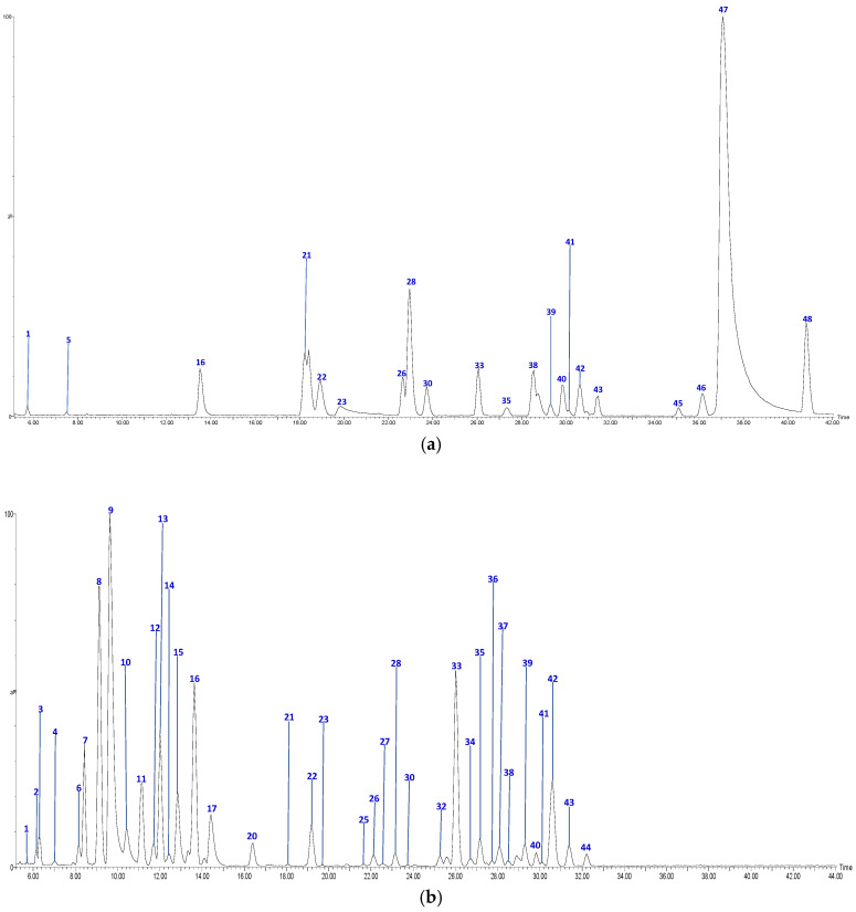
The UPLC-QTOF-ESI MS analysis described in the Materials and Methods section enabled us to identify 48 different compounds, including 4 chalcones, 15 procyanidin oligomers and the 2 flavan-3-ol monomers, 12 flavonols and glycosylated flavonols, 1 glycosylated isoprenoid derivative and 14 hydroxycinnamic acids and related derivatives (HCA), present in Anna and Jonagold Costa Rican apple cultivars. Figure 1 and Figure 2 show the chromatograms of the four samples and Table 2 summarizes the analysis results for the 48 compounds.
Figure 1.
HPLC Chromatograms of M. domestica extracts: (a) Anna skins (b) Anna flesh, in a Phenomenex Luna RP18 C-18 column (150 mm × 4.6 mm × 4 µm) using a Xevo G2-XS QTOF Mass spectrometer (Waters™, Wimslow, UK) in a mass range from 100 to 1500 amu.
Figure 2.
HPLC Chromatograms of M. domestica extracts: (a) Jonagold skins (b) Jonagold flesh, in a Phenomenex Luna RP18 C-18 column (150 mm × 4.6 mm × 4 µm) using a Xevo G2-XS QTOF Mass spectrometer (Waters™, Wilmslow, UK) in a mass range from 100 to 1500 amu.
Table 2.
Profile of the phenolic compounds identified by UPLC-DAD-ESI-MS/MS in Costa Rican apple cultivars 1.
| No | Tentative Identification | Rt (min) | [M-H]− | Formula | MS2 Fragments | Sample 1 |
|---|---|---|---|---|---|---|
| Hydroxycinamic acids | ||||||
| 7 | Sinapic acid hexoside | 8.91 | 385.1169 | C17H22O10 | [385]: 205, 223 | JF |
| 10 | Caffeoylquinic acid (I of II) | 10.36 | 353.0871 | C16H17O9 | [353]: 191, 179 | JF |
| 14 | Caffeoylquinic acid (II of II) | 12.45 | 353.0809 | C16H17O9 | [353]: 191, 180 | JF |
| 17 | p-Coumaroylquinic acid | 14.55 | 337.0912 | C16H18O8 | [337]: 173 | AF, JF |
| 18 | Shikimic acid | 15.06 | 173.0447 | C7H9O5 | [173]: 93, 111 | AF |
| 19 | Feruloylquinic acid (I of III) | 15.75 | 367.0983 | C17H20O9 | [367]: 173, 191 | AS, AF |
| 21 | Feruloylquinic acid (II of III) | 18.62 | 367.1012 | C17H20O9 | [367]: 173, 191 | AS, AF, JS, JF |
| 24 | Methyl-p-coumaroylquinic acid (I of II) | 20.82 | 351.1098 | C17H20O9 | [351]: 177 | AS, AF |
| 25 | Feruloylquinic acid (III of III) | 21.60 | 367.0983 | C17H20O9 | [367]: 173, 191 | AS, AF, JF |
| 29 | Methyl-p-coumaroylquinic acid (II of II) | 23.60 | 351.1098 | C17H19O8 | [351]: 177 | AF |
| 37 | Di-O-acetyl-O-p-coumaroylsucrose (I of II) | 28.14 | 571.1675 | C25H31O15 | [571]: 529, 553 | JF |
| 40 | Di-O-acetyl-O-p-coumaroylsucrose (II of II) | 29.88 | 571.1673 | C25H31O15 | [571]: 529, 554 | JS, JF |
| 43 | Caffeoyl hexoside (I of II) | 31.45 | 341.084 | C15H17O9 | [341]: 161, 179 | AS, AF, JS, JF |
| 45 | Caffeoyl hexoside (II of II) | 34.65 | 341.084 | C15H17O9 | [341]: 161, 179 | AS, AF, JS |
| Chalcones | ||||||
| 33 | Phloretin-pentosilhexoside (I of II) | 26.14 | 567.1725 | C26H31O14 | [567]: 273 | AS, AF, JS, JF |
| 34 | Phloretin-pentosilhexoside (II of II) | 26.95 | 567.1725 | C26H31O14 | [567]: 273 | JF |
| 41 | Phloridzin | 30.00 | 435.1312 | C21H24O10 | [435]: 167, 273 | AS, AF, JS, JF |
| 48 | Phloretin | 40.98 | 273.0757 | C15H13O5 | [273]: 167, 201 | AS, AF, JS |
| Flavonoids | ||||||
| 23 | Quercetin di-hexoside | 19.82 | 625.1378 | C30H25O13 | [625]: 300, 301 | AS, JS, JF |
| 26 | Quercetin-rutinoside | 22.54 | 609.1459 | C27H29O16 | [609]:300, 301 | AS, JS, JF |
| 30 | Quercetin-hexoside (I of III) | 23.78 | 463.0878 | C21H19O12 | [463]: 300, 301 | AS, JS, JF |
| 31 | Quercetin-hexoside (II of III) | 24.95 | 463.0878 | C21H19O12 | [463]: 300, 301 | AS, AF |
| 32 | Quercetin-pentoside | 25.35 | 433.0732 | C20H17O11 | [433]: 300, 301 | AS, AF, JF |
| 35 | Quercetin acetyl hexoside | 27.16 | 505.1002 | C23H22O13 | [505]: 300, 301 | AS, JS, JF |
| 36 | Quercetin-pentosylhexoside | 27.77 | 595.1245 | C26H27O16 | [595]: 300, 301 | JF |
| 38 | Kaempferol-hexoside | 28.63 | 447.0928 | C21H19O11 | [447]: 284.,285 | AS, AF, JS, JF |
| 39 | Quercetin pentosyl hexoside | 29.24 | 587.1043 | C25H26O15 | [595]: 300, 301 | JS, JF |
| 42 | Quercetin-hexoside (III of III) | 31.25 | 463.0834 | C21H20O12 | [463]: 300, 301 | AS, AF, JS, JF |
| 46 | Kaempferol-hexoside | 36.18 | 477.0944 | C21H20O11 | [447]: 284, 285 | AS, AF, JS |
| 47 | Quercetin | 37.19 | 301.0353 | C15H10O7 | [301]: 151, 179, 255, 273, 283 | AS, JS |
| Proantocyanidins | ||||||
| 1 | Procyanidin B-type dimer (I of IV) | 5.74 | 577.1292 | C30H25O12 | [577]: 287, 289, 407, 425, 451, 559 | JS, JF |
| 2 | Procyanidin B-type dimer (II of IV) | 6.12 | 577.1294 | C30H25O12 | [577]: 287, 289, 407, 425, 451, 559 | JF |
| 3 | Procyanidin B-type dimer (III of IV) | 6.57 | 577.1343 | C30H25O12 | [577]: 287, 289, 407, 425, 451, 559 | AS, AF, JF |
| 4 | Procyanidin B-type trimer (I of III) | 7.31 | 865.2004 | C45H37O18 | [865]: 287, 289, 575, 577, 695, 713, 739 | JF |
| 5 | Procyanidin A-type dimer | 7.67 | 591.1147 | C30H24O13 | [575]: 289, 449 | AF, JS |
| 6 | Procyanidin B-type trimer (II of III) | 8.5 | 865.2004 | C45H37O18 | [865]: 287, 289, 575, 577, 695, 713, 739 | AS, JF |
| 8 | Catechin | 9.01 | 289.0708 | C15H14O6 | [289]: 205,245, 271 | AS, JF |
| 9 | Procyanidin B-type dimer (IV of IV) | 9.52 | 577.1392 | C30H25O12 | [577]: 287, 289, 407, 425, 451, 559 | AS, AF, JF |
| 11 | Procyanidin B-type trimer (III of III) | 11.48 | 865.2004 | C45H37O18 | [865]: 287, 289, 575, 577, 695, 713, 739 | AS, JF |
| 12 | Procyanidin tetramer B (I of III) | 11.76 | 1153.2629 | C60H49O24 | [1153]: 287, 289, 575, 577, 863, 865, 983, 1001, 1027, 1135 | JF |
| 13 | Epicatechin | 12.14 | 289.0708 | C15H13O6 | [289]: 205, 245, 271 | AS, JF |
| 15 | Procyanidin B-type tetramer (II of III) | 12.82 | 1153.2704 | C60H49O24 | [1153]: 287, 289, 575, 577, 863, 865, 983, 1001, 1027, 1135 | JF |
| 20 | Procyanidin B-type pentamer (I of III) | 16.55 | 1441.2936 | C75H61O30 | [1441]: 287, 289, 575, 577, 865, 1153, 1315 | JF |
| 22 | Procyanidin B-type pentamer (II of III) | 19.13 | 1441.2939 | C75H61O30 | [1441]: 287, 289, 575, 577, 865, 1153, 1315 | JS, JF |
| 27 | (epi)catechin 3-O-gallate | 22.89 | 609.1459 | C27H29O16 | [441]: 153, 289, 315 | JF |
| 28 | Procyanidin B-type tetramer (III of III) | 23.20 | 1153.2701 | C60H49O24 | [1153]: 287, 289, 575, 577, 863, 865, 983, 1001, 1027, 1135 | AS, JS, JF |
| 44 | Procyanidin B-type pentamer (III of III) | 32.05 | 1441.2931 | C75H61O30 | [1441]: 287, 289, 575, 577, 865, 1153, 1315 | JF |
| Others | ||||||
| 16 | Vomifoliol-pentosilhexoside | 13.97 | 517.2293 | C24H37O12 | [517]: 205, 385 | AS, JS, JF |
1 Anna skin (AS), Anna flesh (AF), Jonagold skin (JS), Jonagold flesh (JF).
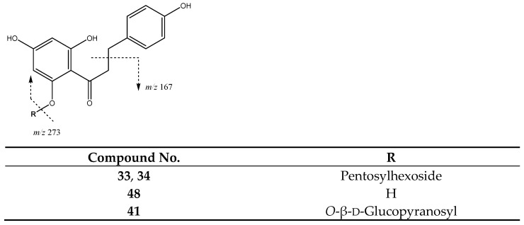
Chalcones constitute one group of compounds found in these fruit samples. For instance, peaks 33 (Rt = 26.14 min) and 34 (Rt = 26.95 min) with [M-H]− at m/z 567.1725 (C26H31O14) were identified as diglycoside derivatives of phloretin. They exhibit a main fragment at m/z 273 due to the loss of glycosides to yield the phloretin ion aglycone, thus these peaks are tentatively assigned to phloretin-pentosylhexoside isomers (Figure 3). In turn, peak 41 (Rt = 30.00 min) with [M-H]− at m/z 435.1312 (C21H23O10) is tentatively identified as phloridzin (phloretin 2′-O-glucose) with main fragments at 273 due to the cleavage of the glycoside [27] and at m/z 167 due to loss of the benzylic group. Finally, peak 48 (Rt = 40.98 min) with [M-H]− at m/z 273.0757 (C15H13O5) is tentatively assigned to the aglycone phloretin, which shows a characteristic main fragment at m/z 167 due to the loss of the benzylic moiety, as previously described [28,29].
Figure 3.
Chalcones structure and main fragments.
Another type of compound previously reported in apple is the glycosylated isoprenoid derivatives. In our study, peak 16 (Rt = 13.97 min) with an [M-H]− at m/z 517.2293 (C24H37O12) was tentatively identified as such, with main fragments at m/z 385 [M-H-132]− and 205 [M-H-312]− corresponding to the loss of a pentoside and a pentosylhexoside (Figure 4). The resulting ion is coincident with vomifoliol, thus allowing the peak to be assigned to a vomifoliol-pentosylhexoside isomer [30].
Figure 4.
Vomifoliol-pentosylhexoside structure and main fragments.
A relevant and abundant group of compounds found in Costa Rican Anna and Jonagold apples is constituted by flavan-3-ols, corresponding to monomers and procyanidin oligomers, including dimers, trimers, tetramers and several pentamers. Firstly, monomers catechin and epicatechin which were present in peaks 8 (Rt = 9.01 min) and 13 (Rt = 12.14 min). As shown in Figure 5, both compounds showed a [M-H]− at m/z 289.0708 (C15H13O6) with main fragments at m/z 245 [M-42-H]− due to retro-Diels-Alder fission (RDA) of ring A, m/z 205 produced by fission of ring A, and m/z 271 due to loss of water [31].
Figure 5.
Flavan-3-ols monomers structure and main fragments.
In addition, an (epi)-catechin 3-O-gallate was present in peak 27 (Rt = 22.89 min) with [M-H] at m/z 609.1459 (Figure 6), with main fragments at m/z 289 due to loss of gallate moiety, m/z 315 and m/z 153 by α-cleavage of carbonyl group [31].
Figure 6.
(Epi)-Catechin 3-O-gallate structure and main fragments.
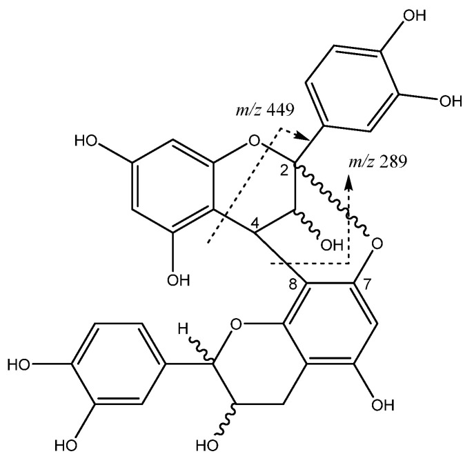
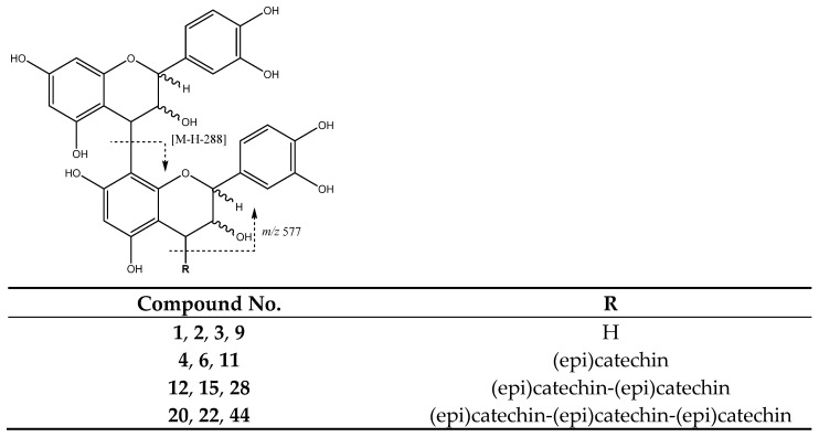
Procyanidin oligomers of monomeric (epi)catechin units can be linked to each other by an interflavonoid C4-C8 link (Figure 7). In the case of procyanidins type A, a linkage in C2-O-C7 is also present [32]. For instance, peak 5 (Rt = 7.67 min) shows a [M-H]− at m/z 575.1185 (C30H23O12) with a main fragment ion at m/z 449 [M-H-126]− due to the elimination of a phloroglucinol molecule from this A-type dimer, and a fragment of m/z 289 that corresponds to the monomer [33].
Figure 7.
Procyanidin A-type dimer structure and main fragments.
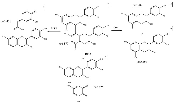
In our study, procyanidin B-type oligomers linked through a C4-C8 single bond are the most abundant group of flavan-3-ols obtained. Among these, procyanidin B dimers are present in peaks 1 (Rt = 5.74 min), 2 (Rt = 6.12 min), 3 (Rt = 6.57 min), and 9 (Rt = 9.52 min) with [M-H]− at m/z 577.1343 (C30H25O12). The main fragments shown by these compounds (Figure 8) were at m/z 559 which originates from water loss and m/z 451 which is a result of the elimination of the phloroglucinol through heterocyclic ring fission (HRF). As well as m/z 425 [M-H-152]− and 407 [M-H-170]− from retro Diels-Alder (RDA), the ion at m/z 289 originates from quinone-methide cleavage (QM) resulting in the ion of the monomer [34].
Figure 8.
Fragmentation pathways of B-type procyanidin dimers: HRF, Heterocyclic ring fusion; RDA, retro-Diels–Alder; QM, quinone methide.
On the other hand, procyanidin B-type trimers (Figure 9) are shown in peaks 4 (Rt = 7.31 min), 6 (Rt = 8.50 min) and 11 (Rt = 11.48 min) with [M-H]− at m/z 865.2004 (C45H37O18). They undergo QM cleavage of the upper interflavanoid bond producing ions of m/z 287 and 577, whereas cleavage of the lower interflavanoid bond forms ions of m/z 289 and 575 [33].
Figure 9.
Proanthocyanidin B-type trimers, tetramers and pentamers structures and main fragments.
In addition, as also shown in Figure 9, peaks 12 (Rt = 11.76 min), 15 (Rt = 12.82 min) and 28 (Rt = 23.20 min) with [M-H]− at m/z 1153.2579 (C60H49O24) correspond to B-type tetramers. Finally, peaks 20 (Rt = 16.55 min), 22 (Rt = 19.13 min) and 44 (Rt = 32.05 min) with [M-H]− at m/z 1441.2936 (C75H61O30) were tentatively assigned to B-type pentamers. For these compounds, main fragment ions were observed from QM cleavage as multiples of the monomer: m/z 289, 577 and 865 for tetramers, and additionally m/z 1153 for pentamers [32].
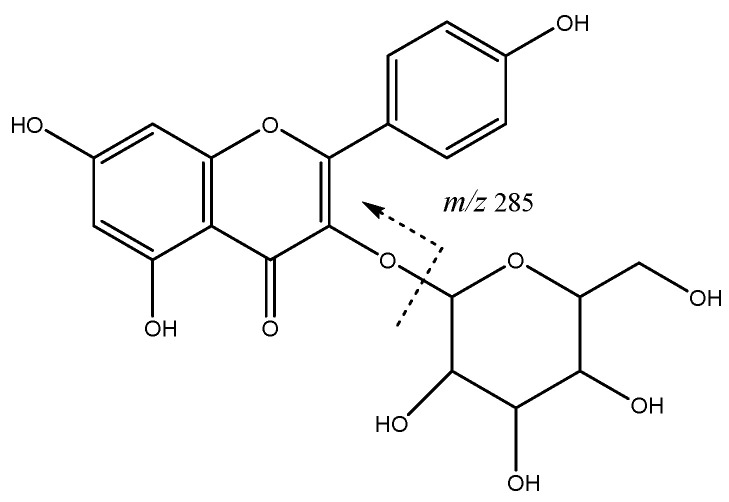
Flavonoids constitute another group of compounds found on these apple extracts. For instance, peaks 38 (Rt = 28.63 min) and 46 (Rt = 31.25 min) with [M-H]- at m/z 447.0928 (C21H19O11) were tentatively assigned to kaempferol hexosides with main fragment at m/z 285 (Figure 10).
Figure 10.
Kaempferol hexoside fragmentation.
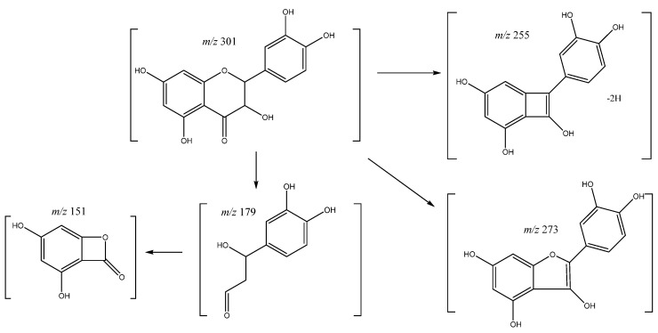
On the other hand, quercetin was assigned to peak 47 (Rt = 37.25) showing a negative molecular ion [M-H]− at m/z 301.0353 (C15H9O7). The main fragment ions (Figure 11) were found at m/z 179 and 151 from retrocyclization pathway [35], m/z 283 [M-18-H]− due to loss of water, m/z 273 [M-28-H]− from the loss of CO, and m/z 255 [M-18-28-H]− due to loss of water and CO [36].
Figure 11.
Quercetin fragmentation pathway.
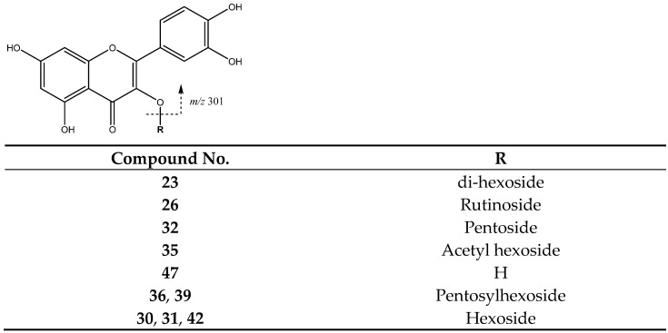
Peaks 30 (Rt = 23.78 min), 31 (Rt = 24.95 min) and 42 (Rt = 31.25 min) with [M-H]− at m/z 463.0875 (C21H19O12) were tentatively identified as quercetin-hexoside isomers. These compounds suffer the loss of the hexoside [M-H-162]− that results in the aglycone at m/z 301 (Figure 12). Meanwhile, peak 23 (Rt = 19.82 min) was tentatively assigned to quercetin di-hexoside with [M-H]− at m/z 625.1378, showing that the loss of the two hexosides delivers the aglycone at m/z 301 [37].
Figure 12.
Flavonol glycosides structure and main fragments.
Peak 32 (Rt = 25.35 min) was assigned to quercetin pentoside at [M-H]− 433.0732 (C20H17O11), elucidated by the fragment of the aglycone due to loss of glycoside [38]. Peaks 36 (Rt = 27.77 min) and 39 (Rt = 29.24 min) had a [M-H]− at m/z 595.1245 (C26H27O16), and were tentatively identified as quercetin-pentosyl-hexoside with the main fragment at m/z 301 due the loss of both glycoside units to yield the quercetin moiety. Peak 26 (Rt = 22.54 min) with [M-H]− at m/z 609.1488 (C27H29O16) was identified as quercetin-rutinoside, showing the loss of the rutinoside moiety [M-H-308]−. Peak 35 (Rt = 29.09 min) with [M-H]− at m/z 505.1025 (C23H21O13) was identified as quercetin-acetylhexoside with fragment at m/z 301 [M-162-H]− due the loss of acetylhexoside moiety.
Phenolic acids and derivatives constitute another group of compounds found in these samples. The smallest acid found was peak 18 (Rt = 15.06 min) corresponding to shikimic acid (Figure 13). Main fragments at m/z 111 and 93 were generated from RDA fission and from subsequent loss of water, respectively.
Figure 13.

Shikimic acid structure and main fragments.
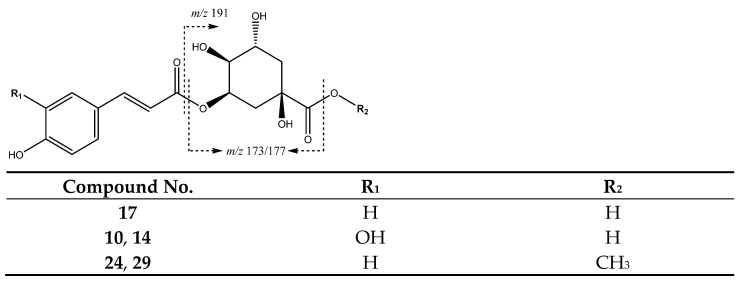
A series of 4-hydroxycinnamic acid derivatives were identified, as summarized in Table 2. For instance, as shown in Figure 14, peak 17 (Rt = 14.55 min) with [M-H]− at m/z 337.0912 (C16H17O8) was assigned to a coumaroylquinic acid with a main fragment at m/z 173 due to the loss of water of the quinic acid ion [34].
Figure 14.
Structures and main fragments of coumaroyl and caffeic acid derivatives.
In turn, peaks 24 (Rt = 20.82 min) and 29 (Rt = 23.60 min) correspond to coumaroylquinic acid methyl esters with [M-H]− 351.1098 (C17H19O8), with a main fragment at m/z 177 due to the loss of CO from the quinic acid moiety. In addition, peaks 10 (Rt = 10.36 min) and 14 (Rt = 12.45 min) correspond to caffeoylquinic acids with a [M-H]− at m/z 353.0809 (C16H17O9) and main fragments at m/z 191 [quinic acid-H]−, and 135 due to the loss of CO2 from the quinic acid ion, as shown in Figure 15 [39].
Figure 15.
Caffeoylquinic acid fragmentation pathway.
Peaks 43 (Rt = 31.45 min) and 45 (Rt = 34.65 min) show [M-H]− at m/z 341.0872 (C15H17O9) and were tentatively identified as caffeoyl-hexoside isomers, which show a main fragment at m/z 161 [M-H-179]− due to the loss of glycoside (Figure 16). Meanwhile, peaks 37 (Rt = 28.14 min) and 40 (Rt = 29.88 min), with [M-H]− at 571.1675 (C25H31O15) were tentatively assigned to di-O-acetyl-O-p-coumaroylsucrose, with main fragments at m/z 553 [M-18-H]− due to loss of water, at m/z 529 [M-42-H]− because of acetyl loss, and at m/z 487 [M-84-H]− due to the loss of both acetyl moieties [40].
Figure 16.
Structure and fragmentation of glycosylated and acetyl-glycosylated derivatives from hydroxycinnamic acids.
Additionally, as shown in Figure 17, peaks 19 (Rt = 15.75 min), 21 (Rt= 18.62 min) and 25 (Rt = 21.60 min), with [M-H]− at 367.1012 (C9H5O3) were identified as feruloylquinic acid isomers, with main fragments at m/z 191 [quinic acid-H]− and m/z 173 [quinic acid-H2O-H]−, as reported for these compounds [41]. Finally, peak 7 was assigned to sinapic acid hexoside showing [M−H]− at m/z 385.1169 (C17H21O10) and main fragments at m/z 223 [M-H-162]− due to the loss of the glycoside unit, at m/z 205 due the additional loss of water, and at m/z 191 due to the loss of the methoxy group from de aglycone.
Figure 17.
Structures and main fragments of sinapic acid and feruloyl quinic acid derivatives.
Regarding the total number of polyphenols in M. domestica samples, the flesh of Jonagold shows the greatest number of compounds and exhibits the highest number of flavan-3-ols as well as being the most abundant in procyanidin tetramers and pentamers. Anna skins contain the second highest number of compounds and show flavonoids as the most abundant group of polyphenols.
When comparing data in the literature regarding compound characterization from apple skins, our results for Anna and Jonagold cultivars are similar to the total number of compounds and diversity in Golden Delicious and Braeburn cultivars from Slovenia [28]. In addition, both Costa Rican cultivars show a greater number and diversity in respect to other cultivars from Brazil and Canada [25,42,43]. In respect to flesh, the Jonagold cultivar is far superior to Anna and other cultivars from the literature, especially regarding proanthocyanidins both in total occurrence and in greater polymerization degree [24,25,44], for instance in procyanidin trimers, tetramers and pentamers found in Costa Rican Jonagold flesh.
In the case of glycosylated flavonoids, our findings show a similar number of compounds to European cultivars [24,28,44] and they indicate more diversity in quercetin derivatives. In respect to the occurrence of hydroxycinnamic acid derivatives, our results are within the range reported for cultivars from South Korea [45] and Europe [46,47,48]. Finally, for the chalcones group, our findings are similar to results reported for cultivars from Canada and China [25,49].
In sum, as within recent studies on other fruits [50], the profiling of polyphenols reveals high diversity in Costa Rican cultivars, which is in agreement with evidence showing that apples’ secondary metabolites profile is greatly influenced by location [44,51] as well as with findings from studies on other species indicating that tropical forests have a greater diversity of secondary metabolites [52]. Thus, the present work can be of interest for further research and future studies should take into consideration the parameters from the cultivars themselves, such as origin, location, soil composition and their relationship with chemical metabolites and bioactivities.
2.3. Antioxidant Activity
The DPPH and ORAC values obtained are summarized in Table 3. All samples show high antioxidant values, with Jonagold flesh presenting the best value with IC50 = 3.96 μg/mL for DPPH and 14.80 mmol Trolox equivalents/g for ORAC, followed by Anna and Jonagold skins, while Anna flesh exhibits the lowest antioxidant activity with IC50 = 11.33 μg/mL for DPPH and 4.53 mmol Trolox equivalents/g for ORAC. Regarding DPPH antioxidant findings from the literature, the results available for extracts show IC50 values for skins ranging between 41.41 and 55.54 µg/mL [53] and 710 µg/mL for flesh [48] in cultivars from India and Portugal, respectively, thus extracts from Anna and Jonagold cultivars show better results for both skins and flesh, as shown in Table 3. Another study on cultivars from Austria reported DPPH values ranging between 2.29 and 7.44 mmol TE/100 g DM [54] with our results for Anna and Jonagold cultivars showing values (2.12–8.05 mmol TE/100 g DM) within that range.
Table 3.
DPPH and ORAC antioxidant activity from the extracts of M. domestica cultivars.
| Sample | DPPH 1,2 | ORAC 1.2 | |
|---|---|---|---|
| IC50 (μg Extract/mL) | (mmol TE/g Extract) | (mmol TE/g Extract) | |
| Anna | |||
| Skin | 6.90 a ± 0.02 | 3.25 a ± 0.01 | 11.19 a ± 0.25 |
| Flesh | 11.33 b ± 0.05 | 1.98 b ± 0.01 | 5.96 b ± 0.23 |
| Jonagold | |||
| Skin | 9.76 c ± 0.17 | 2.30 c ± 0.04 | 7.44 c ± 0.10 |
| Flesh | 3.96 d ± 0.02 | 5.68 d ± 0.03 | 14.80 d ± 0.26 |
1 Values are expressed as mean ± S.D. 2 Different superscript letters in the same column indicate that differences are significant at p < 0.05 using ANOVA with a Tukey post hoc as statistical test. ORAC, oxygen radical absorbance capacity; DPPH, 2,2-diphenyl-1-picrylhidrazyl method.
On the other hand, for ORAC, reports from the literature indicate values in the range of 8.60–44.07 mmol TE/100 g DM for skins and 2.4–42.97 mmol TE/100 g DM for apple flesh in cultivars from Germany [24] and Norway [47]. On the other hand, other studies report values varying between 0.45–10.62 mmol TE/100 g FW for skins and between 0.19 and 2.61 mmol TE/100 g FW for flesh in cultivars from Chile [56] and Italy [55]. Our results for skins (15.53–18.43 mmol TE/100 g DM and 3.10–4.23 mmol TE/100 g FW) and for flesh (6.37–20.99 TE/100 g DM and 1.01–2.94 mmol TE/100 g FW) from Anna and Jonagold cultivars fall within those ranges.
In addition, a correlation analysis was performed among the total phenolic contents (TPC, Table 1) and the antioxidant activity results from ORAC and DPPH methods. A significant negative correlation (p < 0.05) was found between TPC and DPPH results (R = −0.983) as wells as a significant positive correlation (p < 0.05) between TPC and ORAC values (R = 0.980). Therefore, these results align with previous findings reporting a correlation between total polyphenolic contents and different types of antioxidant activities [57].
Finally, the results for Jonagold flesh are of particular importance since there are few reports on the flesh being richer in polyphenols and having higher antioxidant activity than the skin, which points to the interest for further research on biological models. For instance, some studies have described the antioxidant mechanisms associated with proanthocyanidins with an increase in the Nuclear factor E2-related factor 2 (Nrf2) translocation to the nucleus [58], which activates the transcription of genes responsible for maintaining cellular redox homeostasis and protect cells from oxidative damage [59].
2.4. Cytotoxicity
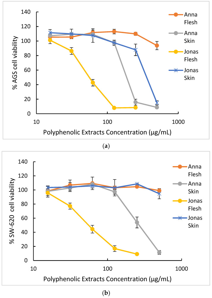
Table 4 summarizes the IC50 values for the cytotoxic effect of M. domestica extracts on different human carcinoma cells related to the digestive tract, namely AGS (gastric adenocarcinoma) and SW-620 (colorectal adenocarcinoma) cell lines, while the dose–response curves are displayed in Figure 18. The development of digestive tract cancers has been associated with lower consumption of vegetables and fruits [60]; in particular, 60% of stomach cancer and 43% of colon cancer are attributed to deficient consumption of vegetables [61]. In Costa Rica, colon cancer is the second most common cancer and gastric cancer has the third and fourth incidence rate in men and women, respectively [62]. Proanthocyanidins found in apples have been associated with exert antitumoral effects reaching and interacting directly with the gastrointestinal cells [63,64]. Thus, it is of interest to evaluate these extracts’ cytotoxicity using as targets these tumoral cancer cell lines.
Table 4.
Cytotoxicity of M. domestica extracts to gastric (AGS) and colon (SW-620) carcinoma cells as well as to Vero non-tumoral cells.
| Sample | IC50 (µg/mL) 1,2 (SI) 3 | ||
|---|---|---|---|
| AGS | SW-620 | Vero | |
| Anna | |||
| Skin | 167.22 a,* ± 10 (3.0) | 295.93 b,# ± 29 (1.7) | > 500 a,≠ |
| Flesh | > 500 b,* | > 500 a,* | > 500 a,* |
| Jonagold | |||
| Skin | 398.44 c,* ± 7 (1.3) | > 500 a,* | 305.72.b,* ± 30 |
| Flesh | 60.03 d,* ± 1.7 (5.1) | 62.41 c,* ± 5.2 (4.9) | > 500 a,# |
1 Different superscript letter in the same column indicates that differences are significant at p < 0.05 using ANOVA with a Tukey post hoc as statistical test. 2 Different superscript signs in the same row indicate that differences are significant at p < 0.05 using ANOVA with a Tukey post hoc as statistical test. 3 Selectivity Index.
Figure 18.
Cytotoxicity dose–response curves of apple extracts on AGS and SW620 tumor cell lines. Results are presented as mean ± SE of three independent experiments. (a) Samples in AGS cells (b) Samples in SW620 cells.
As observed in Table 4, the best cytotoxic effects of Costa Rican M. domestica against AGS and SW-620 cells were observed for Jonagold cultivar, with the flesh sample (IC50 values of 60.0 ± 1.7 and 62.4 ± 5.2 μg/mL, respectively). Anna cultivar skin sample showed a moderate cytotoxic effect against AGS cells (IC50 of 167 ± 10 μg/mL).
The dose–response curves for each extract displayed in Figure 18 confirm Jonagold flesh as the best extract with the highest cytotoxic effect on both AGS and SW620 adenocarcinoma cell lines. In fact, both plots demonstrate a marked slope in the dose–response curves for this extract compared to the other samples. Anna and Jonagold skins show a more moderate cytotoxicity to obtain bioactive compounds, while Anna flesh represents the sample with the lowest cytotoxic effect in both tumoral cell lines tested.
Some studies have evaluated the cytotoxic effect of apples (Malus domestica) in tumoral cell lines and variations were observed for samples of the same species cultivated in different locations. Studies using French apples evaluated the cytotoxic effect against colorectal adenocarcinoma cells (SW-620) and esophageal adenocarcinoma (OE-33) showing 50% of cytotoxicity with similar concentrations of our study (45–60 μg/mL); however, for both studies, the extracts were enriched with a specific type of polyphenol [65,66]. Other studies showed moderate cytotoxic effect with acetone extracts from whole apple extracts from Lithuanian cultivars against the human colon adenocarcinoma cell line (HT-29) and human glioblastoma cell line (U-87), reporting an IC50 of 113.3 μg/mL and 119.7 μg/mL, respectively [67]. A lower cytotoxic effect was reported for acetone and alcoholic extracts from apple (M. domestica) pomace cultivated in India. These Indian apples achieved 50% cytotoxicity only in oral carcinoma (KB) with concentrations of 100 μg/mL, but for cervical squamous cells carcinoma (SiHa) and colorectal adenocarcinoma (HT-29), 50% was not reached even with treatment of 400 μg/mL [68]. On the other hand, studies of Indian M. domestica apples achieved an improved cytotoxic effect using innovative delivering strategies such as silver nanoparticles. This approach permits an IC50 of 10 μg/mL [69] and 33.8 μg/mL [70] to be achieved against breast cancer cells (MFC-7).
The cytotoxic effect in tumor cell lines has been reported for other Malus species, also demonstrating very fluctuating results. Malus sieversii acetone extracts, grown in China, were assessed on breast cancer cell lines (MCF-7 and MAD-MB-231) and showed a very low cytotoxic effect (IC50 of 33.44 mg/mL and 20.94 mg/mL) [71]. Similarly, a methanolic extract of Chinese apples, Malus pumila, were evaluated against cancer colon cells (SW-480), stomach cancer cells (BCG 803) and esophageal cancer cells (CaEs-12) and a weak cytotoxic activity was reported with IC50 varying between 3.5–4.3 mg/mL in all cell lines [72]. In the opposite side, the cytotoxicity of Chinese apples, M. pumila, was evaluated against liver hepatocellular carcinoma (HepG2) and a strong inhibitory grown rate of 50% was achieved with concentration of less than 4 μg/mL for pulp extracts and less than 20 μg/mL for skin extracts [73]. Finally, ornamental crabapple Malus sp. (“red splendor”) has also been studied, and the cytotoxic activity showed values of 48.3 μg/mL, 64.5 μg/mL and 78.9 μg/mL, for SW-480, BCG 803 and CaEs-17, respectively [72].
In addition to the IC50 values used to quantify the cytotoxic effect, Table 4 shows the selectivity index, which is defined as the ratio of IC50 values of non-tumor cells to cancer cells. The highest selectivity index values in this study correspond to the Jonagold flesh sample (5.1 for AGS cells and 4.9 for SW-620 cells) and the Anna skin sample (3.0 for AGS cells). According to previous reports, extracts with SI greater than three are considered to have high selectivity towards cancer cells and suggest a possible therapeutic potential [74,75]. For M. domestica extracts, a comparison of IC50 values of non-tumor cells to cancer cells has been reported previously for breast cancer cell lines. The selectivity ratio in this Indian apple was 2.2, which is a lower value compared to our study, even though the Indian apple extracts were applied to the cells using nanoparticle delivery systems [70].
In addition, correlation analysis was performed between the cytotoxicity results obtained and total phenolic contents. Significant negative correlation (p < 0.05) was found between IC50 cytotoxic values on SW620 cancer cells and TPC (r = −0.908) and between IC50 cytotoxic values on AGS cancer cells and TPC (r = −0.902). Furthermore, correlation analysis performed between the IC50 cytotoxic activity in both adenocarcinoma cell lines (Table 4) and the number of compounds identified for each polyphenol group (Table 2) showed no significant correlation (p < 0.05) with HCA, chalcones or flavonoids for either SW620 or AGS cell lines. In contrast, a significant negative correlation (p < 0.05) with the number of procyanidins was found for cytotoxicity results on both SW620 (r = −0.978) and AGS (r = −0.894) cell lines. These r coefficients represent similar and higher values, respectively, than the ones for TPC, suggesting procyanidin’s major contribution to the cytotoxic activity against both tumor cells.
The predominant role of proanthocyanidins in the cytotoxic effect against tumoral cells has been widely documented for grape seeds extracts [76,77,78,79]. However, not many reports are available for other natural sources; some of the few reports include exotic fruits such as Japanese Quince [80] and Bactris guineensis [81] and other widespread consumed fruits, such as berries [82].
The association of proanthocyanidins and cytotoxic effect in tumoral cells has been linked to the degree of polymerization of these polyphenols. For grapes, grape seeds and pine bark assays in colon cancer cells (HCT116, SW-480, SW-620, HT-29, Caco-2, RKO and LoVo), the anti-proliferative effect positively correlated with an increase in the degree of polymerization [83]. Another report [65] compared two polyphenol-enriched fractions from M. domestica, reporting a 50% inhibition of colorectal carcinoma (SW-620) cell growth with 45 μg/mL of the fraction rich in polymers and no effect in the monomer fraction even at a concentration of 100 μg/mL. Other studies in esophageal gastric adenocarcinoma demonstrated that oligomer procyanidins showed more potent antiproliferative activities that the monomeric and dimeric procyanidins [66]. These reports are consistent with the pattern shown in the present study. The strongest cytotoxic activity, an IC50 of 60 and 63 μg/mL in AGS and SW-620, respectively, was assessed for flesh samples of the Jonagold cultivar (Table 4) which is the one showing an enriched profile of proanthocyanidins oligomers (Table 2), specifically trimers, tetramers and pentamers B-type procyanidins.
The antitumor effect of proanthocyanidins has been associated with an apoptotic induction and a regulation of inflammatory pathways that ends in an inhibition of the tumor cell proliferation [81,84,85]. Some reports from grape seeds and apple procyanidins (M. pumila) have described an induction of cell cycle arrest by down-regulation of cyclin D1, CDK4 and survivin. In addition, these reports describe an induction of apoptosis through an increase in mitochondrial membrane permeability, a cytochrome c release and enhance of caspase 3 and caspase 9 expression and activation, which represents a hallmark of apoptosis [76,85,86]. However, despite these preliminary reports, the specific mechanism has yet to be elucidated. Reports on the bioavailability of procyanidins indicate that these molecules reach the colon almost intact and would interact there with colorectal cancer cells [64] similar to the ones evaluated in this work. In sum, the promising results obtained for Jonagold flesh suggest that there is a need for further studies, for instance in other cancer cell lines, to determine the prospective of these Costa Rican apples as source of enriched proanthocyanidins extracts and their related bioactivities.
3. Materials and Methods
3.1. Materials, Reagents and Solvents
M. domestica fruits of Anna and Jonagold cultivars were acquired in ripe state in late summer from producers in Los Santos, Costa Rica. Cultivars were confirmed with the support of the Costa Rican National Herbarium and vouchers are deposited there. Reagents, such as fluorescein, 2,2-azobis(2-amidinopropane) dihydrochloride (AAPH), 2,2-diphenyl-1-picrylhidrazyl (DPPH), Trolox, gallic acid, and Amberlite XAD-7 resin, fetal bovine serum, glutamine, penicillin, streptomycin, amphotericin B, trypsin–EDTA, were provided by Sigma-Aldrich (St. Louis, MO, USA). Human gastric adenocarcinoma cell line AGS, human colorectal adenocarcinoma SW 620 and monkey normal epithelial kidney cells Vero were obtained from American Type Culture Collection (ATCC, Rockville, MD, USA), while solvents such as acetone, chloroform and methanol were purchased from Baker (Center Valley, PA, USA), while DMSO was acquired from Sigma-Aldrich (St. Louis, MO, USA).
3.2. Phenolic Extracts from Malus Domestica Fruits
M. domestica fruits were rinsed in water, peeled, and both skin and flesh material were frozen at −20 °C and then lyophilized in a Free Zone Cascade Benchtop Freeze Dry System 720401000 (Labconco, Kansas, MO, USA), with Ice Holding Capacity of 4.5 L and Collector Temperature of −105 °C and system vacuum level < 133 × 10−3 mbar. The lyophilized material was preserved at −20 °C until extraction. Freeze-dried samples were extracted under Pressurized Liquid Extraction (PLE) conditions, in a Dionex™ ASE™ 150 Accelerated Solvent Extractor (Thermo Scientific™, Walthman, MA, USA) using methanol:water (70:30) as solvent in a 34 mL cell, at 40 °C. Next, the extract was evaporated under vacuum to eliminate the methanol and the aqueous phase was washed with ethyl acetate and chloroform to remove less polar compounds. Afterwards, the aqueous extract was evaporated under vacuum to eliminate organic solvent residues and was eluted (2 mL/min) in Amberlite XAD7 column (150 mm × 20 mm), starting with 300 mL of water to remove sugars, and then with 200 mL each of methanol:water (80:20) and pure methanol to obtain the polyphenols. Finally, the enriched extract was obtained after evaporating to dryness at 40 °C using a Buchi™ 215 (Flawil, Switzerland) rotavapor.
3.3. Total Phenolic Content
The polyphenolic content was determined as previously reported [87] by a modification of the Folin–Ciocalteu (FC) method [88], whose reagent is composed of a mixture of phosphotungstic and phosphomolybdic acids. Each sample was dissolved in MeOH (0.1% HCl) and combined with 0.5 mL of FC reagent. Afterwards, 10 mL of Na2CO3 (7.5%) were added and the volume was completed to 25 mL with water. Blanks were prepared in a similar way, but using 0.5 mL of MeOH (0.1% HCl) instead of the sample. The mixture was left standing in the dark for 1 h and then the absorbance was measured at 750 nm. Values obtained were extrapolated in a gallic acid calibration curve. Total phenolic content was expressed as mg gallic acid equivalents (GAE)/g sample. Analyses were performed in triplicate.
3.4. UPLC-ESI-MS Analysis
The UPLC-MS system used to analyze the composition of M. domestica extracts consisted of a Xevo G2-XS QTOF (Waters, UK) coupled with an AQUITY H Class UPLC system with quaternary pump. ESI source parameters were set to a capillary voltage of 2 kV, sampling cone of 20 eV, source temperature of 150 °C, and source offset of 10 °C. The desolvation temperature was set at 450 °C, the cone gas flow at 0 L/h and the desolvation gas flow at 900 L/h.
Measurement was performed in MSe high resolution negative mode using an acquisition mass range from 100 m/z to 2000 m/z and a scan rate of 0.5 s, where fragmentation was carried out using Independent Data Acquisition for all eluting compounds with collision energy ramp from 20 V to 30 V storing at the high energy function. Instrument calibration was applied in the mass range of the measurement with sodium formate. Lock mass correction was applied directly to the measurement using leucine enkephalin infusion measured each 30 s during the run. The data was analyzed using MassLynx V4.2 software from Waters.
Separation was carried out on a Luna RP-C18 column (150 mm × 4.6 mm i.d. × 4 µm, Phenomenex, Torrance, CA, USA) with a pre-column filter (Phenomenex, Torrance, CA, USA). Solvents used in the mobile phase were water with 0.1% formic acid (A), methanol with 0,1% formic acid (B) and acetonitrile with 0.1% formic acid (C). Then, 5 μL of sample was injected with a flow rate of 0.5 mL/min at 40 °C. The chromatographic gradient started at 83% A, 12% B and 7% C, changing to 79.2% A, 12% B and 8.8% C at 4.8 min, then to 74% A, 15% B, and 11% C at 14.8 min, then to 0% A, 85% B and 15% C at 48 min, holding it for 10 min. Then, the column was equilibrated for 5 min to initial conditions.
3.5. DPPH Radical-Scavenging Activity
DPPH evaluation was performed as previously reported [89] and was expressed as IC50 (µg/mL), which is the amount of sample required to reach the 50% radical-scavenging activity, and also as mmol of Trolox equivalents (TE)/g extract. Briefly, a solution of 2,2-diphenyl-1-picrylhidrazyl (DPPH) (0.25 mM) was prepared using methanol as solvent. Next, 0.5 mL of this solution was mixed with 1 mL of extract or Trolox at different concentrations, and incubated at 25 °C in the dark for 30 min. DPPH absorbance was measured at 517 nm. Blanks were prepared for each concentration. The percentage of the radical-scavenging activity of the sample or Trolox was plotted against its concentration to calculate IC50 (µg/mL). The samples were analyzed in three independent assays. In order to express the DPPH results as mmol TE/g extract, the IC50 (µg/mL) of Trolox was converted to mmol/mL using Trolox molecular weight (250.29 mg/mmol) and then dividing by the IC50 of each sample.
3.6. ORAC Antioxidant Activity
The Oxygen Radical Absorbance Capacity (ORAC) antioxidant activity was determined following a method previously described [90] using fluorescein as a fluorescence probe. The reaction was performed in 75 mM phosphate buffer (pH 7.4) at 37 °C. The final assay mixture consisted of AAPH (12 mM), fluorescein (70 nM), and either Trolox (1–8 µM) or the extract at different concentrations. Fluorescence was recorded every minute for 98 min in black 96-well untreated microplates (Nunc, Denmark), using a Polarstar Galaxy plate reader (BMG Labtechnologies GmbH, Offenburg, Germany) with 485-P excitation and 520-P emission filters. Fluostar Galaxy software version 4.11-0 (BMG Labtechnologies GmbH, Offenburg, Germany) was used to measure fluorescence. Fluorescein was diluted from a stock solution (1.17 mM) in 75 mM phosphate buffer (pH 7.4), while AAPH and Trolox solutions were freshly prepared. All reaction mixtures were prepared in duplicate and three independent runs were completed for each extract. Fluorescence measurements were normalized to the curve of the blank (no antioxidant). From the normalized curves, the area under the fluorescence decay curve (AUC) was calculated as:
| (1) |
where f0 is the initial fluorescence reading at 0 min and fi is the fluorescence reading at time i. The net AUC corresponding to a sample was calculated as follows:
| Net AUC = AUCantioxidant − AUCblank | (2) |
The regression equation between net AUC and antioxidant concentration was calculated. The ORAC value was estimated by dividing the slope of the latter equation by the slope of the Trolox line obtained for the same assay. Final ORAC values were expressed as mmol of Trolox equivalents (TE)/g of phenolic extract.
3.7. Evaluation of Cytotoxicity of Extracts
3.7.1. Cell Culture
The human gastric adenocarcinoma cell line AGS, the human colorectal adenocarcinoma SW 620 and monkey normal epithelial kidney cells Vero were grown in minimum essential Eagle’s medium (MEM) containing 10% fetal bovine serum (FBS) in the presence of 2 mmol/L glutamine, 100 IUmL−1 penicillin, 100 μg/mL streptomycin and 0.25 μg/mL amphotericin B. The cells were grown in a humidified atmosphere containing 5% CO2 at 37 °C and were sub-cultured by detaching with trypsin–EDTA solution at about 70–80% confluence. For the experiments, 100 μL of a cell suspension of 1.5 × 10⁵ cells/mL were seeded overnight into 96-well plates. The cells were further exposed for 48 h to various concentrations of extracts (50 μL), dissolved in DMSO and diluted with cell culture medium to final concentrations between 15–500 μg/mL. The DMSO concentrations used in the experiments were below of 0.1% (v/v) and control cultures were prepared with the addition of DMSO (vehicle control).
3.7.2. Assessment of Cytotoxicity by MTT Assay
After incubation for 48 h, MTT assays were performed to evaluate the cell viability. The decrease in the viability correlates with the cytotoxic activity of the extract. Briefly, the medium was eliminated, cells were washed twice with 100 μL of PBS and incubated with 100 μL MTT solution (3-(4,5-dimethylthiazolyl-2)-2,5-diphenyltetrazolium bromide, 5 mg/mL in cell culture medium) for 2 h at 37 °C. The formazan crystals formed were dissolved in 100 μL of ethanol 95% and the absorbance was read at 570 nm in a microplate reader. Dose–response curves were established for each extract and the concentration, which is enough to reduce the cell viability by 50% (IC50), was calculated.
In order to evaluate whether the cytotoxicity activity was specific against the cancer cells, a selectivity index (SI) was determined. This index is defined as the ratio of IC50 values of normal epithelial kidney cells (Vero) to cancer cells (AGS or SW620).
3.8. Statistical Analysis
One-way analysis of variance (ANOVA) followed by Tukey’s post hoc test was applied to TPC, DPPH, ORAC and cytotoxicity results, and differences were considered significant at p < 0.05. In order to evaluate whether the total phenolic contents (TPC) contributes to the antioxidant activity evaluated with DPPH and ORAC methodologies, a correlation analysis was carried out as well as cytotoxicity assays. R (version x64 4.1.1) was used as the statistical program.
4. Conclusions
The UPLC-HRMS analysis using the QTOF-ESI MS technique allowed 106 compounds to be characterized in phenolic enriched extracts of skins and flesh of Anna and Jonagold apple cultivars in Costa Rica. Among them, the flesh of the Jonagold cultivar displayed the most abundant number of polyphenols and also exhibited higher and more diversified procyanidin oligomers than cultivars from other countries reported in the literature. Furthermore, this extract also showed the best results for TPC, ORAC and DPPH antioxidant activities as well as for cytotoxicity IC50 values against SW620 and AGS cancer cell lines. In addition, the abundance of procyanidins showed a significant positive correlation (p < 0.05) with the ORAC results and a significant negative correlation (p < 0.05) with DPPH and cytotoxicity towards AGS and SW620 tumor. These findings align with the fact that procyanidin oligomers were more abundant and presented a higher degree of polymerization, including tetramers and pentamers, in the flesh of Jonagold extract, displaying better bioactivity effects. The overall results from this study and particularly the ones obtained for the flesh of Jonagold cultivar, support findings suggesting the importance of considering fruit varieties [91]. As mentioned, the higher degree of polymerization in procyanidins has been linked with anti-inflammatory and anticancer activities [92], therefore additional research would contribute to determining the potential health benefits of these extracts.
Acknowledgments
Authors also thank financial support from the Costa Rica Institute of Technology. Special thanks are due to FRUTALCOOP for providing the apple fruits and to Alonso Quesada from Costa Rican National Herbarium for his support with the vouchers.
Author Contributions
Conceptualization, M.N.-H.; formal analysis, M.N.-H., F.V.-H., S.Q.-M., G.A.-C., K.W.-R., M.I.Q.-F. and A.S.-K.; funding acquisition, M.N.-H. and E.A.-S.; investigation, M.N.-H. and E.A.-S.; methodology, M.N.-H. and A.S.-K.; writing—original draft, M.N.-H., S.Q.-M., G.A.-C., D.A.-C., K.W.-R. and F.V.-H.; writing—review and editing, M.N.-H., E.A.-S., S.Q.-M., G.A.-C., K.W.-R., M.I.Q.-F., D.A.-C., F.V.-H. and A.S.-K. All authors have read and agreed to the published version of the manuscript.
Funding
This work was partially funded by grants from FEES-CONARE (115-B2-657 and ED-3239) and from the University of Costa Rica (115-B0-047 and ED-2033).
Institutional Review Board Statement
Not applicable.
Informed Consent Statement
Not applicable.
Data Availability Statement
The data presented in this study are available within this article.
Conflicts of Interest
The authors declare no conflict of interest.
Sample Availability
Samples of the compounds are not available from the authors.
Footnotes
Publisher’s Note: MDPI stays neutral with regard to jurisdictional claims in published maps and institutional affiliations.
References
- 1.Ahmad Khan M.S., Ahmad I. Herbal Medicine. In: Ahmad Khan M.S., Ahmad I., Chattopadhyay D., editors. New Look to Phytomedicine. Elsevier; Amsterdam, The Netherlands: 2019. pp. 3–13. [DOI] [Google Scholar]
- 2.Boyer J., Liu R.H. Apple Phytochemicals and Their Health Benefits. Nutr. J. 2004;3:5. doi: 10.1186/1475-2891-3-5. [DOI] [PMC free article] [PubMed] [Google Scholar]
- 3.Lavie L., Lavie P. Molecular mechanisms of cardiovascular disease in OSAHS: The oxidative stress link. Eur. Respir. J. 2009;33:1467–1484. doi: 10.1183/09031936.00086608. [DOI] [PubMed] [Google Scholar]
- 4.Mariani E., Polidori M.C., Cherubini A., Mecocci P. Oxidative Stress in Brain Aging, Neurodegenerative and Vascular Diseases: An Overview. J. Chromatogr. B. 2005;827:65–75. doi: 10.1016/j.jchromb.2005.04.023. [DOI] [PubMed] [Google Scholar]
- 5.Vaidya F.U., Chhipa A.S., Sagar N., Pathak C. Oxidative Stress and Inflammation Can Fuel Cancer. In: Maurya P.K., Dua K., editors. Role of Oxidative Stress in Pathophysiology of Diseases. Springer; Singapore: 2020. pp. 229–258. [DOI] [Google Scholar]
- 6.Pignatelli P., Menichelli D., Pastori D., Violi F. Oxidative Stress and Cardiovascular Disease: New Insights. Kardiol. Pol. 2018;76:713–722. doi: 10.5603/KP.a2018.0071. [DOI] [PubMed] [Google Scholar]
- 7.Ivanov A.V., Bartosch B., Isaguliants M.G. Oxidative Stress in Infection and Consequent Disease. Oxid. Med. Cell. Longev. 2017;2017:3496043. doi: 10.1155/2017/3496043. [DOI] [PMC free article] [PubMed] [Google Scholar]
- 8.Koch W. Dietary Polyphenols—Important Non-Nutrients in the Prevention of Chronic Noncommunicable Diseases. A Systematic Review. Nutrients. 2019;11:1039. doi: 10.3390/nu11051039. [DOI] [PMC free article] [PubMed] [Google Scholar]
- 9.Asgary S., Rastqar A., Keshvari M. Functional Food and Cardiovascular Disease Prevention and Treatment: A Review. J. Am. Coll. Nutr. 2018;37:429–455. doi: 10.1080/07315724.2017.1410867. [DOI] [PubMed] [Google Scholar]
- 10.Vinson J.A., Su X., Zubik L., Bose P. Phenol Antioxidant Quantity and Quality in Foods: Fruits. J. Agric. Food Chem. 2011;49:5315–5321. doi: 10.1021/jf0009293. [DOI] [PubMed] [Google Scholar]
- 11.Tungmunnithum D., Thongboonyou A., Pholboon A., Yangsabai A. Flavonoids and Other Phenolic Compounds from Medicinal Plants for Pharmaceutical and Medical Aspects: An Overview. Medicines. 2018;5:93. doi: 10.3390/medicines5030093. [DOI] [PMC free article] [PubMed] [Google Scholar]
- 12.Unusan N. Proanthocyanidins in Grape Seeds: An Updated Review of Their Health Benefits and Potential Uses in the Food Industry. J. Funct. Foods. 2020;67:103861. doi: 10.1016/j.jff.2020.103861. [DOI] [Google Scholar]
- 13.Patocka J., Bhardwaj K., Klimova B., Nepovimova E., Wu Q., Landi M., Kuca K., Valis M., Wu W. Malus Domestica: A Review on Nutritional Features, Chemical Composition, Traditional and Medicinal Value. Plants. 2020;9:1408. doi: 10.3390/plants9111408. [DOI] [PMC free article] [PubMed] [Google Scholar]
- 14.Mariadoss A.V.A., Vinyagam R., Rajamanickam V., Sankaran V., Venkatesan S., David E. Pharmacological Aspects and Potential Use of Phloretin: A Systemic Review. Mini-Rev. Med. Chem. 2019;19:1060–1067. doi: 10.2174/1389557519666190311154425. [DOI] [PubMed] [Google Scholar]
- 15.Eberhardt M., Lee C., Liu R.H. Antioxidant activity of fresh apples. Nature. 2000;405:903–904. doi: 10.1038/35016151. [DOI] [PubMed] [Google Scholar]
- 16.Acquavia M., Pascale R., Foti L., Carlucci G., Scrano L., Martelli G., Brienza M., Coviello D., Bianco G., Lelario F. Analytical Methods for Extraction and Identification of Primary and Secondary Metabolites of Apple (Malus domestica) Fruits: A Review. Separations. 2021;8:91. doi: 10.3390/separations8070091. [DOI] [Google Scholar]
- 17.Feng S., Yi J., Li X., Wu X., Zhao Y., Ma Y., Bi J. Systematic Review of Phenolic Compounds in Apple Fruits: Compositions, Distribution, Absorption, Metabolism, and Processing Stability. J. Agric. Food Chem. 2021;69:7–27. doi: 10.1021/acs.jafc.0c05481. [DOI] [PubMed] [Google Scholar]
- 18.Shehzadi K., Rubab Q., Asad L., Ishfaq M., Shafique B., Ali Nawaz Ranjha M.M., Mahmood S., Mueen-Ud-Din G., Javaid T., Sabtain B., et al. A Critical Review on Presence of Polyphenols in Commercial Varieties of Apple Peel, their Extraction and Health Benefits. Op. Acc. J. Bio. Sci. Res. 2020;6:18. doi: 10.46718/JBGSR.2020.06.000141. [DOI] [Google Scholar]
- 19.Abdelfattah A., Freilich S., Bartuv R., Zhimo V.Y., Kumar A., Biasi A., Salim S., Feygenberg O., Burchard E., Dardick C., et al. Global Analysis of the Apple Fruit Microbiome: Are All Apples the Same? Environ. Microbiol. 2021;23:6038–6055. doi: 10.1111/1462-2920.15469. [DOI] [PMC free article] [PubMed] [Google Scholar]
- 20.Bustos-Segura C., Poelman E.H., Reichelt M., Gershenzon J., Gols R. Intraspecific Chemical Diversity among Neighbouring Plants Correlates Positively with Plant Size and Herbivore Load but Negatively with Herbivore Damage. Ecol. Lett. 2017;20:87–97. doi: 10.1111/ele.12713. [DOI] [PubMed] [Google Scholar]
- 21.Ramírez S.M. Informe Comercio Exterior del Sector Agropecuario 2019–2020. Secretaría Ejecutiva de Planificación Sectorial Agropecuaria; San Jose, Costa Rica: 2021. SEPSA 2021, 2021–003. [Google Scholar]
- 22.Navarro M., Moreira I., Arnaez E., Quesada S., Azofeifa G., Vargas F., Alvarado D., Chen P. Polyphenolic characterization and antioxidant activity of Malus domestica and Prunus domestica cultivars from Costa Rica. Foods. 2018;7:15. doi: 10.3390/foods7020015. [DOI] [PMC free article] [PubMed] [Google Scholar]
- 23.Arnous A., Meyer A.S. Comparison of methods for compositional characterization of grape (Vitis vinifera L.) and apple (Malus domestica) skins. Food Bioprod. Process. 2008;86:79–86. doi: 10.1016/j.fbp.2008.03.004. [DOI] [Google Scholar]
- 24.Kschonsek J., Wolfram T., Stöckl A., Böhm V. Polyphenolic Compounds Analysis of Old and New Apple Cultivars and Contribution of Polyphenolic Profile to the In Vitro Antioxidant Capacity. Antioxidants. 2018;7:20. doi: 10.3390/antiox7010020. [DOI] [PMC free article] [PubMed] [Google Scholar]
- 25.Tsao R., Yang R., Young J.C., Zhu H. Polyphenolic Profiles in Eight Apple Cultivars Using High-Performance Liquid Chromatography (HPLC) J. Agric. Food Chem. 2003;51:6347–6353. doi: 10.1021/jf0346298. [DOI] [PubMed] [Google Scholar]
- 26.Ma Y., Ban Q., Shi J., Dong T., Jiang C.-Z., Wang Q. 1-Methylcyclopropene (1-MCP), Storage Time, and Shelf Life and Temperature Affect Phenolic Compounds and Antioxidant Activity of ‘Jonagold’ Apple. Postharvest Biol. Technol. 2019;150:71–79. doi: 10.1016/j.postharvbio.2018.12.015. [DOI] [Google Scholar]
- 27.Hilt P., Schieber A., Yildirim C., Arnold G., Klaiber I., Conrad J., Beifuss U., Carle R. Detection of Phloridzin in Strawberries (Fragaria × Ananassa Duch.) by HPLC−PDA−MS/MS and NMR Spectroscopy. J. Agric. Food Chem. 2003;51:2896–2899. doi: 10.1021/jf021115k. [DOI] [PubMed] [Google Scholar]
- 28.Zupan A., Mikulic-Petkovsek M., Slatnar A., Stampar F., Veberic R. Individual Phenolic Response and Peroxidase Activity in Peel of Differently Sun-Exposed Apples in the Period Favorable for Sunburn Occurrence. J. Plant. Physiol. 2014;171:1706–1712. doi: 10.1016/j.jplph.2014.08.010. [DOI] [PubMed] [Google Scholar]
- 29.Shao X., Bai N., He K., Ho C.-T., Yang C.S., Sang S. Apple Polyphenols, Phloretin and Phloridzin: New Trapping Agents of Reactive Dicarbonyl Species. Chem. Res. Toxicol. 2008;21:2042–2050. doi: 10.1021/tx800227v. [DOI] [PubMed] [Google Scholar]
- 30.Schwab W., Schreier P. Vomifoliol l-0-β-d-xylopyranosyl-6-o-β-d-glucopyranoside: A disaccharide glycoside from apple fruit. Phytochemistry. 2008;29:161–164. doi: 10.1016/0031-9422(90)89030-D. [DOI] [PubMed] [Google Scholar]
- 31.Hamed A.I., Al-Ayed A.S., Moldoch J., Piacente S., Oleszek W., Stochmal A. Profiles Analysis of Proanthocyanidins in the Argun Nut (Medemia argun -an Ancient Egyptian Palm) by LC-ESI-MS/MS: Analysis of PACs in M. Argun Nut by LC-ESI-MS/MS. J. Mass Spectrom. 2014;49:306–315. doi: 10.1002/jms.3344. [DOI] [PubMed] [Google Scholar]
- 32.Rue E.A., Rush M.D., van Breemen R.B. Procyanidins: A Comprehensive Review Encompassing Structure Elucidation via Mass Spectrometry. Phytochem. Rev. 2018;17:1–16. doi: 10.1007/s11101-017-9507-3. [DOI] [PMC free article] [PubMed] [Google Scholar]
- 33.Karonen M., Loponen J., Ossipov V., Pihlaja K. Analysis of Procyanidins in Pine Bark with Reversed-Phase and Normal-Phase High-Performance Liquid Chromatography–Electrospray Ionization Mass Spectrometry. Anal. Chim. Acta. 2004;522:105–112. doi: 10.1016/j.aca.2004.06.041. [DOI] [Google Scholar]
- 34.Jaiswal R., Karakose H., Ruhmann S., Goldner K., Neumuller M., Treutter D., Kuhnert N. Identification of phenolic compounds in plum fruits (Prunus salicina L. and Prunus domestica L.) by HPLC/MS and characterization of varieties by quantitative phenolic fingerprints. J. Agric. Food Chem. 2013;61:12020–12031. doi: 10.1021/jf402288j. [DOI] [PubMed] [Google Scholar]
- 35.Fabre N., Rustan I., Hoffmann E., Quetin-Leclercq J. Determination of Flavone, Flavonol, and Flavanone Aglycones by Negative Ion Liquid Chromatography Electrospray Ion Trap Mass Spectrometry. J. Am. Soc. Mass Spectrom. 2001;12:707–715. doi: 10.1016/S1044-0305(01)00226-4. [DOI] [PubMed] [Google Scholar]
- 36.Ćirić A., Prosen H., Jelikić-Stankov M., Đurđević P. Evaluation of Matrix Effect in Determination of Some Bioflavonoids in Food Samples by LC–MS/MS Method. Talanta. 2012;99:780–790. doi: 10.1016/j.talanta.2012.07.025. [DOI] [PubMed] [Google Scholar]
- 37.Regueiro J., Sánchez-González C., Vallverdú-Queralt A., Simal-Gándara J., Lamuela-Raventós R., Izquierdo-Pulido M. Comprehensive identification of walnut polyphenols by liquid chromatography coupled to linear ion trap–Orbitrap mass spectrometry. Food Chem. 2014;152:340–348. doi: 10.1016/j.foodchem.2013.11.158. [DOI] [PubMed] [Google Scholar]
- 38.Lin L.-Z., Harnly J.M. A Screening Method for the Identification of Glycosylated Flavonoids and Other Phenolic Compounds Using a Standard Analytical Approach for All Plant Materials. J. Agric. Food Chem. 2007;55:1084–1096. doi: 10.1021/jf062431s. [DOI] [PMC free article] [PubMed] [Google Scholar]
- 39.Zhang J.-Y., Zhang Q., Li N., Wang Z.-J., Lu J.-Q., Qiao Y.-J. Diagnostic Fragment-Ion-Based and Extension Strategy Coupled to DFIs Intensity Analysis for Identification of Chlorogenic Acids Isomers in Flos Lonicerae Japonicae by HPLC-ESI-MSn. Talanta. 2013;104:1–9. doi: 10.1016/j.talanta.2012.11.012. [DOI] [PubMed] [Google Scholar]
- 40.Fujimoto K., Nakamura S., Matsumoto T., Ohta T., Yoshikawa M., Ogawa K., Kashiwazaki E., Matsuda H. Structures of acylated sucroses from the flower buds of Prunus mume. J. Nat. Med. 2014;68:481–487. doi: 10.1007/s11418-014-0818-z. [DOI] [PubMed] [Google Scholar]
- 41.Cádiz-Gurrea M.D.L.L., Fernández-Arroyo S., Joven J., Segura-Carretero A. Comprehensive Characterization by UHPLC-ESI-Q-TOF-MS from an Eryngium bourgatii Extract and Their Antioxidant and Anti-Inflammatory Activities. Food Res. Int. 2013;50:197–204. doi: 10.1016/j.foodres.2012.09.038. [DOI] [Google Scholar]
- 42.Alberti A., Zielinski A.A.F., Couto M., Judacewski P., Mafra L.I., Nogueira A. Distribution of Phenolic Compounds and Antioxidant Capacity in Apples Tissues during Ripening. J. Food Sci. Technol. 2017;54:1511–1518. doi: 10.1007/s13197-017-2582-z. [DOI] [PMC free article] [PubMed] [Google Scholar]
- 43.Khanizadeh S., Tsao R., Rekika D., Yang R., Charles M.T., Vasantha Rupasinghe H.P. Polyphenol Composition and Total Antioxidant Capacity of Selected Apple Genotypes for Processing. J. Food Compost. Anal. 2008;21:396–401. doi: 10.1016/j.jfca.2008.03.004. [DOI] [Google Scholar]
- 44.Łysiak G.P., Michalska-Ciechanowska A., Wojdyło A. Postharvest Changes in Phenolic Compounds and Antioxidant Capacity of Apples Cv. Jonagold Growing in Different Locations in Europe. Food Chem. 2020;310:125912. doi: 10.1016/j.foodchem.2019.125912. [DOI] [PubMed] [Google Scholar]
- 45.Lee J., Jeong M.-C., Ku K.-H. Chemical, Physical, and Sensory Properties of 1-MCP-Treated Fuji Apple (Malus domestica Borkh.) Fruits after Long-Term Cold Storage. Appl. Biol. Chem. 2017;60:363–374. doi: 10.1007/s13765-017-0288-6. [DOI] [Google Scholar]
- 46.Groth S., Budke C., Neugart S., Ackermann S., Kappenstein F.-S., Daum D., Rohn S. Influence of a Selenium Biofortification on Antioxidant Properties and Phenolic Compounds of Apples (Malus domestica) Antioxidants. 2020;9:187. doi: 10.3390/antiox9020187. [DOI] [PMC free article] [PubMed] [Google Scholar]
- 47.Hagen S.F., Borge G.I.A., Bengtsson G.B., Bilger W., Berge A., Haffner K., Solhaug K.A. Phenolic Contents and Other Health and Sensory Related Properties of Apple Fruit (Malus domestica Borkh., Cv. Aroma): Effect of Postharvest UV-B Irradiation. Postharvest Biol. Technol. 2007;45:1–10. doi: 10.1016/j.postharvbio.2007.02.002. [DOI] [Google Scholar]
- 48.Pires T.C.S.P., Dias M.I., Barros L., Alves M.J., Oliveira M.B.P.P., Santos-Buelga C., Ferreira I.C.F.R. Antioxidant and Antimicrobial Properties of Dried Portuguese Apple Variety (Malus Domestica Borkh. Cv Bravo de Esmolfe) Food Chem. 2018;240:701–706. doi: 10.1016/j.foodchem.2017.08.010. [DOI] [PubMed] [Google Scholar]
- 49.Xiao Z., Zhang Y., Chen X., Wang Y., Chen W., Xu Q., Li P., Ma F. Extraction, Identification, and Antioxidant and Anticancer Tests of Seven Dihydrochalcones from Malus ‘Red Splendor’ Fruit. Food Chem. 2017;231:324–331. doi: 10.1016/j.foodchem.2017.03.111. [DOI] [PubMed] [Google Scholar]
- 50.Navarro-Hoyos M., Arnáez-Serrano E., Quesada-Mora S., Azofeifa-Cordero G., Wilhelm-Romero K., Quirós-Fallas M.I., Alvarado-Corella D., Vargas-Huertas F., Sánchez-Kopper A. Polyphenolic QTOF-ESI MS Characterization and the Antioxidant and Cytotoxic Activities of Prunus domestica Commercial Cultivars from Costa Rica. Molecules. 2021;26:6493. doi: 10.3390/molecules26216493. [DOI] [PMC free article] [PubMed] [Google Scholar]
- 51.McGhie T.K., Hunt M., Barnett L.E. Cultivar and Growing Region Determine the Antioxidant Polyphenolic Concentration and Composition of Apples Grown in New Zealand. J. Agric. Food Chem. 2005;53:3065–3070. doi: 10.1021/jf047832r. [DOI] [PubMed] [Google Scholar]
- 52.Coll Aráoz M.V., Mercado M.I., Grau A., Catalán C.A.N. Intraspecific Variation of Sesquiterpene Lactones Associated to a Latitudinal Gradient in Smallanthus macroscyphus (Heliantheae: Asteraceae) Chemoecology. 2016;26:143–151. doi: 10.1007/s00049-016-0213-1. [DOI] [Google Scholar]
- 53.Vineetha V.P., Girija S., Soumya R.S., Raghu K.G. Polyphenol-rich apple (Malus domestica L.) peel extract attenuates arsenic trioxide induced cardiotoxicity in H9c2 cells via its antioxidant activity. Food Funct. 2014;5:502–511. doi: 10.1039/c3fo60470e. [DOI] [PubMed] [Google Scholar]
- 54.Oszmiański J., Lachowicz S., Gamsjäger H. Phytochemical analysis by liquid chromatography of ten old apple varieties grown in Austria and their antioxidative activity. Eur. Food Res. Technol. 2020;246:437–448. doi: 10.1007/s00217-019-03411-z. [DOI] [Google Scholar]
- 55.Giomaro G., Karioti A., Bucchini A., Giamperi L., Ricci D., Fraternale D. Polyphenols profile and antioxidant activity of skin and pulp of a rare apple from Marche region (Italy) Chem. Central J. 2014;10:8–45. doi: 10.1186/1752-153X-8-45. [DOI] [PMC free article] [PubMed] [Google Scholar]
- 56.Yuri J.A., Neira A., Fuentes M., Saez B., Razmilic I. A New Sunburned Apple Category Browning under Conventional and Organic Management: Phenolic Compounds and Antioxidant Capacity in Cold Storage. J. Appl. Bot. Food Qual. 2019;92:288–297. doi: 10.5073/JABFQ.2019.092.040. [DOI] [Google Scholar]
- 57.Lizcano L.J., Bakkali F., Ruiz-Larrea B., Ruiz-Sanz J.I. Antioxidant activity and polyphenol content of aqueous extracts from Colombia Amazonian plants with medicinal use. Food Chem. 2010;119:1566–1570. doi: 10.1016/j.foodchem.2009.09.043. [DOI] [Google Scholar]
- 58.Rodríguez-Ramiro I., Ramos S., Bravo L., Goya L., Martín M.Á. Procyanidin B2 Induces Nrf2 Translocation and Glutathione S-Transferase P1 Expression via ERKs and P38-MAPK Pathways and Protect Human Colonic Cells against Oxidative Stress. Eur. J. Nutr. 2012;51:881–892. doi: 10.1007/s00394-011-0269-1. [DOI] [PubMed] [Google Scholar]
- 59.Ohnuma T., Matsumoto T., Itoi A., Kawana A., Nishiyama T., Ogura K., Hiratsuka A. Enhanced Sensitivity of A549 Cells to the Cytotoxic Action of Anticancer Drugs via Suppression of Nrf2 by Procyanidins from Cinnamomi Cortex Extract. Biochem. Biophys. Res. Commun. 2011;413:623–629. doi: 10.1016/j.bbrc.2011.09.014. [DOI] [PubMed] [Google Scholar]
- 60.Turati F., Rossi M., Pelucchi C., Levi F., La Vecchia C. Fruit and vegetables and cancer risk: A review of 722 southern European studies. Br. J. Nutr. 2015;113:S102–S110. doi: 10.1017/S0007114515000148. [DOI] [PubMed] [Google Scholar]
- 61.La Vecchia C., Altieri A., Tavani A. Vegetables, fruit, antioxidants and cancer: A review of Italian studies. Eur. J. Nutr. 2001;40:261–267. doi: 10.1007/s394-001-8354-9. [DOI] [PubMed] [Google Scholar]
- 62.World Health Organization (WHO) Cancer Today-Population fact Sheets-Costa Rica. [(accessed on 25 November 2021)]. Available online: https://gco.iarc.fr/today/data/factsheets/populations/188-costa-rica-fact-sheets.pdf.
- 63.Rauf A., Imran M., Abu-Izneid T., Patel S., Pan X., Naz S., Sanches Silva A., Saeed F., Rasul Suleria H.A. Proanthocyanidins: A Comprehensive Review. Biomed. Pharmacother. 2019;116:108999. doi: 10.1016/j.biopha.2019.108999. [DOI] [PubMed] [Google Scholar]
- 64.Williamson G., Manach C. Bioavailability and Bioefficacy of Polyphenols in Humans. II. Review of 93 Intervention Studies. Am. J. Clin. Nutr. 2005;81:243S–255S. doi: 10.1093/ajcn/81.1.243S. [DOI] [PubMed] [Google Scholar]
- 65.Gosse F., Guyot S., Roussi S., Lobstein A., Fischer B., Seiler N., Raul F. Chemopreventive properties of apple procyanidins on human colon cancer-derived metastatic SW620 cells and in a rat model of colon carcinogenesis. Carcinogenesis. 2005;26:1291–1295. doi: 10.1093/carcin/bgi074. [DOI] [PubMed] [Google Scholar]
- 66.Pierini R., Kroon P.A., Guyot S., Ivory K., Johnson I.T., Belshaw N.J. Procyanidin Effects on Oesophageal Adenocarcinoma Cells Strongly Depend on Flavan-3-Ol Degree of Polymerization. Mol. Nutr. Food Res. 2008;52:1399–1407. doi: 10.1002/mnfr.200700513. [DOI] [PubMed] [Google Scholar]
- 67.Butkeviciute A., Petrikaite V., Jurgaityte V., Liaudanskas M., Janulis V. Antioxidant, Anti-Inflammatory, and Cytotoxic Activity of Extracts from Some Commercial Apple Cultivars in Two Colorectal and Glioblastoma Human Cell Lines. Antioxidants. 2021;10:1098. doi: 10.3390/antiox10071098. [DOI] [PMC free article] [PubMed] [Google Scholar]
- 68.Rana S., Kumar S., Rana A., Padwad Y., Bhushan S. Biological Activity of Phenolics Enriched Extracts from Industrial Apple Pomace. Ind. Crops Prod. 2021;160:113158. doi: 10.1016/j.indcrop.2020.113158. [DOI] [Google Scholar]
- 69.Lokina S., Stephen A., Kaviyarasan V., Arulvasu C., Narayanan V. Cytotoxicity and Antimicrobial Activities of Green Synthesized Silver Nanoparticles. Eur. J. Med. Chem. 2014;76:256–263. doi: 10.1016/j.ejmech.2014.02.010. [DOI] [PubMed] [Google Scholar]
- 70.Mariadoss A., Ramachandran V., Shalini V., Agilan B., Franklin J.H., Sanjay K., Alaa Y.G., Tawfiq M.A., Ernest D. Green synthesis, characterization and antibacterial activity of silver nanoparticles by Malus domestica and its cytotoxic effect on (MCF-7) cell line. Microb. Pathog. 2019;135:103609. doi: 10.1016/j.micpath.2019.103609. [DOI] [PubMed] [Google Scholar]
- 71.Li C.X., Zhao X.H., Zuo W.F., Zhang T.L., Zhang Z.Y., Chen X.S. Phytochemical Profiles, Antioxidant, and Antiproliferative Activities of Four Red-fleshed Apple Varieties in China. J. Food Sci. 2020;85:718–726. doi: 10.1111/1750-3841.15056. [DOI] [PubMed] [Google Scholar]
- 72.Han M., Li G., Liu X., Li A., Mao P., Liu P., Li H. Phenolic Profile, Antioxidant Activity and Anti-Proliferative Activity of Crabapple Fruits. Hortic. Plant. J. 2019;5:155–163. doi: 10.1016/j.hpj.2019.01.003. [DOI] [Google Scholar]
- 73.Liang X., Zhu T., Yang S., Li X., Song B., Wang Y., Lin Q., Cao J. Analysis of Phenolic Components and Related Biological Activities of 35 Apple (Malus pumila Mill.) Cultivars. Molecules. 2020;25:4153. doi: 10.3390/molecules25184153. [DOI] [PMC free article] [PubMed] [Google Scholar]
- 74.Mahavorasirikul W., Viyanant V., Chaijaroenkul W., Itharat A., Na-Bangchang K. Cytotoxic Activity of Thai Medicinal Plants against Human Cholangiocarcinoma, Laryngeal and Hepatocarcinoma Cells in Vitro. BMC Complementary Altern. Med. 2010;10:55. doi: 10.1186/1472-6882-10-55. [DOI] [PMC free article] [PubMed] [Google Scholar]
- 75.Adams L.S., Phung S., Yee N., Seeram N.P., Li L., Chen S. Blueberry Phytochemicals Inhibit Growth and Metastatic Potential of MDA-MB-231 Breast Cancer Cells through Modulation of the Phosphatidylinositol 3-Kinase Pathway. Cancer Res. 2010;70:3594–3605. doi: 10.1158/0008-5472.CAN-09-3565. [DOI] [PMC free article] [PubMed] [Google Scholar]
- 76.Chatelain K., Phippen S., McCabe J., Teeters C.A., O’Malley S., Kingsley K. Cranberry and Grape Seed Extracts Inhibit the Proliferative Phenotype of Oral Squamous Cell Carcinomas. Evid. Based Complementary Altern. Med. 2011;2011:467691. doi: 10.1093/ecam/nen047. [DOI] [PMC free article] [PubMed] [Google Scholar]
- 77.Kaur M., Agarwal R., Agarwal C. Grape Seed Extract Induces Anoikis and Caspase-Mediated Apoptosis in Human Prostate Carcinoma LNCaP Cells: Possible Role of Ataxia Telangiectasia Mutated–P53 Activation. Mol. Cancer Ther. 2006;5:1265–1274. doi: 10.1158/1535-7163.MCT-06-0014. [DOI] [PubMed] [Google Scholar]
- 78.Kaur M., Mandair R., Agarwal R., Agarwal C. Grape Seed Extract Induces Cell Cycle Arrest and Apoptosis in Human Colon Carcinoma Cells. Nutr. Cancer. 2008;60:2–11. doi: 10.1080/01635580802381295. [DOI] [PMC free article] [PubMed] [Google Scholar]
- 79.Dinicola S., Cucina A., Pasqualato A., D’Anselmi F., Proietti S., Lisi E., Pasqua G., Antonacci D., Bizzarri M. Antiproliferative and Apoptotic Effects Triggered by Grape Seed Extract (GSE) versus Epigallocatechin and Procyanidins on Colon Cancer Cell Lines. Int. J. Mol. Sci. 2012;13:651–664. doi: 10.3390/ijms13010651. [DOI] [PMC free article] [PubMed] [Google Scholar]
- 80.Owczarek K., Hrabec E., Fichna J., Sosnowska D., Koziołkiewicz M., Szymański J., Lewandowska U. Flavanols from Japanese Quince (Chaenomeles japonica) Fruit Suppress Expression of Cyclooxygenase-2, Metalloproteinase-9, and Nuclear Factor-KappaB in Human Colon Cancer Cells. Acta Biochim. Pol. 2017;64:567–576. doi: 10.18388/abp.2017_1599. [DOI] [PubMed] [Google Scholar]
- 81.Quesada M.S., Azofeifa G., Campone L., Pagano I., Pérez A.M., Cortés C., Rastrelli L., Quesada S. Bactris guineensis (Arecaceae) Extract: Polyphenol Characterization, Antioxidant Capacity and Cytotoxicity against Cancer Cell Lines. J. Berry Res. 2020;10:329–344. doi: 10.3233/JBR190449. [DOI] [Google Scholar]
- 82.Minker C., Duban L., Karas D., Järvinen P., Lobstein A., Muller C.D. Impact of Procyanidins from Different Berries on Caspase 8 Activation in Colon Cancer. Oxid. Med. Cell. Longev. 2015;2015:154164. doi: 10.1155/2015/154164. [DOI] [PMC free article] [PubMed] [Google Scholar]
- 83.Lizarraga D., Lozano C., Briedé J.J., van Delft J.H., Touriño S., Centelles J.J., Torres J.L., Cascante M. The Importance of Polymerization and Galloylation for the Antiproliferative Properties of Procyanidin-Rich Natural Extracts: Antiproliferative Properties of Natural Extracts. FEBS J. 2007;274:4802–4811. doi: 10.1111/j.1742-4658.2007.06010.x. [DOI] [PubMed] [Google Scholar]
- 84.Yang N., Gao J., Hou R., Xu X., Yang N., Huang S. Grape Seed Proanthocyanidins Inhibit Migration and Invasion of Bladder Cancer Cells by Reversing EMT through Suppression of TGF-β Signaling Pathway. Oxid. Med. Cell. Longev. 2021;2021:5564312. doi: 10.1155/2021/5564312. [DOI] [PMC free article] [PubMed] [Google Scholar]
- 85.Liu W., Qi Y., Liu L., Tang Y., Wei J., Zhou L. Suppression of Tumor Cell Proliferation by Quinine via the Inhibition of the Tumor Necrosis Factor Receptor-Associated Factor 6-AKT Interaction. Mol. Med. Rep. 2016;14:2171–2179. doi: 10.3892/mmr.2016.5492. [DOI] [PubMed] [Google Scholar]
- 86.Miura T., Chiba M., Kasai K., Nozaka H., Nakamura T., Shoji T., Kanda T., Ohtake Y., Sato T. Apple Procyanidins Induce Tumor Cell Apoptosis through Mitochondrial Pathway Activation of Caspase-3. Carcinogenesis. 2008;29:585–593. doi: 10.1093/carcin/bgm198. [DOI] [PubMed] [Google Scholar]
- 87.Navarro M., Sanchez F., Murillo R., Martín P., Zamora W., Monagas M., Bartolomé B. Phenolic assesment of Uncaria tomentosa L. (Cat’s Claw): Leaves, stem, bark and wood extracts. Molecules. 2015;20:22703–22717. doi: 10.3390/molecules201219875. [DOI] [PMC free article] [PubMed] [Google Scholar]
- 88.Singleton V., Rossi J. Colorimetry of total phenolics with phosphomolybdic-phosphotungstic acid reagents. Am. J. Enol. Vitic. 1965;16:144–158. [Google Scholar]
- 89.Navarro M., Arnaez E., Moreira I., Quesada S., Azofeifa G., Wilhelm K., Vargas F., Chen P. Polyphenolic Characterization, Antioxidant, and Cytotoxic Activities of Mangifera indica Cultivars from Costa Rica. Foods. 2019;8:384. doi: 10.3390/foods8090384. [DOI] [PMC free article] [PubMed] [Google Scholar]
- 90.Navarro M., Arnaez E., Moreira I., Hurtado A., Monge D., Monagas M. Polyphenolic Composition and Antioxidant Activity of Uncaria tomentosa Commercial Bark Products. Antioxidants. 2019;8:339. doi: 10.3390/antiox8090339. [DOI] [PMC free article] [PubMed] [Google Scholar]
- 91.Scalzo J., Politi A., Pellegrini N., Mezzetti B., Battino M. Plant genotype affects total antioxidant capacity and phenolic contents in fruit. Nutrition. 2005;21:207–213. doi: 10.1016/j.nut.2004.03.025. [DOI] [PubMed] [Google Scholar]
- 92.Monagas M., Hernandez B., Garrido I., Martin-Álvarez P.J., Gomez C., Bartolomé B. Quality assessment of commercial dietary antioxidant products from Vitis vinifera L. grape seeds. Nutr. Cancer. 2005;53:244–254. doi: 10.1207/s15327914nc5302_13. [DOI] [PubMed] [Google Scholar]
Associated Data
This section collects any data citations, data availability statements, or supplementary materials included in this article.
Data Availability Statement
The data presented in this study are available within this article.


