Abstract
Background
C-reactive protein (CRP) is an independent biomarker of systemic inflammation and a predictor of future cardiovascular disease (CVD). More than just a pure bystander, CRP directly interacts with endothelial cells to decrease endothelial nitric oxide synthase (eNOS) expression and bioactivity, decrease nitric oxide (NO) production, and increase the release of vasoconstrictors and adhesion molecules. Race is significantly associated with CRP levels and CVD risks. With aerobic exercise, the vessel wall is exposed to chronic high laminar shear stress (HiLSS) that shifts the endothelium phenotype towards an anti-inflammatory, antioxidant, antiapoptotic, and antiproliferative environment. Thus, the purpose of this study was to assess the racial differences concerning the CRP-induced effects in endothelial cells and the potential role of HiLSS in mitigating these differences.
Methods
Human umbilical vein endothelial cells (HUVECs) from four African American (AA) and four Caucasian (CA) donors were cultured and incubated under the following conditions: (1) static control, (2) CRP (10 μg/mL, 24 hours), (3) CRP receptor (FcγRIIB) inhibitor followed by CRP stimulation, (4) HiLSS (20 dyne/cm2, 24 hours), and (5) HiLSS followed by CRP stimulation.
Results
AA HUVECs had significantly higher FcγRIIB receptor expression under both basal and CRP incubation conditions. Blocking FcγRIIB receptor significantly attenuated the CRP-induced decrements in eNOS expression only in AA HUVECs. Finally, HiLSS significantly counteracted CRP-induced effects.
Conclusion
Understanding potential racial differences in endothelial function is important to improve CVD prevention. Our results shed light on FcγRIIB receptor as a potential contributor to racial differences in endothelial function in AA.
1. Introduction
Endothelial dysfunction plays an integral role in the progression of cardiovascular disease (CVD) and is characterized by a reduction in nitric oxide (NO) that promotes a proinflammatory and proatherogenic environment [1–4]. It is well documented that African Americans (AA), regardless of admixture, exhibit a higher prevalence, an earlier onset, and greater severity of CVD [5], with approximately twice the rate in essential hypertension (HT) compared to Caucasians (CA) [6]. Furthermore, the elevated levels of systemic inflammation and endothelial dysfunction observed in AAs compared to CAs may contribute to the higher prevalence of cardiovascular risk factors and the pathogenesis of HT in this population.
Several large-scale studies, including the Dallas Heart Study, have reported racial differences in plasma C-reactive protein (CRP) levels, with AA exhibiting significantly higher median CRP levels compared to CA [7]. Current evidence has demonstrated that plasma CRP level is not only a prognostic biomarker of CVD but also has a direct effect on endothelial cells (ECs) by reducing endothelial NO synthase (eNOS) mRNA and protein expression necessary for NO production [4, 8–10]. CRP has also been associated with endothelial dysfunction, exhibited as diminished large artery compliance in AA but not in CA [11].
Furthermore, gene knock-out and pharmacological inhibition experiments have provided evidence that CRP effects are mediated by FcγRIIB (CD32) and FcγRI (CD64) receptors [12–14]. Tanigaki et al. showed that CRP leads to the phosphorylation of FcγRIIB's cytoplasmic ITIM that in turn results in the recruitment and activation of SHIP1 in endothelium to attenuate the downstream PI3K signaling and inactivation of eNOS [15, 16]. The FcγRIIB receptor is necessary for the CRP antagonism of eNOS, exhibited in blocking IgG antibodies preventing the CRP-induced effects.
Laminar shear stress (LSS), a frictional force exerted by the blood on the endothelium, is a major determinant of vessel diameter [17, 18] and vascular remodeling [19, 20]. Aerobic exercise elevates LSS, compared to resting physiological levels [21, 22]. In addition, chronic exposure to high LSS (HiLSS; > 10 dyne/cm2) shifts EC phenotype towards an anti-inflammatory, antioxidant, antiapoptotic, and antiproliferative environment [23, 24]. Other benefits of HiLSS include a robust increase in eNOS protein expression and NO production [25–27] unlike static conditions and resting physiological levels of shear stress (0-4 dyne/cm2), which have the opposite effects [28].
Racial differences in endothelial function are well established. Yet, research in this area has largely been conducted at the physiologic level. Mechanistic research on the effect of CRP on the endothelium is still lacking. Therefore, the purpose of this study was to assess CRP as a potential mediator of racial differences in endothelial function. Additionally, examine the effects of HiLSS using primary ECs from AA and CA donors.
2. Materials and Methods
2.1. Cell Culture
Primary human umbilical vein ECs (HUVECs) were purchased from Lonza Walkersville Inc. (Walkersville, MD) and chosen for this study to obtain normal, naive ECs and minimize the possibility of preexistent factors influencing endothelial function (age, CVD risk factor exposure, etc.) [29]. Cells from four AA and four CA donors were cultured and treated under identical conditions, and experiments were performed at 80-90% confluency in duplicates.
Ethical concerns pertaining to human cell culture work are important and not overlooked by our laboratory. The University of Illinois at Chicago Office for the Protection of Research Subjects determined that this work does not meet the definition of human subject research as defined by 45 CFR 46.102(f). Further, Lonza Walkersville, Inc. accepts tissue only if consent for research has been obtained. Audits are frequently conducted to ensure appropriate operational procedures, and compliance with the Protection of Human Subjects regulations, of consent processes and receipt of any de-identified demographic or medical history information from donors.
2.2. Laminar Shear Stress Protocol
Confluent monolayers were exposed to an arterial level of unidirectional HiLSS (20 dyne/cm2) for 24 hours with a rotating cone-in-plate instrument at a 0.5° angle, designed for 100 mm tissue culture dish [26, 30]. Using a cone-and-plate device provides a unidirectional laminar flow with a moving upper conical boundary and thus does not generate any pressure gradients that could alter cell function [31, 32].
2.3. Materials
Recombinant human CRP was purchased from EDM Millipore. The known effects of CRP on endothelial cells have been ascribed to endotoxins and Azide. All CRP preparations were purified under sterile conditions using endotoxin-removal columns (Pierce Biochemicals), and CRP was used only if the concentration of endotoxin was ≤0.125 EU/mL. All cell culture media was endotoxin-free. Dialysis was used for Azide removal using 1 liter of PBS buffer with the buffer being changed three times. In 4 major cohort studies performed in the United States, the quintile distributions of CRP for men and for women not taking hormone replacement were < 0.5, 0.5 to 1.0, 1.0 to 2.0, 2.0 to 4.0, and > 4.0 mg/L [33], with a 10 μg/mL (10 mg/L) level indicative of an acute infection or trauma [33]. Therefore, a 10 μg/mL dose of CRP was used for 24 hours to examine the CRP-induced effects in these experiments. For inhibitory experiments, HUVECs were pretreated with a concentration of 100 μg/mL of FcγRIIB antibody (AB; IgG) from R&D Systems (Minneapolis, MN) for 1 hr. This dose was chosen based on preliminary serial concentration experiments.
2.4. Western Blot
Immediately following treatment, both the static and HiLSS culture dishes were harvested for protein analysis as previously described [34]. Aliquots of cell lysate were separated by NuPAGE (Bis-Tris) gels and transferred to polyvinylidene fluoride (PVDF) membranes, which were blocked with 5% nonfat dry milk dissolved in Tris-Buffered Saline and then incubated overnight with primary antibodies at 4°C. Immunoreactive proteins were detected by chemiluminescence with Thermo Scientific SuperSignal (Pierce Biotechnology, IL). Primary antibodies included anti-eNOS, FcγRIIB, and anti-β-actin (Santa Cruz Biotechnology, CA). Western blot densitometry analyses were completed using the ImageJ software to quantify protein expression levels. Western blot data are represented as bar graphs to reflect the relative expression normalized to β-actin protein expression, which was used as an internal control.
2.5. Nitrate/Nitrite (NOx) Measurements
Following treatment, cell supernatant was collected and stored immediately at –80°C. NOx assay kit (abcam, Cambridge, MA) was used to measure NOx levels. The NOx colorimetric kit utilizes total nitrate/nitrite (NO end-products) quantification via Griess reagent. This method has been validated to reflect NOx in biological samples with the intra/interassay CV < 5% for NOx in the observed ranges [35].
2.6. Statistical Analysis
All variables were checked for normal distribution with the Shapiro-Wilk test, and descriptive statistics were performed. A one-way (within race/ethnicity and within experimental conditions) and two-way ANOVA was used to examine any race by shear stress interaction effects. Post hoc adjustments for multiple comparisons were done using the Bonferroni's test. Analysis was performed using SPSS version 21.0 (SPSS Inc., Chicago, IL). Data are expressed as mean ± SE and the level of significance set at p ≤ 0.05.
3. Results
Under basal and CRP conditions, AA HUVECs expressed significantly higher levels of FcγRIIB receptors than CA (Figure 1, p < 0.001). Blocking FcγRIIB receptor attenuated CRP-induced racial difference in eNOS protein expression (Figure 2, p < 0.01). CRP significantly reduced NOx production in AA and CA HUVECs (Figure 3, p < 0.001). Blocking the FcγRIIB receptor reduced the CRP-induced blunting of NOx production, but the change was not statistically significant.
Figure 1.
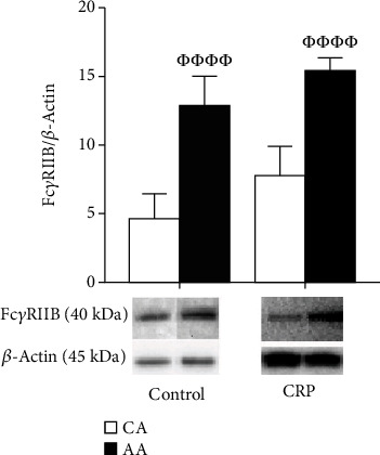
Racial difference in FcγRIIB receptor protein expression. HUVECs from CA and AA were incubated with CRP (10 μg/mL) for 24 hr (mean ± SE). ΦΦΦΦp < 0.001, compared to CA.
Figure 2.
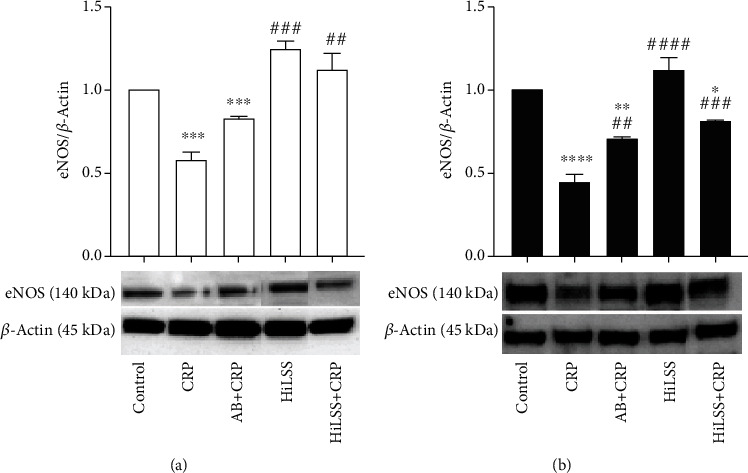
eNOS protein expression under different conditions. Effects of HiLSS application and CRP incubation were evaluated in (a) CA HUVECs and (b) AA HUVECs (mean ± SE). ∗p < 0.05, ∗∗p < 0.01, ∗∗∗p < 0.005, ∗∗∗∗p < 0.001, compared to control. ## p < 0.01, ### p < 0.005, #### p < 0.001, compared to CRP.
Figure 3.
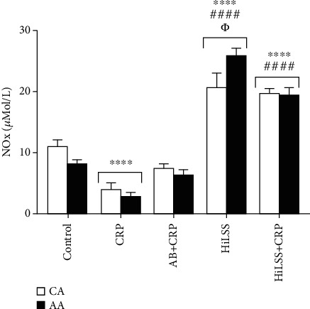
Nitrate/nitrite (NOx) levels under different conditions in CA and AA HUVECs (mean ± SE). ∗∗∗∗p < 0.001, compared to control. #### p < 0.001, compared to CRP. Φp < 0.05, between racial groups.
HiLSS significantly mitigated CRP-induced attenuation of eNOS expression in both racial groups (Figure 4, p < 0.001). Within each racial group, applying HiLSS prior to CRP incubation resulted in the greatest attenuation of CRP induced effects on eNOS (Figure 2, CA: p < 0.01, AA: p < 0.005). Moreover, a robust downregulation in the expression of FcγRIIB receptor was observed following HiLSS (Figure 5, p < 0.001). Exposing HUVECs to HiLSS prior to CRP incubation attenuated CRP-induced upregulation of FcγRIIB receptor (Figure 5, p < 0.001). Furthermore, only HiLSS condition resulted in higher production in NOx in the culture media in AA compared to CA (Figure 3, p < 0.05).
Figure 4.
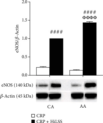
The effect of HiLSS on eNOS protein expression after CRP preincubation. HUVECs from CA and AA were preincubated with CRP (10 μg/mL) for 24 hr followed by static control or HiLSS (20 dyne/cm2) for another 24 hr (mean ± SE). #### p < 0.001, compared to CRP. ΦΦΦΦp < 0.001, compared to CA.
Figure 5.
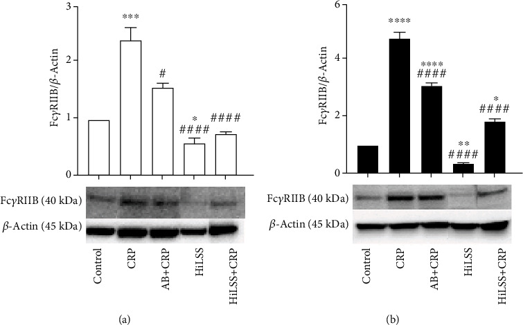
FcγRIIB receptor protein expression under different conditions. Effects of HiLSS application and CRP incubation were evaluated in (a) CA HUVECs and (b) AA HUVECs (mean ± SE). ∗p < 0.05, ∗∗p < 0.01, ∗∗∗p < 0.005, ∗∗∗∗p < 0.001, compared to control. # p < 0.05, ## p < 0.01, #### p < 0.001, compared to CRP.
4. Discussion
The key findings of this study suggest that HUVECs obtained from AA and CA donors respond differently to CRP, with AA HUVECs exhibiting higher FcγRIIB receptor expression under basal and CRP conditions, and being more responsive to HiLSS than CA. HiLSS reversed the detrimental effects of CRP on eNOS in ECs. Furthermore, FcγRIIB receptor mediates the CRP-induced detrimental effects on endothelial function. To our knowledge, this is the first study to evaluate racial differences in CRP-induced effects in primary ECs and the effect of HiLSS.
Numerous epidemiological studies provide solid evidence that serum CRP levels are higher in AA compared to CA with similar CVD risks [7, 36, 37]. The inverse association of CRP and endothelial function was first shown when acetylcholine-induced brachial artery dilation was reduced in patients with elevated levels of serum CRP [38, 39] suggesting that CRP might affect vasodilatory capacity, potentially by affecting NO bioavailability. NO is one of the most important vasodilators released by the endothelium, and reduced bioavailability is a hallmark of endothelial dysfunction in AA in vitro [40]. The importance of NO is not limited to vasodilation properties as it inhibits inflammation, platelet aggregation, and smooth muscle cell proliferation [41–43].
Inflammation largely contributes to endothelial dysfunction and reduces NO bioavailability in healthy people [44]. In vivo, AA have higher levels of inflammation, specifically CRP, than their CA peers in healthy [11] and diseased cohorts. AA diabetic patients with microalbuminuria, a population associated with higher levels of CRP, showed a blunted response to vasodilators due to decreased levels of NO production [38]. In vitro studies have shown that HUVECs obtained from AA exhibit higher oxidative stress levels, higher inflammation and adhesion molecule expression, and a greater degree of fibrinolytic potential compared to CA [40, 45, 46]. There is a reduction in NO bioavailability accompanied by an increased production of superoxide (O2−) and peroxynitrite (ONOO−) in AA HUVECs. If these results translate to humans, they may contribute to the higher prevalence and severity of endothelial dysfunction and hypertension in AA [40]. In addition, our group has shown that AA HUVECs produce more endothelial microparticles (an emerging biomarker of endothelial dysfunction) compared to CA HUVECs which correlates with inflammation, in vivo and in vitro [47]. In our study, AA HUVECs exhibited significantly greater expression of FcγRIIB receptor. Inferring this to humans, CRP-induced effect may converge with other mechanisms, such as oxidative stress, to promote endothelial dysfunction and vascular diseases in AA.
It has been reported that CRP binding to Fcγ receptors, FcγRI, and FcγRIIB on ECs decreases eNOS and prostacyclin and increases IL-8 and adhesion molecules [48]. Blocking FcγRI and FcγRIIB by antibodies or transfection with small interference RNA effectively attenuated NF-κB activity and inhibited VCAM-1 and ICAM-1 upregulation [49]. Our investigation shows that blocking FcγRIIB inhibited the CRP-induced suppression of eNOS protein expression in AA HUVECs. Similar effects were found in other studies by attenuating the decrease in eNOS expression and the increase in IL-8 after CRP incubation [14, 15]. Thus, Fcγ receptors serve as a mediators of CRP-induced biological effects in HUVECs.
It is undeniable that social determinants contribute to racial disparities in HT and CVD. However, it is imperative to determine biological mediators of racial disparity in CVD. The current study provides preliminary evidence that higher levels of FcγRIIB receptor expression contributes to, at least partially, the greater CRP-induced biological effects in AA HUVECs. The novelty of our study is that the synergistic effects of increased CRP receptors and greater response to CRP may explain the racial differences in endothelial function. This may be particularly relevant for clinical studies involving chronic inflammation or hypertension in AA cohorts. In addition, the pre-IgG incubation eliminated, to an extent, the CRP-induced effects. Despite the functional similarities between CRP and IgG, our data suggest that the CRP detrimental effect on eNOS is only partially mediated by the Fcγ receptor pathways.
An important role of the endothelium in the vasculature is sensing blood flow to regulate vascular function. HiLSS induces an atheroprotective phenotype by altering EC gene expression profile and initiating endothelial quiescence [50]. DNA microarray data have shown that HiLSS (12 dyne/cm2, 24 hours) downregulates genes related to inflammation and proliferation in ECs [50]. On the other hand, low LSS (< 5 dyne/cm2) displays an atherogenic phenotype [28]. Recently, it has been shown that AA HUVECs respond differently to HiLSS, evidenced by a significantly larger reduction in NADPH oxidase subunit expression and a greater production of antioxidants [25].
Our results support the hypothesis that HiLSS can attenuate the CRP-induced proatherogenic effects and eliminate the racial differences in suppressed eNOS expression in HUVECs response to CRP. AA ECs may be more responsive to HiLSS, as evidenced by higher NOx production. In addition, the application of HiLSS prior to CRP incubation alleviated eNOS expression for both racial groups. However, compared to CA, AA HUVECs exhibited a greater recovery of eNOS. Based on our findings and others, translating this to human, it appears that the endothelium, in terms of endothelial dysfunction, AA may benefit more from high levels of blood flow generated by aerobic exercise compared to CA. Potential mechanisms remain to be investigated.
Our data also suggests that HiLSS maintains FcγRIIB receptor expression in HUVECs at lower levels compared to the static condition. The inhibitory effect of HiLSS on FcγRIIB receptor expression and function may contribute to the antiatherogenic effect of laminar flow. This may help regulate endothelial function, cell survival, and integrity under inflammatory conditions to protect the endothelium from moving toward a proatherogenic status.
4.1. Limitations
Although our observations are novel, the results and conclusion drawn from our study should be interpreted with caution for several reasons. Long-term influences of risk factors, social determinants of CVD, and other physiological changes cannot be applied in this cell model. This highly controlled in vitro model does not prove a causal relationship between higher expression of CRP receptors and CVD prevalence in AA. However, this highly controlled in vitro model does eliminate the uncontrolled physiological confounding variables found in human subjects. Based on the outcomes of this study, it is reasonable to suggest that greater CRP receptor expression is a potential mechanism that may explain racial differences. When compared to human studies, the sample size of our experiments is smaller, but cell culture studies inherently have less random error because it eliminates complex physiology and other confounding variables that allow for the smaller sample size to be analyzed. Previously in our lab, a sample size that has produced positive results entailed an N = 4 donors for each group and thus deemed appropriate to follow suit for this study [25, 47, 51, 52]. Future studies should examine Fcγ receptor expression in ECs isolated from human vessels where CVD is evident. Second, although we used HUVECs in our study, there are different EC types throughout the human body where Fcγ receptor expression and activities might differ between racial groups. Lastly, while the majority of CRP is produced systemically by the liver, there is data showing that human aortic ECs (HAECs) synthesize and secrete CRP locally [53]. However, the amount of CRP produced by ECs is relatively much less than that from the liver. We are unaware of any reports of this in HUVECs, nor did we measure CRP mRNA/protein in our experiments.
5. Conclusion
A better understanding of the racial differences in endothelial function will help us improve hypertension prevention and treatment strategies. Our results highlight FcγRIIB receptor as a novel therapeutic target, particularly in AA. Future studies are needed to investigate the variance of the FcγRIIB expression in other types of ECs. Additionally, there is a need for translational research to elucidate the role of different Fcγ receptors in AA and CA with elevated CRP levels. Uncovering the clinical consequences of Fcγ receptors on the racial differences in endothelial function could result in a more individualized prevention/treatment approach.
Acknowledgments
This study was funded by an American College of Sports Medicine (ACSM) Doctoral Student Grant (Chenyi Ling), internal funds of the Vascular Health Laboratory (Dr. Michael D. Brown, PI), and NIH/NHLBI Grant RO1 HL085497 (PI: Michael D. Brown, PhD).
Data Availability
The data used to support the findings of this study are available from the corresponding author upon request, after publication.
Consent
All authors have given consent for publication of this work.
Disclosure
The findings from this project are not being considered for publication by another journal at this time.
Conflicts of Interest
The authors declare that there is no conflict of interest.
Authors' Contributions
Chenyi Ling and Michael D. Brown designed the experiments. Chenyi Ling, Heather Grimm, and Mark D. Cook coordinated and performed the experiments. Chenyi Ling, Heather Grimm, Mark D. Cook, and Michael D. Brown analyzed and interpreted the data. Chenyi Ling, Maitha Aldokhayyil, and Dulce Gomez were involved in manuscript writing and editing. All authors reviewed and revised the final manuscript. Study finding and supervision were carried out by Michael D. Brown.
References
- 1.Verma S., Anderson T. J. Fundamentals of endothelial function for the clinical cardiologist. Circulation . 2002;105(5):546–549. doi: 10.1161/hc0502.104540. [DOI] [PubMed] [Google Scholar]
- 2.Cai H., Harrison D. G. Endothelial dysfunction in cardiovascular diseases: the role of oxidant stress. Circulation Research . 2000;87(10):840–844. doi: 10.1161/01.RES.87.10.840. [DOI] [PubMed] [Google Scholar]
- 3.Calles-Escandon J., Cipolla M. Diabetes and endothelial dysfunction: a clinical perspective. Endocrine Reviews . 2001;22(1):36–52. doi: 10.1210/edrv.22.1.0417. [DOI] [PubMed] [Google Scholar]
- 4.Scirica B. M., Morrow D. A., Jialal I. Is C-reactive protein an innocent bystander or proatherogenic culprit? The verdict is still out. Circulation . 2006;113(17):2128–2151. doi: 10.1161/CIRCULATIONAHA.105.611350. [DOI] [PubMed] [Google Scholar]
- 5.Flack J. M., Ferdinand K. C., Nasser S. A. Epidemiology of hypertension and cardiovascular disease in African Americans. Journal of Clinical Hypertension . 2003;5(1):5–11. doi: 10.1111/j.1524-6175.2003.02152.x. [DOI] [PMC free article] [PubMed] [Google Scholar]
- 6.WRITING GROUP MEMBERS, Lloyd-Jones D., Adams R. J., et al. Heart disease and stroke statistics—2010 update. Circulation . 2010;121(7):e46–e215. doi: 10.1161/CIRCULATIONAHA.109.192667. [DOI] [PubMed] [Google Scholar]
- 7.Khera A., McGuire D. K., Murphy S. A., et al. Race and gender differences in C-reactive protein levels. Journal of the American College of Cardiology . 2005;46(3):464–469. doi: 10.1016/j.jacc.2005.04.051. [DOI] [PubMed] [Google Scholar]
- 8.Venugopal S. K., Devaraj S., Yuhanna I., Shaul P., Jialal I. Demonstration that C-reactive protein decreases eNOS expression and bioactivity in human aortic endothelial cells. Circulation . 2002;106(12):1439–1441. doi: 10.1161/01.CIR.0000033116.22237.F9. [DOI] [PubMed] [Google Scholar]
- 9.Hein T. W., Singh U., Vasquez-Vivar J., Devaraj S., Kuo L., Jialal I. Human C-reactive protein induces endothelial dysfunction and uncoupling of eNOS in vivo. Atherosclerosis . 2009;206(1):61–68. doi: 10.1016/j.atherosclerosis.2009.02.002. [DOI] [PMC free article] [PubMed] [Google Scholar]
- 10.Michell B. J., Chen Z. P., Tiganis T., et al. Coordinated Control of Endothelial Nitric-oxide Synthase Phosphorylation by Protein Kinase C and the cAMP-dependent Protein Kinase. The Journal of Biological Chemistry . 2001;276(21):17625–17628. doi: 10.1074/jbc.C100122200. [DOI] [PubMed] [Google Scholar]
- 11.Sharma D., DasMahapatra P., Fernandez C., et al. The association of c-reactive protein with arterial compliance in asymptomatic young adults: the Bogalusa heart study. Journal of Human Hypertension . 2013;27(4):256–260. doi: 10.1038/jhh.2012.34. [DOI] [PubMed] [Google Scholar]
- 12.Xing D., Hage F. G., Chen Y. F., et al. Exaggerated neointima formation in human C-reactive protein transgenic mice is IgG Fc receptor type I (FcγRI)-dependent. The American journal of pathology . 2008;172(1):22–30. doi: 10.2353/ajpath.2008.070154. [DOI] [PMC free article] [PubMed] [Google Scholar]
- 13.Yuasa T., Kubo S., Yoshino T., et al. Deletion of Fcγ receptor IIB renders H-2b mice susceptible to collagen-induced arthritis. The Journal of experimental medicine . 1999;189(1):187–194. doi: 10.1084/jem.189.1.187. [DOI] [PMC free article] [PubMed] [Google Scholar]
- 14.Devaraj S., du Clos T. W., Jialal I. Binding and internalization of C-reactive protein by Fcgamma receptors on human aortic endothelial cells mediates biological effects. Arteriosclerosis, thrombosis, and vascular biology . 2005;25(7):1359–1363. doi: 10.1161/01.ATV.0000168573.10844.ae. [DOI] [PubMed] [Google Scholar]
- 15.Tanigaki K., Mineo C., Yuhanna I. S., et al. C-reactive protein inhibits insulin activation of endothelial nitric oxide synthase via the immunoreceptor tyrosine-based inhibition motif of FcγRIIB and SHIP-1. Circulation Research . 2009;104(11):1275–1282. doi: 10.1161/CIRCRESAHA.108.192906. [DOI] [PMC free article] [PubMed] [Google Scholar]
- 16.Tanigaki K., Sundgren N., Khera A., Vongpatanasin W., Mineo C., Shaul P. W. Fcγ receptors and ligands and cardiovascular disease. Circulation Research . 2015;116(2):368–384. doi: 10.1161/CIRCRESAHA.116.302795. [DOI] [PMC free article] [PubMed] [Google Scholar]
- 17.LaBarbera M. Principles of design of fluid transport systems in zoology. Science . 1990;249(4972):992–1000. doi: 10.1126/science.2396104. [DOI] [PubMed] [Google Scholar]
- 18.Langille B., O'Donnell F. Reductions in arterial diameter produced by chronic decreases in blood flow are endothelium-dependent. Science . 1986;231(4736):405–407. doi: 10.1126/science.3941904. [DOI] [PubMed] [Google Scholar]
- 19.Zarins C. K., Zatina M. A., Giddens D. P., Ku D. N., Glagov S. Shear stress regulation of artery lumen diameter in experimental atherogenesis. Journal of Vascular Surgery . 1987;5(3):413–420. doi: 10.1016/0741-5214(87)90048-6. [DOI] [PubMed] [Google Scholar]
- 20.Epstein F. H., Gibbons G. H., Dzau V. J. The emerging concept of vascular remodeling. New England Journal of Medicine . 1994;330(20):1431–1438. doi: 10.1056/NEJM199405193302008. [DOI] [PubMed] [Google Scholar]
- 21.Woodman C. R., Price E. M., Laughlin M. H. Shear stress induces eNOS mRNA expression and improves endothelium-dependent dilation in senescent soleus muscle feed arteries. Journal of Applied Physiology . 2005;98(3):940–946. doi: 10.1152/japplphysiol.00408.2004. [DOI] [PubMed] [Google Scholar]
- 22.Cheng C. P., Herfkens R. J., Taylor C. A. Comparison of abdominal aortic hemodynamics between men and women at rest and during lower limb exercise. Journal of Vascular Surgery . 2003;37(1):118–123. doi: 10.1067/mva.2002.107. [DOI] [PubMed] [Google Scholar]
- 23.Wasserman S. M., Mehraban F., Komuves L. G., et al. Gene expression profile of human endothelial cells exposed to sustained fluid shear stress. Physiological Genomics . 2002;12(1):13–23. doi: 10.1152/physiolgenomics.00102.2002. [DOI] [PubMed] [Google Scholar]
- 24.Wasserman S. M., Topper J. N. Adaptation of the endothelium to fluid flow: in vitro analyses of gene expression and in vivo implications. Vascular Medicine . 2004;9(1):35–45. doi: 10.1191/1358863x04vm521ra. [DOI] [PubMed] [Google Scholar]
- 25.Feairheller D., Park R., Rizzo V., Kim B., Brown M. D. Racial differences in the responses to shear stress in human umbilical vein endothelial cells. Vascular health and risk management . 2011;7:425–431. doi: 10.2147/VHRM.S22435. [DOI] [PMC free article] [PubMed] [Google Scholar]
- 26.Go Y.-M., Park H., Maland M. C., Jo H. [52] In vitro system to study role of blood flow on nitric oxide production and cell signaling in endothelial cells. Methods in Enzymology . 1999;301:513–522. doi: 10.1016/S0076-6879(99)01114-3. [DOI] [PubMed] [Google Scholar]
- 27.Park J.-Y., Farrance I. K. G., Fenty N. M., et al. NFKB1 promoter variation implicates shear-induced NOS3 gene expression and endothelial function in prehypertensives and stage I hypertensives. American Journal of Physiology-Heart and Circulatory Physiology . 2007;293(4):H2320–H2327. doi: 10.1152/ajpheart.00186.2007. [DOI] [PMC free article] [PubMed] [Google Scholar]
- 28.Malek A. M., Alper S. L., Izumo S. Hemodynamic shear stress and its role in atherosclerosis. JAMA . 1999;282(21):2035–2042. doi: 10.1001/jama.282.21.2035. [DOI] [PubMed] [Google Scholar]
- 29.Krzywanski D. M., Moellering D. R., Westbrook D. G., et al. Endothelial cell bioenergetics and mitochondrial DNA damage differ in humans having African or West Eurasian maternal ancestry. Circulation Genomic and precision medicine . 2016;9(1):26–36. doi: 10.1161/CIRCGENETICS.115.001308. [DOI] [PMC free article] [PubMed] [Google Scholar]
- 30.Sorescu G. P., Sykes M., Weiss D., et al. Bone Morphogenic Protein 4 Produced in Endothelial Cells by Oscillatory Shear Stress Stimulates an Inflammatory Response. The Journal of Biological Chemistry . 2003;278(33):31128–31135. doi: 10.1074/jbc.M300703200. [DOI] [PubMed] [Google Scholar]
- 31.Thoumine O., Nerem R. M., Girard F. R. Oscillatory shear stress and hydrostatic pressure modulate cell-matrix attachment proteins in cultured endothelial cells. In Vitro Cellular & Developmental Biology-Animal . 1995;31(1):45–54. doi: 10.1007/BF02631337. [DOI] [PubMed] [Google Scholar]
- 32.Thoumine O., Ziegler T., Girard P. R., Nerem R. M. Elongation of confluent endothelial cells in culture: the importance of fields of force in the associated alterations of their cytoskeletal structure. Experimental Cell Research . 1995;219(2):427–441. doi: 10.1006/excr.1995.1249. [DOI] [PubMed] [Google Scholar]
- 33.Ridker P. M. Clinical application of C-reactive protein for cardiovascular disease detection and prevention. Circulation . 2003;107(3):363–369. doi: 10.1161/01.CIR.0000053730.47739.3C. [DOI] [PubMed] [Google Scholar]
- 34.Dimmeler S., Haendeler J., Nehls M., Zeiher A. M. Suppression of apoptosis by nitric oxide via inhibition of interleukin-1β–converting enzyme (ICE)-like and cysteine protease protein (CPP)-32–like proteases. The Journal of experimental medicine . 1997;185(4):601–608. doi: 10.1084/jem.185.4.601. [DOI] [PMC free article] [PubMed] [Google Scholar]
- 35.Bryan N. S., Grisham M. B. Methods to detect nitric oxide and its metabolites in biological samples. Free radical biology and medicine . 2007;43(5):645–657. doi: 10.1016/j.freeradbiomed.2007.04.026. [DOI] [PMC free article] [PubMed] [Google Scholar]
- 36.Wee C. C., Mukamal K. J., Huang A., Davis R. B., McCarthy E. P., Mittleman M. A. Obesity and C-reactive protein levels among white, black, and Hispanic US adults. Obesity . 2008;16(4):875–880. doi: 10.1038/oby.2008.7. [DOI] [PMC free article] [PubMed] [Google Scholar]
- 37.Fox E. R., Benjamin E. J., Sarpong D. F., et al. Epidemiology, heritability, and genetic linkage of C-reactive protein in African Americans (from the Jackson Heart Study) The American Journal of Cardiology . 2008;102(7):835–841. doi: 10.1016/j.amjcard.2008.05.049. [DOI] [PMC free article] [PubMed] [Google Scholar]
- 38.Jawa A., Nachimuthu S., Pendergrass M., Asnani S., Fonseca V. Impaired vascular reactivity in African-American patients with type 2 diabetes mellitus and microalbuminuria or proteinuria despite angiotensin-converting enzyme inhibitor therapy. The Journal of Clinical Endocrinology & Metabolism . 2006;91(1):31–35. doi: 10.1210/jc.2005-1632. [DOI] [PubMed] [Google Scholar]
- 39.Davignon J., Ganz P. Role of endothelial dysfunction in atherosclerosis. Circulation . 2004;109(23_suppl_1):p. III-27-III–32. doi: 10.1161/01.CIR.0000131515.03336.f8. [DOI] [PubMed] [Google Scholar]
- 40.Kalinowski L., Dobrucki I. T., Malinski T. Race-specific differences in endothelial function. Circulation . 2004;109(21):2511–2517. doi: 10.1161/01.CIR.0000129087.81352.7A. [DOI] [PubMed] [Google Scholar]
- 41.Janssens S., Flaherty D., Nong Z., et al. Human endothelial nitric oxide synthase gene transfer inhibits vascular smooth muscle cell proliferation and neointima formation after balloon injury in rats. Circulation . 1998;97(13):1274–1281. doi: 10.1161/01.CIR.97.13.1274. [DOI] [PubMed] [Google Scholar]
- 42.Coleman J. W. Nitric oxide in immunity and inflammation. International Immunopharmacology . 2001;1(8):1397–1406. doi: 10.1016/S1567-5769(01)00086-8. [DOI] [PubMed] [Google Scholar]
- 43.Radomski M. W., Palmer R. M. J., Moncada S. Endogenous nitric oxide inhibits human platelet adhesion to vascular endothelium. Lancet . 1987;330(8567):1057–1058. doi: 10.1016/S0140-6736(87)91481-4. [DOI] [PubMed] [Google Scholar]
- 44.Clapp B., Hingorani A., Kharbanda R., et al. Inflammation-induced endothelial dysfunction involves reduced nitric oxide bioavailability and increased oxidant stress. Cardiovascular Research . 2004;64(1):172–178. doi: 10.1016/j.cardiores.2004.06.020. [DOI] [PubMed] [Google Scholar]
- 45.Feairheller D. L., Park J. Y., Sturgeon K. M., et al. Racial differences in oxidative stress and inflammation: in vitro and in vivo. Clinical and Translational Science . 2011;4(1):32–37. doi: 10.1111/j.1752-8062.2011.00264.x. [DOI] [PMC free article] [PubMed] [Google Scholar]
- 46.Frist S. T., Taylor H. A., Jr., Kirk K. A., et al. Expression of PAI-1, t-PA and u-PA in cultured human umbilical vein endothelial cells derived from racial groups. Thrombosis Research . 1995;77(3):279–290. doi: 10.1016/0049-3848(95)91615-R. [DOI] [PubMed] [Google Scholar]
- 47.Brown M. D., Feairheller D. L., Thakkar S., Veerabhadrappa P., Park J.-Y. Racial differences in tumor necrosis factor-α-induced endothelial microparticles and interleukin-6 production. Vascular health and risk management . 2011;7:541–550. doi: 10.2147/VHRM.S22930. [DOI] [PMC free article] [PubMed] [Google Scholar]
- 48.Jialal I., Devaraj S., Venugopal S. K. C-reactive protein: risk marker or mediator in atherothrombosis? Hypertension . 2004;44(1):6–11. doi: 10.1161/01.HYP.0000130484.20501.df. [DOI] [PubMed] [Google Scholar]
- 49.Devaraj S., Davis B., Simon S. I., Jialal I. CRP promotes monocyte-endothelial cell adhesion via Fcγ receptors in human aortic endothelial cells under static and shear flow conditions. American Journal of Physiology-Heart and Circulatory Physiology . 2006;291(3):H1170–H1176. doi: 10.1152/ajpheart.00150.2006. [DOI] [PubMed] [Google Scholar]
- 50.Okita K., Nishijima H., Murakami T., et al. Can exercise training with weight loss lower serum C-reactive protein levels? Arteriosclerosis, thrombosis, and vascular biology . 2004;24(10):1868–1873. doi: 10.1161/01.ATV.0000140199.14930.32. [DOI] [PubMed] [Google Scholar]
- 51.Feairheller D. L., Diaz K. M., Sturgeon K. M., Williamson S. T., Brown M. D. Racial differences in the time-course oxidative stress responses to acute exercise. Journal of exercise physiology online/American Society of Exercise Physiologists . 2011;14:49–59. [PMC free article] [PubMed] [Google Scholar]
- 52.Grimm H., Kretzschmar J., Cook M. D., Brown M. D. The effects of exercise, aspirin, and celecoxib in an atherogenic environment. Medicine and science in sports and exercise . 2018;50(10):2033–2039. doi: 10.1249/MSS.0000000000001657. [DOI] [PubMed] [Google Scholar]
- 53.Venugopal S. K., Devaraj S., Jialal I. Macrophage Conditioned Medium Induces the Expression of C-Reactive Protein in Human Aortic Endothelial Cells: Potential for Paracrine/Autocrine Effects. The American journal of pathology . 2005;166(4):1265–1271. doi: 10.1016/S0002-9440(10)62345-0. [DOI] [PMC free article] [PubMed] [Google Scholar]
Associated Data
This section collects any data citations, data availability statements, or supplementary materials included in this article.
Data Availability Statement
The data used to support the findings of this study are available from the corresponding author upon request, after publication.


