

CONFLICT OF INTEREST
None.
ETHICAL APPROVAL
Patient consent was obtained for reporting and publication.
Sir,
Coronavirus disease‐19 (COVID‐19) caused by severe acute respiratory syndrome coronavirus 2 (SARS‐CoV‐2) remains a global threat. Vaccination is one of the most effective interventions to overcome it. 1 Various immune‐mediated diseases (IMD) flares or new disease onset after SARS‐CoV2‐vaccination have been reported. 2 We report a case of systemic lupus erythematosus (SLE) following COVID‐19 vaccination with Covishield in a 22‐year‐old female who presented with pain in right knee while climbing up and down the stairs after 2 weeks of receiving first dose of vaccine. She was thought to have chondromalacia and treated with physiotherapy and analgesics for 3 weeks which relieved symptoms up to 80%.
While on this treatment, she received second dose of vaccine, about 2 months after the first dose. She then developed on and off fever for 15 days (99–100°F). After ten days, she developed polyarthralgia (small as well as large joints), bipedal edema, cutaneous rash over fingertips, and petechiae over lower limb (Figure 1).
FIGURE 1.
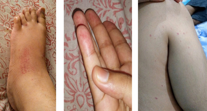
Pedal edema, cutaneous rash, and petechiae
Patient has history of infective jaundice (non‐B) at the age of 9 years. She received HPV vaccine at the age of 17 years. Her vaccination is completed up to date without any adverse reaction. Her younger sister is on levothyroxine (50 µg daily) for autoimmune thyroiditis. History of allergic rhinitis and asthma is present in paternal grandmother. There is no other significant genetic, autoimmune, medical, or surgical history in the family. Patient has no history of any addictions and no known drug allergy.
On general examination, left cervical lymph node and mild liver enlargement were observed. Neck and abdominal ultrasound examination revealed bilateral cervical lymphadenopathy, (Level 1) and mild hepatomegaly, respectively. Serial investigations (Table 1) revealed diagnosis of SLE. Anti‐nuclear antibody (ANA) immunoblot test showed positive antigens for dsDNA, nucleosomes, histones, and AMA m2. Anti‐nuclear antibodies by indirect immunofluorescence (ANA‐IIF) were strongly positive (titer 1:320). Immunoglobulin values were raised (total serum IgG 28 g/L, total serum IgM 5 g/L, total serum IgA 5.5 g/L). Total serum IgE was 119.9 IU/ml. Hemogram showed hemoglobin (Hb) 9.3 g/dL, hematocrit (PCV) 26.8%, RBC count 3.67 million/mm3, MCV 73.2 Fl, MCH 25.5 pg, MCHC 34.9%, platelet count 134 × 103/UL, total WBC count (TLC) 4640/mm3, neutrophils 60%, lymphocytes 32.9%, erythrocyte sedimentation rate (ESR) 92 mm/h, CRP 2.8 mg/L. Direct Coombs test (DCT) was weakly positive. D‐Dimer, serum ferritin, aPTT, 24‐h urinary protein, and blood urea nitrogen levels were 5.1 μg/ml, 173.37 μg/L, 45.5 s, 300 mg/24 h, and 22.5 mg/dl, respectively. Urine routine showed 1+ albuminuria and 3‐4 RBC per high power field. Chest X‐ray was normal.
TABLE 1.
Laboratory values at first visit and after 1 month of follow‐up
| Laboratory investigations | First visit | Follow‐up (1 month later) |
|---|---|---|
| Hemoglobin (g/dl) | 9.3 | 10 |
| Platelet count (×103/UL) | 134 | 194 |
| Total leucocyte count (per cm) | 4640 | 13,500 |
| Lymphocytes (%) | 32.9 | 26 |
| Neutrophils (%) | 60 | 70 |
| Serum creatinine(mg/dl) | 1.2 | 0.67 |
| Serum urea (mg/dl) | 22.5 | 24 |
| LDH (units/L) | 454 | 185 |
| Urine albumin | 1+ | Trace |
| D‐Dimer (µg/ml) | 5.1 | 0.53 |
| ESR (mm/h) | 92 | 63 |
| aPTT (s) | 45.5 | 35 |
| Spot albumin creatinine ratio (mg/mmol) | – | 34.9 |
| Direct coombs test (DCT) | Weakly positive | – |
She was diagnosed to have SLE with anemia of chronic diseases and started on prednisolone (50 mg daily), hydroxychloroquine (400 mg daily), mycophenolate mofetil (2 g daily), furosemide (20 mg daily), telmisartan (20 mg daily), folic acid, calcium, and vitamin D3. Fundoscopy was performed before initiating hydroxychloroquine. Follow‐up after a month showed significant improvement. Pedal edema, petechiae, and rash subsided. Based on the response, steroid dose was tapered.
Different types of vaccines have been developed for the prevention of COVID‐19. 3 Adverse reactions after different types of vaccines may occur due to interactions between susceptibility of the host and some of the vaccine components. Molecular mimicry is one of the implicated mechanisms for such reactions. 2
SLE, a chronic multisystemic autoimmune disease more common in females, is associated with autoantibodies (eg, ANA, antidsDNA, etc.) against different autoantigens forming immune complexes. Inadequate removal of these complexes from the host triggers inflammatory response which causes tissue damage. Vaccines against COVID have led to flares of IMD like pericarditis, neuropathy, sarcoidosis, and myasthenia gravis. 4 SLE has been reported after SARS‐CoV‐2 infection, 5 but there are no published data on the onset of SLE after receiving vaccine against COVID‐19.
Patil S, Patil A. Systemic lupus erythematosus after COVID‐19 vaccination: A case report. J Cosmet Dermatol. 2021;20:3104–3105. 10.1111/jocd.14386
DATA AVAILABILITY STATEMENT
The data that support the findings of this study are available from the corresponding author upon reasonable request.
References
- 1. Velikova T, Georgiev T. SARS‐CoV‐2 vaccines and autoimmune diseases amidst the COVID‐19 crisis. Rheumatol Int. 2021;41:509‐518. [DOI] [PMC free article] [PubMed] [Google Scholar]
- 2. Segal Y, Shoenfeld Y. Vaccine‐induced autoimmunity: the role of molecular mimicry and immune crossreaction. Cell Mol Immunol. 2018;15:586‐594. [DOI] [PMC free article] [PubMed] [Google Scholar]
- 3. Wang J, Peng Y, Xu H, Cui Z, Williams RO III. The COVID‐19 vaccine race: challenges and opportunities in vaccine formulation. AAPS PharmSciTech. 2020;21:225. [DOI] [PMC free article] [PubMed] [Google Scholar]
- 4. Watad A, De Marco G, Mahajna H, et al. Immune‐mediated disease flares or new‐onset disease in 27 subjects following mRNA/DNA SARS‐CoV‐2 vaccination. Vaccines (Basel). 2021;29(9):435. [DOI] [PMC free article] [PubMed] [Google Scholar]
- 5. Zamani B, Masoud S, Taba M, Shayestehpour M. Systemic lupus erythematosus manifestation following COVID‐19: a case report. J Med Case Rep. 2021;15:29. [DOI] [PMC free article] [PubMed] [Google Scholar]
Associated Data
This section collects any data citations, data availability statements, or supplementary materials included in this article.
Data Availability Statement
The data that support the findings of this study are available from the corresponding author upon reasonable request.