In response to a routine request for the resupply of figures during the original peer review process, incorrect images were unintentionally submitted and published in the July 2019 article by Upparahalli Venkateshaiah and colleagues (1). The correct versions of Figure 2A and Figure 4A are included here. The authors would like to apologize to the readership for the error.
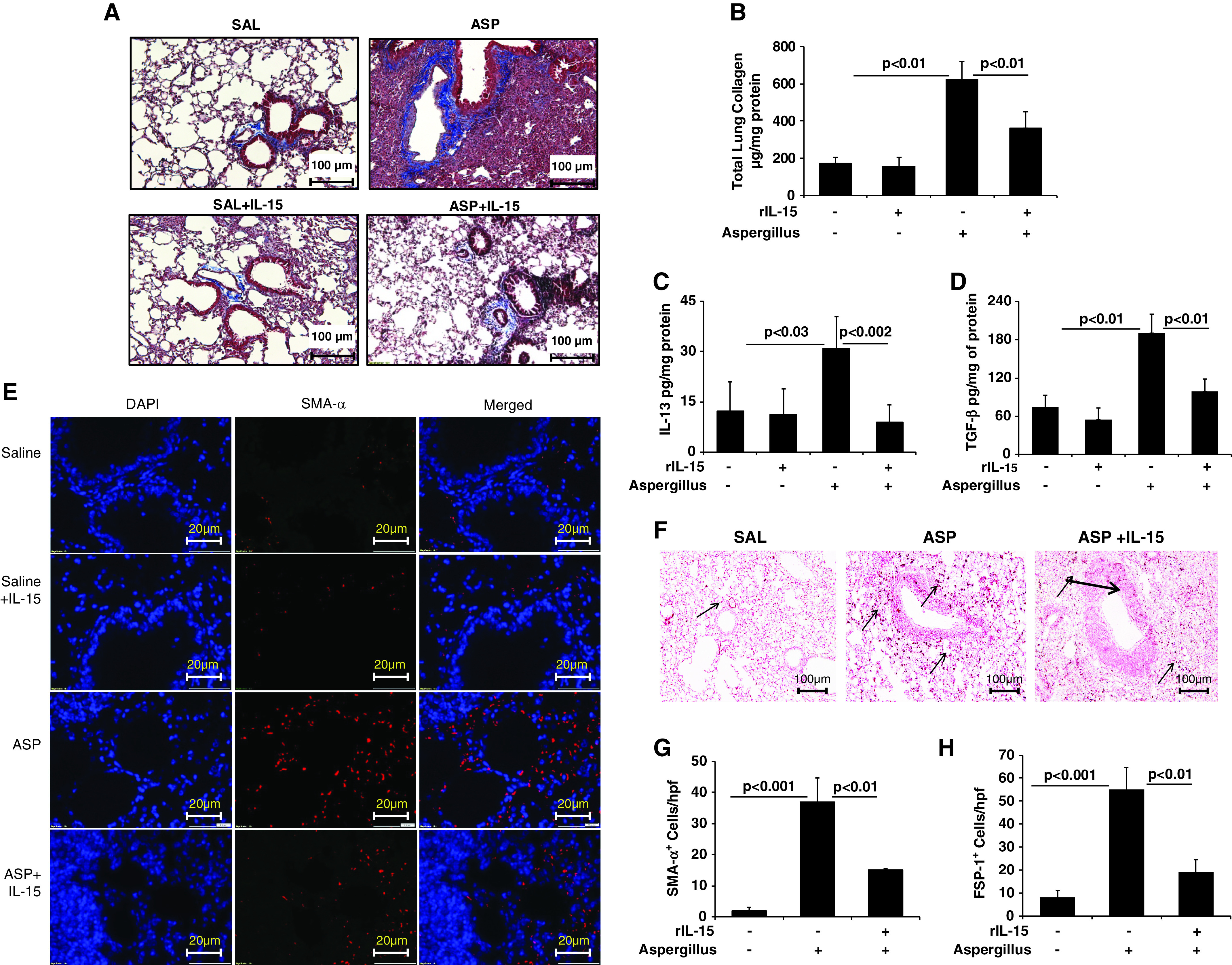
Figure 2.

Consequences of rIL-15 pretreatment in Aspergillus-induced bronchial fibrosis in mice. (A) Representative light-microscopy photomicrographs of lung sections with Masson’s trichrome staining show perivascular and peribronchiolar collagen accumulation after 3 weeks of saline or Aspergillus challenge in mice treated with and without rIL-15. (B) Total collagen expression in saline- and rIL-15–treated, Aspergillus-challenged mice. ELISA results for profibrotic IL-13 and TGF-β1 expression in saline- and rIL-15–treated, Aspergillus-challenged mice are shown. (C and D) Immunofluorescence staining revealed α-SMA+ cells in Aspergillus- and rIL-15–treated, Aspergillus-challenged mice. (E) Very few eosinophils are seen in the saline-treated (for 3 wk) mice. (F) Arrows indicated FSP1+ cell expression in Aspergillus and rIL-15–treated Aspergillus-challenged mice. Quantitation of α-SMA+ and FSP1+ cells is shown in saline-, (G and H) Aspergillus-, and rIL-15–treated Aspergillus-challenged mice. Data are expressed as mean ± SD, n = 12 mice/group. Scale bars: 100 μm and 20 μm. ASP = Aspergillus; FSP1 = fibroblast-specific protein 1; rIL-15 = recombinant IL-15; SAL = saline.
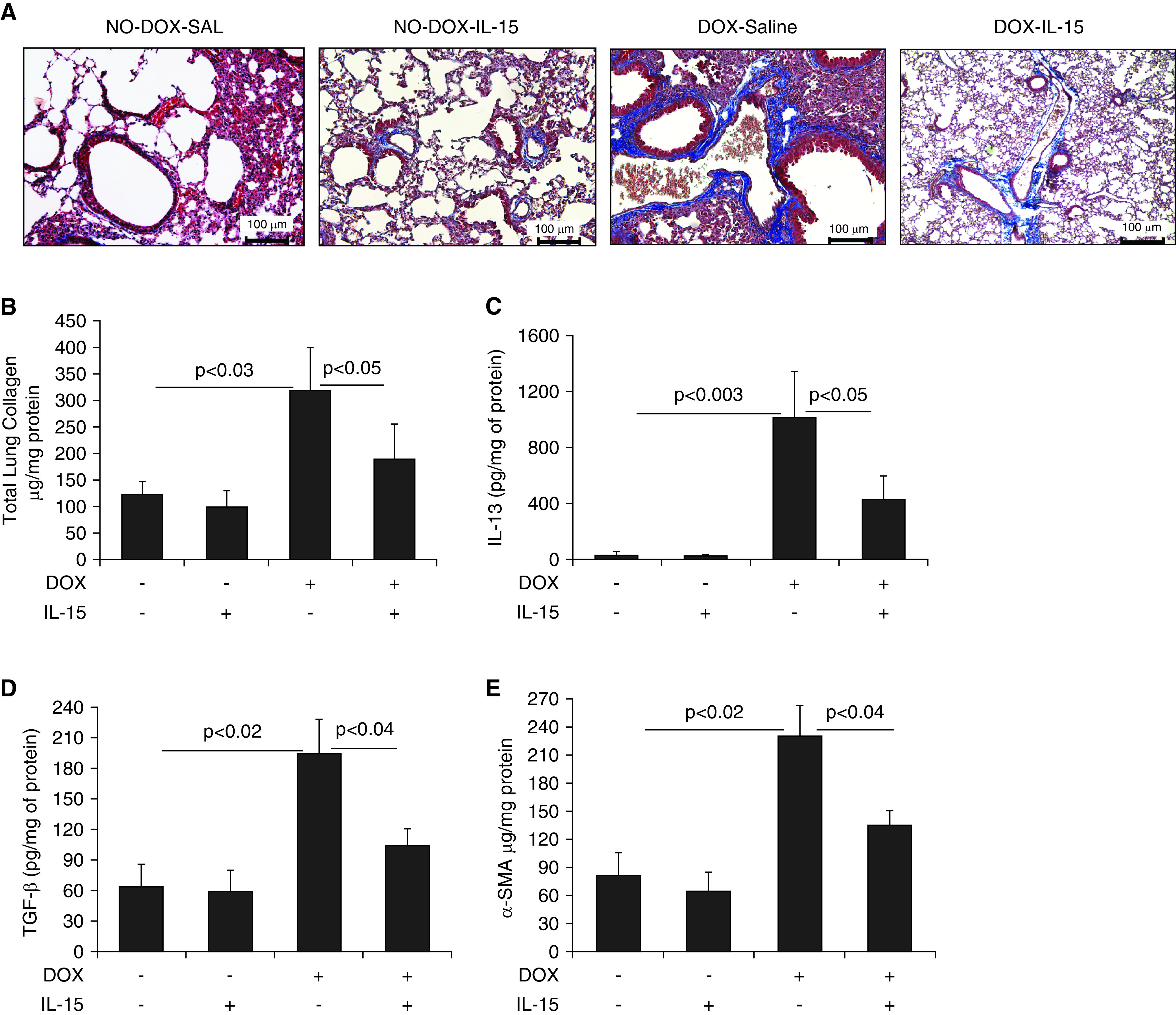
Figure 4.

Pharmacological delivery of IL-15 significantly reduces IL-13–induced collagen accumulation and profibrotic cytokines in the lungs of CC10–IL-13 transgenic mice. (A) Light-microscopy photomicrographs of Masson’s trichome–stained lung sections from saline- or IL-15–treated, no-DOX– or DOX-exposed CC10–IL-13 bitransgenic mice. (B–E) Total lung collagen (B) and the profibrotic cytokines IL-13 (C), TGF-β1 (D), and α-SMA (E) in saline- and IL-15–treated no-DOX– and DOX-exposed IL-13 bitransgenic mice are shown. Data are expressed as mean ± SD, n = 12 mice/group. Scale bars: 100 μm.
Reference
- 1. Upparahalli Venkateshaiah S, Niranjan R, Manohar M, Verma AK, Kandikattu HK, Lasky JA, Mishra A. Attenuation of allergen-, IL-13–, and TGF-α–induced lung fibrosis after the treatment of rIL-15 in mice. Am J Respir Cell Mol Biol . 2019;61:97–109. doi: 10.1165/rcmb.2018-0254OC. [DOI] [PMC free article] [PubMed] [Google Scholar]


