



Abstract
BACKGROUND:
Although development of reliable diagnostic criteria for temporomandibular disorders (TMD) have operationalized identification of a subgroup with myofascial pain (mTMD), causal mechanisms remain elusive.
OBJECTIVES:
This study examines masticatory muscle activity (MMA) in more homogenous research subgroups of mTMD.
METHODS:
Data from an existing case-control study of women were used to subcategorize mTMD cases based on joint pain with palpation to isolate muscle-only pain (M-pain) vs muscle and joint pain (MJ-pain). Differences in laboratory indicators of MMA, specifically research diagnostic criteria for sleep bruxism (SB) and high background EMG activity, and other clinical and sociodemographic indicators were examined between groups.
RESULTS:
Compared to controls, the MJ-pain subgroup did not show elevated background EMG or sleep bruxism. In contrast, the M-pain subgroup showed significantly higher background EMG and a trend toward elevated prevalence of sleep bruxism.
CONCLUSIONS:
These results may explain why it has been difficult for studies of SB in mixed TMD and even mTMD samples to find a consistent positive association, since a positive association may be limited to mTMD without joint pain. The subcategorizing of mTMD based on joint pain with palpation (i.e., M-pain, MJ-pain) appears to reveal subgroups with relatively high and low sleep masticatory muscle-specific activity. Findings need replication in a larger study with updated mTMD diagnostic criteria, but may prove useful for understanding mechanism of pain maintenance in mTMD with and without joint pain.
Keywords: Myofascial TMD, masticatory muscle activity, sleep bruxism, PSG, EMG, muscle-only pain
BACKGROUND
Despite increased research, the etiology and mechanisms of myofascial TMD remain elusive.1 One area of interest has been the role of masticatory muscle activity (MMA) in the onset and maintenance of TMDs, such as the suspected role of sleep bruxism as a risk factor for TMD.2–4 This area of research has been compromised by construct and measurement challenges for both TMD and MMA. For example, bruxism itself has been a difficult construct to define 5–8 which may have contributed to decades of conflicting study results with exciting findings based on self-reported measures of sleep bruxism 9 that fail to replicate in studies using more valid laboratory measures.10, 11
In addition to issues on measurement of MMA, use of case definitions for TMDs have also limited research interpretations. Although the development of the research diagnostic criteria (RDC/TMD)12 and more recently the diagnostic criteria (DC/TMD)13 for TMDs have increased the reliability of research on clinical presentations of TMD including the myofascial subtype (mTMD), the composition of case groups used in research studies are phenotypically heterogeneous, potentially contributing to inconsistent findings. For example, the first polysomnography study examining sleep bruxism in TMD cases and controls included a mixed TMD sample of articular and myofascial presentations. It is unclear if a systematic RDC/TMD examination was conducted to classify patients, but the study found no association between TMD status and sleep bruxism.14 A larger study by the same group, using RDC/TMD criteria for myofascial pain specifically, i.e., mTMD, did find that sleep bruxism presented a small risk of mTMD.15 It may be possible that the narrowed case definition may have improved the study’s ability to find an association. However, the sample size of this second study15 was still relatively small and the prevalence of SB using PSG was much higher than reported in other studies, calling into question the validity of study procedures and/or recruitment methods.
The most recent study improved on the limitations of previous investigations by including a larger sample size, RDC/TMD criteria for mTMD and polysomnography using various levels of rhythmic masticatory muscle activity including research diagnostic criteria for sleep bruxism (RDC/SB).16 It found no mTMD elevation of sleep bruxism or subthreshold sleep bruxism compared to controls.10 However, this research group did examine background sleep EMG (i.e., resting state hypertonicity during non-bruxism sleep periods) and found elevated levels among the mTMD group compared to controls, suggesting a more complex relationship between MMA than captured by SB.17 Although there have been other PSG studies focused on bruxism, these sample on SB rather than on TMD and to our knowledge, no other laboratory PSG studies of TMD with control groups have been conducted. Despite this scarcity of evidence, SB and MMA continue to be widely considered as risk factors for TMD largely based on research using self-reported measures of SB which have not been found to correlate with valid PSG measures.11 However, these PSG-based studies on the risk of SB for mTMD clearly yielded mixed results10, 15 and the most robust of the two yielded null results for SB.10
The above noted studies also highlight that case group definitions matter. Although the (R)DC/TMD Axis I groupings differentiate between clinically meaningful phenotypes,18, 19 the presence of some joint pain is not excluded between different Axis I groups. Therefore, unless specifically measured, joint pain may be present to an unknown degree in case groups defined even by Group I mTMD criteria. For example, individuals meeting RDC/TMD criteria often meet multiple Axis I Groups, with one study reporting that 83% of participants met more than one Group criteria and 91% of those meeting Group I criteria met Group III criteria.20 Additionally, in a longitudinal study of mTMD,21 approximately 66% of participants meeting Group I criteria at baseline also met criteria for Group III disorders. When studied over a 5 year period, mTMD was the most chronic subtype but co-occurrence of Group III was associated with increased chronicity vs remission of mTMD. This evidence suggests that co-occurring joint pain may be clinically relevant and phenotypically important for uncovering mechanisms of myofascial pain.
Although any chronic pain involves a complex interplay of peripheral and central mechanisms, the composition of joint and muscle pain in a case group is potentially important given that mechanisms involved in chronic joint pain are presumably different from mechanisms driving muscle pain.1 For example, arthritic pain is associated with an inflammatory nociceptive process1 while pain of the masticatory muscles has not been to date conclusively associated with inflammation in the muscles, although pain in joint-adjoining muscles may be induced by joint inflammatory nociceptive processes when these are present.22 As described above, defining a case group at the mTMD level includes a mixture of joint and muscle pain even if clinical judgement determined the pain is primarily muscle-based. As a result, it is possible that even the mTMD classification of TMD may produce too heterogeneous a group for studies on causal mechanisms because of the potential presence of both muscle and joint pain. To improve on the internal validity of mechanistic studies on mTMD, not the diagnostic criteria in general, the present study aims to examine the utility of isolating primarily muscle-related pain in mTMD to elucidate the role of MMA in the etiology and/or pathogenesis of mTMD. The overriding hypothesis is that reducing phenotypic variability in TMDs may reveal pain mechanisms that would otherwise be obscured when studying a phenotypically heterogeneous sample, even within a relatively homogeneous mTMD sample.
METHODS
Sample
To test our hypotheses, data were drawn from an existing case control study of myofascial TMD which included a sleep study with laboratory-based estimates of research diagnostic criteria for sleep bruxism (SB) and background sleep EMG.10 Cases and controls were women recruited from the New York University Dental Orofacial Pain Clinic or via electronic advertisements in facility elevators based on whether or not they had facial pain. Treating orofacial pain clinicians screened out pain patients whose pain was primarily neuropathic in nature or not primarily of myogenic origin. Participants were enrolled without knowledge of their masticatory muscle activity. Women were excluded if they had experienced dental work in the last 48 hours or had ever experienced a physical trauma involving the face. All subjects underwent informed consent procedures before enrollment and the study underwent all ethical reviews via the NYU Medical School Institution Review Board, IRB#07–303, including final approval on February 23, 2018 for continuing data analysis. Below key points relevant to the present analysis are summarized. Additional details on study procedures can be found in the publications of the primary results of the study.10, 11, 17
Sleep Study Procedures
Participants were asked to spend two nights in an NYU affiliated sleep laboratory to monitor their sleep quality, breathing and masticatory muscle activity via polysomnography (PSG) and electromyography (EMG). Interviews were conducted before and after each night of sleep. The first night of sleep study was intended to acclimate the participant to the new environment. Data were used on the second night of the sleep study except in 10 cases when a second night was not completed and thus the first night was analyzed.
Measures
Diagnosis of Myofascial TMD at Study Enrollment
All participants underwent facial examination by a research clinician to determine if they met criteria for mTMD based on Research Diagnostic Criteria for Temporomandibular and Muscle Disorder (RDC/TMD) Group I (myofascial pain) at the time of enrollment. Standardization of the diagnostic criteria was ensured by use of an external orofacial pain specialist with use of RDC/TMD training materials to train a foreign-trained research clinician. In addition, the research clinician was locally supervised by an RDC/TMD trained orofacial pain specialist. To further ensure reliability and guard against examiner drift in standards, reliability checks were implemented throughout the study period by re-examination of a subset of just-enrolled participants. Over the course of multiple reliability test sessions, reliability between the external examiner and research clinician was perfect at the level of Group I diagnosis.
The clinical research examination included palpation of 20 facial muscle sites in the masseter and temporalis, as well as the lateral pole and posterior attachment of the temporomandibular joint (TMJ). Participants were classified as mTMD positive if they self-reported pain in the orofacial region, reported three or more tender points with palpation out of the 20 muscle sites palpated per criteria, and reported elicited and spontaneous pain on the same side of the face. In addition, for study purposes, to ensure that pain was primarily myofascial in origin, clinical judgment that the pain was primarily myogenous rather than of joint origin was required. This was determined by severity of pain on palpation of muscles and joints, as well as participant self-report. The investigators’ previous experience suggested that intraoral palpation was less reliable; therefore, at least one extra oral tender point was also required. Control participants were individuals who did not present with a facial pain problem, had a maximum of 1 painful site on palpation (muscle or joint) examination, and no history of spontaneous pain in the face in the last month.
Subcategorization of mTMD (M-pain vs MJ-pain) to reduce phenotypic heterogeneity
To further subcategorize the mTMD group as either ‘muscle only pain’ (M-pain) or ‘muscle and joint pain’ (MJ-pain), the ratings of pain intensity with palpation recorded during the RDC/TMD clinical research examination were analyzed post-hoc. Individuals who reported any pain on palpation of the TMJ sites on either side of the face were characterized as MJ-pain. Individuals with no pain on palpation of the TMJ, on either the left or the right side of the face, were categorized as M-pain.
Sleep Bruxism
Clinical research diagnostic criteria were used for Sleep Bruxism (RDC/SB)16 which require frequent episodes of rhythmic masticatory muscle activity (RMMA) observed in sleep laboratories. Specifically, sleep bruxism was defined as >4 RMMA episodes per sleep hour OR >25 bursts per sleep hour.10 PhD-level Staff experienced in scoring sleep studies were trained to acceptable levels of reliability by the laboratory staff of the first author of the RDC/SB.16
High Background EMG
Background sleep EMG was intended to measure typical resting muscle activity during sleep, not bruxism activity. For consistency, activity in the right masseter muscle activity was scored. Background sleep EMG was measured as EMG root mean square (RMS) remaining after activity attributable to sleep bruxism and movement artifacts were removed.5 Due to skewness (data not shown), and for parsimony, high background EMG was further dichotomized as the 4th quartile of background EMG (value of 1) vs. 1–3rd quartiles (value of 0). In other contexts, this may be said to define muscle “hypertonicity” at rest.
Observed Sleep Time
To ensure that individuals in the both subgroups of mTMD had equivalent opportunity to engage in SB, select PSG sleep measures were examined. Specifically, total sleep time in minutes and sleep efficiency (i.e., total time asleep divided by total time in bed) were examined.
Interview Items before and after sleep study
A number of items were obtained just before and after the night of the laboratory sleep study. Current facial pain intensity on a 0 to 10 scale (10 representing pain as bad as it could be) was obtained on the night just prior to the sleep study. After waking on the morning after the sleep study, participants were again asked to rate their current facial pain intensity on the same scale.
Other Measures of Interest
Other relevant demographic and clinical characteristics were obtained via the main study interview after enrollment. Spontaneous pain was measured in various ways on a 0–10 scale (10 representing pain as bad as it could be) and is here reported as characteristic pain intensity (CPI), the average of current, worst and average pain intensity in the last 6 months (0–10 scale). In addition to exploring pain intensity on palpation on a 0–10 scale (10=worst pain) across all palpated muscle and joint sites, masseter pain on palpation was averaged across masseter sites: masseter origin, body and insertion (0–10 scale). Duration of facial pain was operationalized as years since onset of facial pain. Given the diverse sample, the distributions of demographic variables were statistically examined and each variable was included in the most parsimonious form. Specifically, variables included age in years, education in years, Hispanic ethnicity (vs non-Hispanic) and income as >$15,000 vs less.
Sleep impairments and fatigue are key features of fibromyalgia,23 a widespread pain condition often comorbid with mTMD.24 Myofascial pain is common to both conditions while joint pain is not. To ensure that any differences in sleep bruxism or background EMG were not a result of sleep disturbances related to comorbid fibromyalgia, it was examined as a covariate. Fibromyalgia status was determined via research diagnosis using the American College of Rheumatology (ACR) 1990 criteria25 assessed during clinical research examination of 11 or more tender points with palpation and participant report of four quadrant pain.10
Statistical Analysis
First, demographic and clinical characteristics of each subcategory of mTMD by location of pain (M-pain vs MJ-pain) were examined and differences tested using Chi-squared, Fisher’s exact tests, T-test or Wilcoxon rank sum test procedures where appropriate. Differences in the proportion of bruxism and high background EMG were explored between mTMD subgroups and between each subgroup and the control group. Secondly, to examine differences between mTMD subgroups (M-pain vs MJ-pain) on MMA and relevant covariates, a series of logistic regression models were run only among the mTMD cases to predict the odds of muscle-only pain (M-pain vs. MJ-pain).
RESULTS
Characteristics of M-pain vs MJ-pain mTMD Groups
TMJ pain with palpation was common among the mTMD group with 73% reporting some joint pain. A total of 34 women with mTMD were classified as M-pain (27%) and 91 were classified as MJ-pain based on the presence or absence of joint pain on palpation. No significant differences were found between M-pain and MJ-pain groups on demographic variables or comorbid fibromyalgia (Table 1). However, characteristic pain intensity was significantly higher among the MJ-pain group (Table 2). Similarly, muscle pain across most palpation sites was consistently higher for the MJ-pain group compared to the M-pain group (detailed data not shown) including average pain intensities across masseter palpation sites (Table 2).
Table 1.
Participants’ Sociodemographic Characteristics by mTMD Subgroups and Controls
| mTMD | Controls (N=46) | |||||
|---|---|---|---|---|---|---|
|
|
||||||
| M-pain (N=34) | MJ-pain (N=91) | |||||
|
|
|
|
||||
| N | Measure | N | Measure | N | Measure | |
|
|
|
|
||||
| Age (years) | ||||||
| Mean (SD) | 34 | 40.3 (15.5) | 90 | 40.3 (14.7) | 46 | 36.1 (13.5) |
| Median | 36.5 | 38 | 31.2 | |||
| Hispanic (%)* | 5 | 16 | 22 | 25 | 9 | 20 |
| Education | ||||||
| Mean (SD) | 34 | 15.4 (2.3) | 88 | 15.5 (2.3) | 46 | 15.7 (2.3) |
| Median | 16 | 16 | 16 | |||
| Income (%>$15,000) | 24 | 71 | 65 | 72 | 32 | 70 |
| Comorbid Fibromyalgia (%) | 6 | 18 | 19 | 21 | -- | -- |
mTMD= myofascial temporomandibular disorder; M-pain= mTMD with muscle only pain; MJ-pain= mTMD with muscle and joint pain
Comparisons across groups (Fishers exact test for categorical variables or Wilcoxon rank-sum tests for continuous variables) did not detect differences at p<.05.
Discrepancies in percentages are due to missing values and rounding. Number of missing values per variable: Age=1, Hispanic=6, Education=3, Income=1, Fibromyalgia=2.
Table 2.
Pain, mTMD Symptoms and other Clinical Features by mTMD Subcategorization
| mTMD | |||||
|---|---|---|---|---|---|
|
|
|||||
| M-pain | MJ-pain | ||||
|
|
|
||||
| N | Measure | N | Measure | p-value | |
|
|
|
|
|||
| Years since FP onset | |||||
| Mean (SD) | 34 | 11.3 (12.5) | 90 | 10.3 (9.8) | 0.63 |
| Median | 6.2 | 7.1 | |||
| Characteristic pain intensity | |||||
| Mean (SD) | 34 | 4.7 (1.6) | 90 | 5.5 (1.2) | 0.02 |
| Median | 4.7 | 5.7 | |||
| Average Pain Across Masseter Sites | |||||
| Mean (SD) | 34 | 2.1 (1.3) | 91 | 2.6 (1.7) | 0.07 |
| Median | 1.7 | 2.3 | 0.04 † | ||
| Pain Intensity before Sleep | |||||
| Mean (SD) | 33 | 1.6 (2.1) | 88 | 2.5 (2.3) | 0.07 |
| Median | 1 | 2 | 0.04 † | ||
| Pain Intensity after Sleep | |||||
| Mean (SD) | 32 | 1.8 (2.4) | 89 | 2.7 (2.3) | 0.06 |
| Median | 1 | 2 | .01 † | ||
| Persistent Pain (vs. Recurrent) (%) | 34 | 38 | 90 | 53 | 0.16 |
| mTMD Jaw-related symptoms (%) | |||||
| Jaw lock | 53 | 50 | 0.84 | ||
| Jaw lock interferes with eating | 29 | 28 | 1.0 | ||
| Jaw click/pop on opening/chewing | 71 | 83 | 0.14 | ||
| Grating or grinding noise | 45 | 45 | 1.0 | ||
| Morning jaw ache/stiffness | 82 | 88 | 0.56 | ||
| Noises/ringing in ear | 35 | 59 | 0.03 | ||
| Bite feels uncomfortable or unusual | 61 | 70 | 0.39 | ||
| Total number of symptoms (Mean (SD)) | 34 | 3.7 ( 2.1) | 91 | 4.1 (1.6) | 0.25 |
| Activities Affected by Facial Pain (%) | |||||
| Chewing | 71 | 79 | 0.33 | ||
| Drinking | 3 | 6 | 1.0 | ||
| Exercising | 6 | 10 | 0.73 | ||
| eating hard foods | 94 | 87 | 0.35 | ||
| eating soft foods | 6 | 4 | 0.67 | ||
| smiling/laughing | 29 | 33 | 0.83 | ||
| sexual activity | 15 | 14 | 1.0 | ||
| cleaning teeth or face | 26 | 22 | 0.64 | ||
| Yawning | 74 | 71 | 1.0 | ||
| Swallowing | 0 | 8 | 0.19 | ||
| Talking | 26 | 31 | 0.67 | ||
| having your usual appearance | 29 | 22 | 0.49 | ||
| Total number of activities (Mean (SD)) | 34 | 3.8 (1.9) | 91 | 3.8 (2.0) | 0.92 |
mTMD= myofascial temporomandibular disorder; M-pain= mTMD with muscle only pain; MJ-pain= mTMD with muscle and joint pain.
P-values provided for differences in means between M-pain vs MJ-pain based on T-tests for means or Fisher’s Exact Tests for difference in frequencies.
Wilcoxon Rank-Sum Test used for differences in medians when variable not normally distributed and results differed appreciably from T-test;
Bold values are significantly different at p<.05.
Masticatory Muscle Activity differences between mTMD Subgroups and Controls
Sleep study data were collected on most participants yielding MMA data on all but 1 participant classified as M-pain and 3 control participants (N=170; 34 M-pain; 90 MJ-pain; 46 controls). Both sleep bruxism and high background EMG were more frequently observed in the M-pain group compared to the MJ-pain subgroup (Figures 1 and 2).
Figure 1.
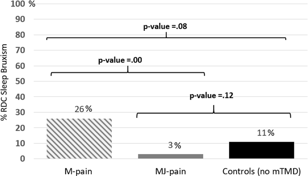
Comparison of Percent RDC Sleep Bruxism Across Groups
P-values for comparison of sleep bruxism between mTMD subgroups and controls are based on Fisher’s exact tests. Sleep bruxism was measured using polysomnography-based Research Diagnostic Criteria for sleep bruxism. mTMD= myofascial temporomandibular disorder; M-pain= mTMD with muscle only pain; MJ-pain= mTMD with muscle and joint pain; Controls= no mTMD.
Figure 2.

Comparison of Percent High Background EMG Across Groups
P-values for comparison of high background EMG between mTMD subgroups and controls were based on Fisher’s exact tests. High background EMG was defined as the 4th quartile of the EMG root mean squared remaining after activity attributable to sleep bruxism and movement artifacts were removed. mTMD= myofascial temporomandibular disorder; M-pain= mTMD with muscle only pain; MJ-pain= mTMD with muscle and joint pain; Controls= no mTMD.
Masticatory Muscle Activity in the M-pain Subgroup vs Controls
Sleep bruxism was twice as frequent in the M-pain subgroup compared to controls although this was just shy of reaching significance (p=.08) (Figure 1). Of note, mean values of average pain across palpated masseter sites were lower among those SB positive group within the M-pain group (SB+=1.2, SB-=2.4, T-test p-value=.02).
High background EMG was significantly elevated among the M-pain group compared to controls, with nearly half the subgroup experiencing high background EMG (p=.00) (Figure 2). Although most pain ratings were higher among those with high background EMG within the M-pain group, none reached statistical significance (data not shown).
Masticatory Muscle Activity in the MJ-pain Subgroup vs Controls
In contrast to the results observed in the M-pain subgroup, sleep bruxism appeared to be lower among the MJ-pain group compared to the control groups but differences failed to reach statistical significance (p=.12) (Figure 1). High background EMG was not significantly different between the MJ-pain subgroup and controls (Figure 2).
Predictors of the Muscle-only Pain vs the Muscle and Joint Pain mTMD Subgroup
mTMD-specific symptoms by M-pain vs MJ-pain
Of the jaw related TMD symptoms explored and the potential activities affected by facial pain, ear noises was the only statistically significant difference between the mTMD subgroups, with a higher proportion of the MJ-pain subgroup reporting ringing or hearing noises in their ear (Table 2). Other trends towards significance in the MJ-pain group were found for trouble swallowing, jaw clicks or pops on opening or while chewing and self-reports of more persistent facial pain. The small sample size of the M-pain group likely limited the ability to examine relevant symptom patterns.
Masticatory Muscle Activity Predicts M-pain
Masticatory muscle activity variables were examined as predictors of M-pain compared to the MJ-pain group among mTMD positive women through a series of logistic regression models. Each of the MMA indicators, i.e., sleep bruxism and high background EMG were significant and independent predictors of M-pain status, suggesting that these measures are capturing different constructs as intended (Table 3). This withstood adjustments for characteristic pain intensity which was higher among the MJ-pain group. We also explored adjusting for other pain intensity measures which were anchored to the night of the sleep study or average palpated pain across masseter sites in various transformations, and results were similar. Years since onset of facial pain, fibromyalgia and sociodemographic variables were not significant predictors (Tables 1 and 2) and they were not added to the model as potential confounders.
Table 3.
Logistic Regression Models Predicting M-Pain Subgroup Among Women with mTMD
| Unadjusted Models | Adjusted Model | |||
|---|---|---|---|---|
|
|
|
|||
| OR | 95% CI | OR | 95% CI | |
|
|
||||
| Sleep Bruxism | 10.44 | 2.63, 41.51 | 11.12 | 2.56, 48.37 |
| High Background EMG | 3.11 | 1.35, 7.18 | 3.78 | 1.51, 9.45 |
| Characteristic Pain Intensity | 0.76 | 0.60, 0.97 | 0.84 | 0.64, 1.09 |
| Pain Intensity Before Sleep | 0.83 | 0.68, 1.02 | ||
| Pain Intensity After Sleep | 0.82 | 0.66, 1.01 | ||
| Average Pain Across Masseter Sites | 0.76 | 0.56, 1.02 | ||
| N | 124 | |||
| Likelihood Ratio Chi-Squared test | 23.72 | |||
| P-value | 0.00 | |||
OR=odds ratio; mTMD= myofascial temporomandibular disorder; M-pain= mTMD without joint pain; All pain variables were on a 0–10 scale; Unadjusted Models include separate models for single variables; The Adjusted Model includes masticatory muscle activity variables adjusted for the most stable and significant pain variable-characteristic pain intensity. Alternative analyses substituted the other pain variables for characteristic pain intensity, in various transformations, and yielded similar results regarding masticatory muscle activity. Sociodemographic variables including age were not significant predictors of M-pain and therefore were not included.
CONCLUSIONS
mTMD may be too heterogeneous for some mechanistic studies
The present study indicates that MMA differs among subgroups of mTMD positive women based on presence or absence of joint pain on palpation. This suggests that one possible reason for inconsistent and null findings on the relationship between MMA and mTMD and difficulties in identifying specific etiological factors in general may be that studies have used mixed TMD case groups. Even limiting to only mTMD may be introducing too much heterogeneity in the study of pain mechanisms.
By reducing heterogeneity via sub-classification of mTMD based on presence or absence of TMJ pain on palpation, it appears that, for mTMD with joint pain, SB frequency may be either similar to controls or possibly less frequent than in controls. So at best, MMA may be most relevant to those mTMD cases without a joint pain component which, in mixed mTMD samples, and definitely in general TMD samples would bias results towards the null. At worst, SB may have an opposite relationship within subgroups, where results were suggestive of lower SB among the MJ-pain group compared to controls. If replicated, these results suggest that research with mixed groups is biased and stymies not only understanding of the SB hypothesis, but discovery of new lines of inquiry on specific mechanisms for the range of facial pain phenotypes.
What may be driving the observed differences in MMA between M-pain and MJ-pain mTMD subgroups?
Null results on SB and background MMA with the MJ-pain group compared to controls along with higher pain reports suggest that lower muscle activity may be a result of attempts to avoid joint pain. This inhibition would be consistent with the pain adaptation model.26 The presence of TMJ pain may indicate that an inflammatory mechanism may differentiate these subgroups. The alternative mechanism in the subgroup with muscle-only pain may be related directly to MMA, or to an alternative mechanisms that does not limit MMA. This is consistent with the psychophysiological model proposed by Laskin in 1969.27 What is clear is that MMA may have a different relationship to the mechanisms driving muscle-only pain than to joint pain.
The parent study examined rank-ordered masseter resting background EMG and found significant differences between cases and controls.17 The present study, using the dichotomized version of high background EMG, found significant differences between mTMD groups stratified by joint pain in particular the M-pain group versus controls. This indicates hypertonicity in the masseter muscle of mTMD patients without joint pain. Therefore, the construct of resting background EMG continues to warrant further study in the etiology of mTMD, particularly among individuals without joint pain. Given sample size limitations, results should be interpreted with caution until replicated.
Limitations
A number of potential limitations should be examined when interpreting these results. First, this sample used now outdated RDC/TMD criteria which had been found to have acceptable reliability for Group 1 (a or b), 28 but have since been updated in DC/TMD. Second, the study was not ideally powered for stratification by joint pain (i.e., M-pain, MJ-pain comparisons) since it was based on secondary data analysis from a study not designed for this purpose. Despite the unbalanced sample size between mTMD subgroups, particularly the small M-pain subgroup, we were able to identify statistically significant results. Null results were primarily limited to the larger subgroup where power would be less of a concern.
Third, the definition of subgroups, specifically of the presence of joint pain was based solely on pain reported during joint palpation. To our knowledge, this is the first attempt, to subcategorizing mTMD cases in this manner; therefore, despite positive findings, we cannot rule out measurement error or guarantee complete homogeneity of subgroups. In addition, although not a limit to internal validity, the data did not include a full workup of all the Axis I Groups, limiting the authors’ ability to describe co-occurring Group II and III diagnoses. Therefore, these analyses and/or alternate methods of identifying myofascial pain stratified by joint pain should be replicated using full DC/TMD criteria with the additional considerations discussed below.
Finally, only chronic cases of mTMD were included and thus, because onset and maintenance factors may differ, results cannot address onset factors. Relatedly, subjects were not followed over time to establish stability of mTMD or MMA status. mTMD status at the single point in time or its stratification by joint pain based on palpation (i.e. M-pain vs MJ-pain) at one point may not relate to the predominant status of the individuals if followed over time. Similarly, one rating of MMA via polysomnography may not represent the dominant pattern. Despite these potential limitations, we were able to detect differences of considerable magnitude and potential clinical importance.
Future directions
Future studies should replicate these findings by first using updated criteria for myofascial TMD. Second, studies attempting to replicate or extend these findings should properly power for stratification of mTMD by joint pain, including a thorough examination of joint pain and or joint health, and establish reliability of such sub-categorizations for research purposes. Little is still known about the natural history of TMDs. How an individual may progress from one TMD subtype to another and why is unclear. To further examine the complex relationship between MMA and pain onset and maintenance, longitudinal studies should explore MMA at repeated time points as well as the potential TMD joint to muscle pain progression and vice versa.
Moreover, theories of muscle and related tissue injury/nociceptive markers should be explored, potentially drawing from the animal literature on joint vs. muscle pain mechanisms. In addition to peripheral mechanisms of nociception, central mechanisms of endogenous pain modulation should also be explored. In fact, perhaps this subcategorization can shed light on other inconsistent and/or null findings using quantitative sensory testing. For example, mixed results on temporal summation and conditioned pain modulation in mTMD29 may be resolved by breaking apart an mTMD group into subgroups with and without some joint pain.
Intervention research may also benefit from examining the M-pain and MJ-pain differences in efficacy and effectiveness. This dichotomy may help elucidate the mechanisms via which the treatment modality may be relieving pain or improving outcomes. One common treatment, oral appliances continue to be a first line treatment for TMDs30, 31 despite mixed evidence on efficacy.32 Predictions on expected treatment response by the M-pain/ MJ-pain dichotomy can be made that may help advance identification of specific subgroups of mTMD patients for whom appliances are likely to be more or less effective. For example, do oral appliances help in mTMD by unloading the joint or simply serve as a reminder not to clench and thus facilitate reduction of MMA? Uncovering these mechanisms of action can lead to significant breakthroughs in understanding the underlying causes of mTMD pain maintenance and lead to improved treatment options for the given phenotype.
It is worth noting that simply addressing comorbid TMDs in research on mTMD would not adequately address the implications of the present study, because all the Axis I Groups are utilitarian entities, not based on discrete etiological origins. In fact, the comorbidity of various Axis I diagnoses seems to exclude this likelihood. Research on mechanisms of TMDs should not be obscured by reification of these diagnostic categories. It is simpler for research replication to focus on powering mTMD positive groups with and without joint pain rather than measure full criteria for the various TMDs. Such clarity ensures that non-overlapping phenotypic groups are properly powered whereas simply adjusting for comorbidity, given the discussed prevalence of meeting criteria for multiple TMDs, would not.
The RDC/TMD and DC/TMD criteria have been extremely useful for clinical purposes, allowing interpretation of research findings, and the application of this research for clinical practice. Although the present results may eventually have implications for the next evolution of diagnostic criteria, such recommendations would be premature and not the authors’ intention. Instead, the authors recommend that research on pain onset and maintenance mechanisms should re-examine the definition of case groups based on the aims of the given study, always beginning with clear connection to currently agreed-upon diagnostic criteria. For example, start with DC/TMD Axis I diagnoses and then further subcategorize or measure relevant features for adequate stratification. Recommendations for updates to the diagnostic criteria will only be possible once well designed and powered research with phenotypically homogeneous case definitions can uncover clinically meaningful differences leading to advances in treatment approaches. This utility can then guide the updates to the clinical entities. Mechanistic studies should build upon the knowledge base that generated these reliable and useful diagnostic systems, but should not be limited by these systems. Good mechanistic studies should be designed with case groups appropriate for the given research question. Such studies combined with treatment outcome studies may lead to even better diagnostic criteria. Homogeneity of case groups in these studies may be defined in a number of ways; these findings, if replicated, suggest that in the study of MMA, the presence or absence of joint pain among mTMD patients may be one meaningful approach.
Summary
The present findings suggests that the subcategorizing of mTMD based on presence of joint pain with palpation (i.e., M-pain, MJ-pain) isolates unique patterns of MMA and thus may prove useful in mechanistic studies. This study does not reveal what the causal role of MMA in the M-pain group may be or confirm that it plays any causal role at all. However, it finds a differential relationship by M-pain vs MJ-pain which may help explain inconsistent results from studies to date and, if replicated and employed in mechanistic studies, may help uncover what potential role MMA may play in the onset and/or maintenance of mTMD.
ACKNOWLEDGEMENTS
This work was supported in part by NIH grants R01DE018569 and R01DE024522–01S1.
Footnotes
Conflict of interest statement: The authors have no conflicts of interest.
REFERENCES
- 1.Cairns BE. Pathophysiology of TMD pain--basic mechanisms and their implications for pharmacotherapy. J Oral Rehabil. 2010; 37: 391–410. [DOI] [PubMed] [Google Scholar]
- 2.Le Resche L, Truelove EL, Dworkin SF. Temporomandibular disorders: a survey of dentists’ knowledge and beliefs. J Am Dent Assoc. 1993; 124: 90–94, 97–106. [DOI] [PubMed] [Google Scholar]
- 3.Glaros AG, Glass EG, McLaughlin L. Knowledge and beliefs of dentists regarding temporomandibular disorders and chronic pain. J Orofac Pain. 1994; 8: 216–222. [PubMed] [Google Scholar]
- 4.Tegelberg A, Wenneberg B, List T. General practice dentists’ knowledge of temporomandibular disorders in children and adolescents. Eur J Dent Educ. 2007; 11: 216–221. [DOI] [PubMed] [Google Scholar]
- 5.Lobbezoo F, Ahlberg J, Glaros AG, Kato T, Koyano K, Lavigne GJ et al. Bruxism defined and graded: an international consensus. J Oral Rehabil. 2013; 40: 2–4. [DOI] [PubMed] [Google Scholar]
- 6.Raphael KG, Santiago V, Lobbezoo F. Is bruxism a disorder or a behaviour? Rethinking the international consensus on defining and grading of bruxism. J Oral Rehabil. 2016; 43: 791–798. [DOI] [PMC free article] [PubMed] [Google Scholar]
- 7.Raphael KG, Santiago V, Lobbezoo F. Bruxism is a continuously distributed behaviour, but disorder decisions are dichotomous (Response to letter by Manfredini, De Laat, Winocur, & Ahlberg (2016)). J Oral Rehabil. 2016; 43: 802–803. [DOI] [PMC free article] [PubMed] [Google Scholar]
- 8.Lobbezoo F, Ahlberg J, Raphael KG, Wetselaar P, Glaros AG, Kato T et al. International consensus on the assessment of bruxism: Report of a work in progress. J Oral Rehabil. 2018; 0. [DOI] [PMC free article] [PubMed] [Google Scholar]
- 9.Manfredini D, Lobbezoo F. Relationship between bruxism and temporomandibular disorders: a systematic review of literature from 1998 to 2008. Oral Surg Oral Med Oral Pathol Oral Radiol Endo. 2010; 109: e26–50. [DOI] [PubMed] [Google Scholar]
- 10.Raphael KG, Sirois DA, Janal MN, Wigren PE, Dubrovsky B, Nemelivsky LV et al. Sleep bruxism and myofascial temporomandibular disorders: a laboratory-based polysomnographic investigation. J Am Dent Assoc. 2012; 143: 1223–1231. [DOI] [PMC free article] [PubMed] [Google Scholar]
- 11.Raphael KG, Janal MN, Sirois DA, Dubrovsky B, Klausner JJ, Krieger AC et al. Validity of self-reported sleep bruxism among myofascial temporomandibular disorder patients and controls. J Oral Rehabil. 2015; 42: 751–758. [DOI] [PMC free article] [PubMed] [Google Scholar]
- 12.Dworkin S, LeResche L. Research diagnostic criteria for temporomandibular disorders: review, criteria, examinations and specifications, critique. J Craniomand Disord. 1992; 6. [PubMed] [Google Scholar]
- 13.Schiffman E, Ohrbach R. Executive summary of the Diagnostic Criteria for Temporomandibular Disorders for clinical and research applications. J Am Dent Assoc. 2016; 147: 438–445. [DOI] [PMC free article] [PubMed] [Google Scholar]
- 14.Rossetti LM, Rossetti PH, Conti PC, de Araujo Cdos R. Association between sleep bruxism and temporomandibular disorders: a polysomnographic pilot study. Cranio. 2008; 26: 16–24. [DOI] [PubMed] [Google Scholar]
- 15.Rossetti LM, Pereira de Araujo Cdos R, Rossetti PH, Conti PC. Association between rhythmic masticatory muscle activity during sleep and masticatory myofascial pain: a polysomnographic study. J Orofac Pain. 2008; 22: 190–200. [PubMed] [Google Scholar]
- 16.Lavigne GJ, Rompre PH, Montplaisir JY. Sleep bruxism: validity of clinical research diagnostic criteria in a controlled polysomnographic study. J Dent Res. 1996; 75: 546–552. [DOI] [PubMed] [Google Scholar]
- 17.Raphael KG, Janal MN, Sirois DA, Dubrovsky B, Wigren PE, Klausner JJ et al. Masticatory muscle sleep background electromyographic activity is elevated in myofascial temporomandibular disorder patients. J Oral Rehabil. 2013; 40: 883–891. [DOI] [PMC free article] [PubMed] [Google Scholar]
- 18.Dworkin SF, LeResche L. Research diagnostic criteria for temporomandibular disorders: review, criteria, examinations and specifications, critique. J Craniomand Disord. 1992; 6: 301–355. [PubMed] [Google Scholar]
- 19.Schiffman E, Ohrbach R, Truelove E, Look J, Anderson G, Goulet JP et al. Diagnostic Criteria for Temporomandibular Disorders (DC/TMD) for Clinical and Research Applications: recommendations of the International RDC/TMD Consortium Network* and Orofacial Pain Special Interest Group. J Orofac Pain. 2014; 28: 6–27. [DOI] [PMC free article] [PubMed] [Google Scholar]
- 20.Schiffman EL, Truelove EL, Ohrbach R, Anderson GC, John MT, List T et al. The Research Diagnostic Criteria for Temporomandibular Disorders. I: overview and methodology for assessment of validity. J Orofac Pain. 2010; 24: 7–24. [PMC free article] [PubMed] [Google Scholar]
- 21.Rammelsberg P, LeResche L, Dworkin S, Mancl L. Longitudinal outcome of temporomandibular disorders: a 5-year epidemiologic study of muscle disorders defined by research diagnostic criteria for temporomandibular disorders. J Orofac Pain. 2003; 17: 9–20. [PubMed] [Google Scholar]
- 22.Bliddal H, Curatolo M. Clinical Manifestations of Muscle and Joint Pain. In: Graven-Nielsen T, Arendt-Nielsen L, Mense S, eds. Fundamentals of Musculoskeletal Pain. Seattle: IASP Press; 2008: 239. [Google Scholar]
- 23.Wu YL, Chang LY, Lee HC, Fang SC, Tsai PS. Sleep disturbances in fibromyalgia: A meta-analysis of case-control studies. J Psychosom Res. 2017; 96: 89–97. [DOI] [PubMed] [Google Scholar]
- 24.Costa YM, Conti PC, de Faria FA, Bonjardim LR. Temporomandibular disorders and painful comorbidities: clinical association and underlying mechanisms. Oral Surg Oral Med Oral Pathol Oral Radiol. 2017; 123: 288–297. [DOI] [PubMed] [Google Scholar]
- 25.Wolfe F, Smythe HA, Yunus MB, Bennett RM, Bombardier C, Goldenberg DL et al. The American College of Rheumatology 1990 Criteria for the Classification of Fibromyalgia. Report of the Multicenter Criteria Committee. Arthritis Rheum. 1990; 33: 160–172. [DOI] [PubMed] [Google Scholar]
- 26.Lund JP, Donga R, Widmer CG, Stohler CS. The pain-adaptation model: a discussion of the relationship between chronic musculoskeletal pain and motor activity. Can J Physiol Pharmacol. 1991; 69: 683–694. [DOI] [PubMed] [Google Scholar]
- 27.Laskin DM. Etiology of the pain-dysfunction syndrome. J Am Dent Assoc. 1969; 79: 147–153. [DOI] [PubMed] [Google Scholar]
- 28.Look JO, Schiffman EL, Truelove EL, Ahmad M. Reliability and Validity of Axis I of the Research Diagnostic Criteria for Temporomandibular Disorders (RDC/TMD) with Proposed Revisions. J Oral Rehabil. 2010; 37: 744–759. [DOI] [PMC free article] [PubMed] [Google Scholar]
- 29.La Touche R, Paris-Alemany A, Hidalgo-Perez A, Lopez-de-Uralde-Villanueva I, Angulo-Diaz-Parreno S, Munoz-Garcia D. Evidence for Central Sensitization in Patients with Temporomandibular Disorders: A Systematic Review and Meta-analysis of Observational Studies. Pain Pract. 2018; 18:388–409. [DOI] [PubMed] [Google Scholar]
- 30.NIH. Temporomandibular Joint and Muscle Disorders. National Institiute of Dental and Craniofacial Research and Office of Research on Women’s Health. http://www.nidcr.nih.gov/oralhealth/Topics/TMJ/TMJDisorders.htm. 2013. Accessed 18 April 2017. [Google Scholar]
- 31.Glass EG, Glaros AG, McGlynn FD. Myofascial pain dysfunction: treatments used by ADA members. Cranio. 1993; 11: 25–29. [DOI] [PubMed] [Google Scholar]
- 32.Al-Ani MZ, Davies Stephen J, Gray Robin JM, Sloan P, Glenny A-M. Stabilisation splint therapy for temporomandibular pain dysfunction syndrome. Cochrane Database of Syst Rev. 2009. [DOI] [PubMed] [Google Scholar]