

Abstract
A 21-year-old otherwise healthy male sustained a nondisplaced, intertrochanteric fracture of the left femur after being “rear-ended” by a motor vehicle while riding his bicycle. His fracture was managed with protected weight-bearing and progressive mobilization. No traction was utilized. The patient had an excellent clinical outcome at two-year follow-up, reporting modified Harris Hip Score 85, Hip Outcome Score-Activities of Daily Living 88, Hip Outcome Score-Sport Specific 89, and International Hip Outcome Tool-33 of 77.
Conclusion
Nonsurgical treatment, consisting of restricted weight-bearing, for non-displaced intertrochanteric femur fracture in young, healthy patients can provide a successful result.
Level of Evidence: V
Keywords: conservative management, hip, trauma, orthopaedic surgery
Introduction
Less than 3% of all intertrochanteric femur fractures occur in patients under age 50.1 The infrequent nature of this injury, as well as the “universal agreement” among orthopaedic surgeons in the 1960’s that all intertrochanteric femur fractures are best treated by internal fixation2 has resulted in a dearth of literature on conservative treatment of intertrochanteric femur fractures, particularly in younger patients. Surgical treatment of intertrochanteric femur fractures in young patients has significant risks, with reported complication rates ranging from 9-57%.1,3 Although displaced intertrochanteric femur fractures are unlikely to achieve a successful result with conservative treatment, a nondisplaced intertrochanteric femur fracture has greater stability, with an increased likelihood of achieving union in acceptable alignment.4 Therefore, the rationale for conservative treatment of stable intertrochanteric femur fractures in young patients is avoidance of risks associated with surgical treatment, the fracture is more likely to heal in acceptable position, higher likelihood of an successful clinical outcome,5–7 and the complications of non-surgical treatment seen in the elderly8 are less likely to be experienced by younger patients. The purpose of this study was to report on successful conservative treatment of a nondisplaced, intertrochanteric fracture in a young patient.
Statement of Informed Consent
The patient consented to publication of his case, and Institutional Review Board (IRB) approval was obtained for this study.
Case Report
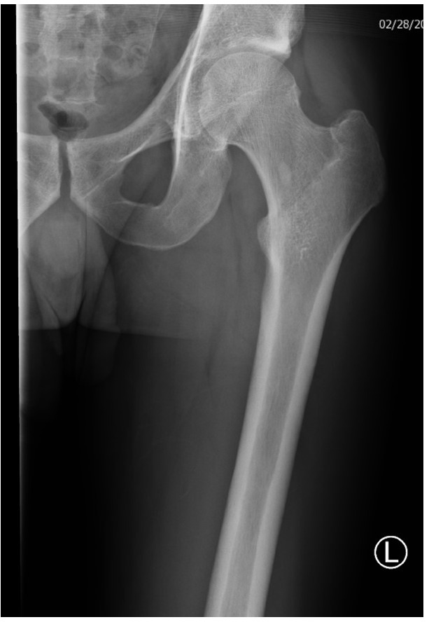
A 21-year old male was thrown from his bicycle when he was “rear-ended” by a motor vehicle and landed on the left side of his body. He had immediate onset of pain in his left hip with the inability to ambulate. He was transported to the emergency room for evaluation, where x-rays (Figures 1.1 and 1.2) and a CT scan were performed (Figure 1.3). These demonstrated a left nondisplaced, intertrochanteric femur fracture. The remainder of his evaluation was unremarkable, except for a small laceration at his left elbow.
Figure 1.1.

X-ray A/P view of the Left Hip, day of injury.
Figure 1.2.

X-ray, Cross Table Lateral view of the Left Hip, day of injury.
Figure 1.3.

3-D Reconstructed Computed Tomography- Frontal view of the Left Hip, day of injury.
Examination of the left lower extremity demonstrated superficial abrasions on his left buttock and lateral aspect of his left knee. He was tender to palpation over the greater trochanter with mild swelling. He was able to actively perform logroll with his left leg and tolerated gentle passive flexion and abduction of the hip without significant discomfort. He was neurovascularly intact.
The patient was counseled about treatment options for this injury, including fixation with a sliding hip screw and conservative treatment, and a joint decision was made to proceed with nonsurgical treatment. He was admitted to the hospital with non-weightbearing restrictions in place for the left lower extremity. Enoxaparin and TED stockings/foot pumps were initially prescribed for DVT prophylaxis. However the patient was switched to Rivaroxaban 10 mg daily after he did not tolerate the needle sticks. No traction devices were utilized. He worked with physical therapy and occupational therapy daily. On hospital day one, he transferred from bed to a chair with a walker and assistance. By hospital day four, he was able to ambulate to the bathroom with a walker and no assistance. By day seven he could independently transfer out of bed, ambulate over 150 feet with crutches, achieved acceptable pain control with oral analgesics, and was discharged to home with in-home physical therapy appointments three times per week.
He was subsequently evaluated at two weeks following his injury and continued to experience mild pain localized to his left hip. X-rays demonstrated blurring of the fracture line and no interval displacement. He was advanced to toe-touch weightbearing. At the six-week evaluation, he had full strength and x-rays demonstrated near-complete healing of the fracture (Figures 2.1 and 2.2). Weight-bearing was advanced, and he was instructed to wean off his crutches. At the three-month evaluation, he reported ability to walk long distances without pain and had returned to swimming and bicycling. He was cleared to begin running and return to sport.
Figure 2.1.
X-ray A/P view of the Left Hip, 6 weeks post-injury.
Figure 2.2.

X-ray, Cross Table Lateral view of the Left Hip, 6 weeks post-injury.
At the two-year follow-up visit, he could perform all activities without limitation. He reported rare discomfort on the lateral aspect of his left hip, managed with stretching and use of a foam roller. X-rays demonstrated complete healing and remodeling of his fracture with a well-preserved joint space (Figures 3.1 and 3.2). Patient-reported outcome scores were obtained: modified Harris Hip Score (mHHS) was 86, Hip Outcome Score for Activities of Daily Living (HOS-ADL) was 88 and Sport Specific (HOS-SS) was 89 (Sport-Specific) International Hip Outcome Tool-33 score (IHOT-33) was 77.
Figure 3.1.

X-ray, A/P view of the Left Hip, 2 years post-injury.
Figure 3.2.

X-ray, Cross Table Lateral view of the Left Hip, 2 years post-injury.
Discussion
This case report describes successful conservative treatment of a stable, nondisplaced intertrochanteric femur fracture in an otherwise healthy 21-year-old male. Historically, conservative treatment for intertrochanteric fractures involved bedrest and traction for 6-12 weeks, followed by advancing to rehabilitation. Because these fractures are uncommon in younger patients, the reports of conservative treatment of intertrochanteric femur fractures has overwhelmingly consisted of elderly patient populations.9 In older patients, multiple studies have demonstrated early reduction and internal fixation improves patient comfort, facilitates nursing care, facilitates early mobilization, and decreases the duration of hospitalization.8,10–13 Conversely, other studies have reported acceptable results with nonsurgical treatment of intertrochanteric femur fractures in elderly patients.5-7,13–15
Horn and Wang reported on their experience treating 170 patients with intertrochanteric fractures and over age 50 with traction. Ambulation was restored in all patients who were previously able to ambulate and there were low reported complication rates (5.3% mortality, 3% pneumonia and 2% decubitus ulceration).5 Scott reported on treatment of intertrochanteric femur fractures with either traction or surgery over a two year period. The non-surgical treatment has a higher prevalence of good outcomes and there was greater mortality observed in the surgical group.14 Bong and colleagues reported on 150 unstable intertrochanteric fractures treated by one of three methods: traction, surgery with medial displacement of the shaft and fixation with a McLaughlin pin and side plate, or valgus osteotomy followed by a McLaughlin pin and plate. The authors found that patients who were younger, previously active, and had adequate nursing care responded the best to conservative management, however similar functional outcomes were noted in all three groups.7
The results of surgical treatment of intertrochanteric femur fracture in young patients, to our knowledge, is limited to two case series. Hwang et al reported on 66 intertrochanteric femur fractures in patients under 40 years old treated with a dynamic hip screw or gamma nail.3 The patients were 70% male and 71% of injuries occurred through high-energy trauma. There were no reported non-unions and radiographic fracture union was achieved a mean of 70.5 days (range 31-213 days). Nine percent of the patients in this study sustained a complication, including infection, aspiration pneumonia, and hardware loosening.
Robinson et al reported on the results of operative treatment of intra- and extracapsular hip fractures in patients under age 50.1 Internal fixation of extracapsular hip fractures was performed using a Richards compression screw with a three- or four-hole plate. Among patients with extracapsular hip fractures, 57% of patients experienced a complication, including fixation failure, infection, adult respiratory distress, and pneumonia.15
There are several notable aspects of this case. The risks of surgery were avoided, and the patient did not sustain a complication resulting from the non-surgical treatment. The patient was hospitalized for only seven days, which was significantly less than 6 weeks of greater reported by previous studies of conservatively treated patients. No traction was required since a non-displaced intertrochanteric femur fracture is a stable pattern injury. Patient reported outcome scores were collected and demonstrated an objectively successful result. Our patient scored above the Patient Acceptable Symptomatic State score (PASS) as determined by Chahal and Maxwell.16,17
There are limitations to this case report. The outcome scores utilized were not available at the time of the Robinson and Hwang studies and we cannot directly compare the clinical outcome of our patient to these historical, surgically treated cohorts. The mHHS, HOS-ADL, HOS-SS, and IHOT-3318 were originally developed to evaluate the outcomes of hip arthroscopy and have not been validated in a hip fracture population. However, since hip arthroscopy is typically performed on young patients, we believe these scores are the most relevant functional outcome scores available for this patient.
In conclusion, we report on a case of a nondisplaced intertrochanteric femur fracture managed with conservative treatment with restricted weight-bearing and achieving an excellent result. While successful conservative treatment of this injury is reported, operative treatment remains the standard of care for many patients with similar injuries, including unstable intertrochanteric femur fractures and in patients over the age of 40.
References
- 1.Robinson CM, Court-Brown CM, McQueen MM, Christie J. Hip fractures in adults younger than 50 years of age: Epidemiology and results. Clin Orthop Relat Res. 1995. pp. 238–246. [PubMed]
- 2.Charnley J. The Closed Treatment of Common Fractures. 4th ed. Edenburgh. Cambridge, UK: Cambridge University Press; 1968. Pertrochanteric Fractures of the Neck of the Femur; p. 160. [Google Scholar]
- 3.Hwang LC, Lo WH, Chen WM, Lin CF, Huang CK, Chen CM. Intertrochanteric fractures in adults younger than 40 years of age. Arch Orthop Trauma Surg. 2001;121(3):123–126. doi: 10.1007/s004020000190. doi: [DOI] [PubMed] [Google Scholar]
- 4.EVANS EM. The treatment of trochanteric fractures of the femur. J Bone Joint Surg Br. 1949;31(2):190–203. doi: 10.1302/0301-620x.31b2.190. doi: [DOI] [PubMed] [Google Scholar]
- 5.Horn JS, Wang YC. The mechanism, traumatic anatomy, and non-operative treatment of intertrochanteric fracture of the femur. Br J Surg. 1964;51(8):574–580. doi: 10.1002/bjs.1800510806. doi: [DOI] [PubMed] [Google Scholar]
- 6.Frew JFM. Colleges and Reports and Universities Councils Associations Great Britain: Conservative Management of Intertrochanteric Fractures. J Bone Jt Surg. 1972;54(4):748–749. [Google Scholar]
- 7.Bong SC, Lau HK, Leong JCY, Fang D, Lau MT. The treatment of unstable intertrochanteric fractures of the hip: A prospective trial of 150 cases. Injury. 1981;13(2):139–146. doi: 10.1016/0020-1383(81)90049-8. doi: [DOI] [PubMed] [Google Scholar]
- 8.Wirtz D, Kohlhof H. The geriatric patient: special aspects of peri-operative management. EFORT open Rev. 2019;4(6):240–247. doi: 10.1302/2058-5241.4.180087. doi: [DOI] [PMC free article] [PubMed] [Google Scholar]
- 9.Chlebeck JD, Birch CE, Blankstein M, Kristiansen T, Bartlett CS, Schottel PC. Nonoperative Geriatric Hip Fracture Treatment Is Associated With Increased Mortality: A Matched Cohort Study. J Orthop Trauma. 2019;33(7):346–350. doi: 10.1097/BOT.0000000000001460. doi: [DOI] [PubMed] [Google Scholar]
- 10.Ragi V, Sridevi A. Comparison of conservative and internal fixation with dynamic hip screw methods in management of intertrochanteric fractures of the femur. Int Arch Intergrated Med. 2016;3(3):70–75. [Google Scholar]
- 11.Park C-G, Yoon T-R, Park K-S. Outcomes of Internal Fixation with Compression Hip Screws in Lateral Decubitus Position for Treatment of Femoral Inter-trochanteric Fractures. Hip Pelvis. 2018;30(4):254. doi: 10.5371/hp.2018.30.4.254. doi: [DOI] [PMC free article] [PubMed] [Google Scholar]
- 12.Baumgaertner M, Curtin S. Surgery LM-TJ of B& J. JM, 1995. Value of the Tip-Apex Distance in Predicting Failure of Fixation of Peritrochanteric Fractures of the Hip. [DOI] [PubMed]
- 13.Chowdary SD, Kiran CR, Lalki C. Comparative Study of Management of Intertrochanteric Fractures (Type 3 and 4 Boyd and Griffin Classification) By Dynamic Hip Screw or Proximal Femoral Nail. J Evid Based Med Healthc. 2017;4(47):2876–2883. doi: 10.18410/jebmh/2017/571. doi: [DOI] [Google Scholar]
- 14.Scott J. The treatment of trochanteric fractures. J Bone Joint Surg Br. 1951;33B(4):508–512. doi: 10.1016/0002-9610(47)90309-7. doi: [DOI] [PubMed] [Google Scholar]
- 15.Cleveland M, Bosworth DM, Thompson F., Wilson H. A Ten-Year of Intertrochanteric Femur. J Bone Joint Surg Am. 1959;41(8):1399–1408. [PubMed] [Google Scholar]
- 16.Chahal J, Van Thiel GS, Mather RC, et al. The Patient Acceptable Symptomatic State for the Modified Harris Hip Score and Hip Outcome Score among Patients Undergoing Surgical Treatment for Femoroacetabular Impingement. Am J Sports Med. 2015;43(8):1844–1849. doi: 10.1177/0363546515587739. doi: [DOI] [PubMed] [Google Scholar]
- 17.Maxwell S, Pergaminelis N, Renouf J, Tirosh O, Tran P. Identification of a Patient Acceptable Symptomatic State Score for the International Hip Outcome Tool in People Undergoing Hip Arthroscopy. Arthrosc -J Arthrosc Relat Surg. 2018;34(11):3024–3029. doi: 10.1016/j.arthro.2018.06.049. doi: [DOI] [PubMed] [Google Scholar]
- 18.Harris WH. Traumatic arthritis of the hip after dislocation and acetabular fractures: Treatment by mold arthroplasty. An end-result study using a new method of result evaluation. J Bone Joint Surg Am. 1969;51(4):737–755. doi: 10.2106/00004623-196951040-00012. doi: [DOI] [PubMed] [Google Scholar]