Abstract
Background and Aims
The efficacy of targeted programmed cell death 1/programmed death ligand 1 (PD-1/PD-L1) monoclonal antibodies (mAbs) has been confirmed in many solid malignant tumors. The overexpression of PD-1/PD-L1 serves as a biomarker to predict prognosis and clinical progression. However, the role of PD-1 in patients with hepatitis B virus-related hepatocellular carcinoma (HBV-HCC) remains indeterminate. Given that HBV is the most important cause for HCC, this study aimed to investigate the prognostic and clinicopathological value of PD-1 in HBV-HCC via a meta-analysis.
Methods
We searched PubMed, Embase, Scopus, the Cochrane Library, Web of Science and Google Scholar up to January 2021 for studies on the correlation between clinicopathology/prognosis and PD-1 in patients with HBV-HCC. The pooled hazard ratios (HRs) and 95% confidence intervals (CIs) were calculated to investigate the prognostic significance of PD-1 expression. The odds ratios (ORs) and 95% CIs were determined to explore the association between PD-1 expression and clinicopathological features.
Results
Our analysis included seven studies with 658 patients, which showed that high PD-1 expression was statistically correlated with poorer overall survival (HR=2.188, 95% CI: [1.262–3.115], p<0.001) and disease-free survival (HR=2.743, 95% CI: [1.980–3.506], p<0.001). PD-1 overexpression was correlated with multiple tumors (OR=2.268, 95% CI: [1.209–4.257], p=0.011), high level of alpha fetoprotein (AFP; OR=1.495, 95% CI: [1.005–2.223], p=0.047) and advanced Barcelona Clinic Liver Cancer (BCLC) stage (OR=3.738, 95% CI: [2.101–6.651], p<0.001).
Conclusions
Our meta-analysis revealed that the high level of PD-1 expression was associated with multiple tumors, high level of AFP and advanced BCLC stage. It significantly predicted a poor prognosis of HBV-HCC, which suggests that anti-PD-1 therapy for HBV-HCC patients is plausible.
Keywords: Hepatocellular carcinoma, Hepatitis B virus, Programmed cell death protein 1, Prognosis, Clinicopathology
Introduction
Hepatocellular carcinoma (HCC) is one of the most common cancers and the sixth leading cause of cancer-related deaths worldwide.1 Although the development of healthcare and living standards have altered the etiology of HCC, most existing HCC cases are still associated with chronic hepatitis B virus (HBV) infection, especially in Asia and Africa.2,3 More than half of the patients have missed the opportunity to accept surgical section at first diagnosis, which makes locoregional therapies and adjuvant therapies necessary.4,5
Immunotherapy is a promising treatment of malignant tumors and programmed cell death 1/programmed death ligand 1 (PD-1/PD-L1) is the most commonly used target.6,7 Belonging to the B7-CD28 of the immunoglobulin superfamily, PD-1 is a type I transmembrane glycoprotein, with a molecular weight of 50∼55 kDa. It is an important immunosuppressive receptor, mainly expressed in activated T cells, B cells, natural killer cells, monocytes and mesenchymal stem cells. Under physiological conditions, PD-1 recognizes antigens via T cell receptors and regulates the function of peripheral T cells as a part of the immune response modulation to allogenic materials or autoantigens, and prevents immune-related diseases.8 However, the tumor environment induces the up-regulation of PD-1 molecules in infiltrating T cells. Tumor cells demonstrate a high expression of the PD-1 ligands PD-L1 and PD-L2, which may lead to the continuous activation of the PD-1 pathway in the tumor microenvironment (TME) and the inhibition of T cell function, so that tumor cells can escape immune surveillance.9,10 PD-L1 monoclonal antibodies (mAbs) can block this pathway and partially restore T cell function, allowing them to play defensive roles in eliminating tumor cells.11 The efficacy of targeted PD-L1 mAb has been confirmed in breast cancer, melanoma, lung cancer and gastric cancer, wherein it stimulates inherent immune function, prolongs survival time and stabilizes disease progression.12,13 Additionally, the overexpression of PD-1/PD-L1 has been found in the tumors mentioned above and has served as a biomarker to predict tumor prognosis and clinical progression.14–16
In recent years, clinical trials of immunotherapy in HCC have been in full swing but the effects are still controversial.17 This difference might lie in the complex microenvironment in HCC and the presence of multiple antigens.18–20 Considering the high infection rate of HBV among HCC patients and the interaction between HBV and the immune system, it is necessary to analyze the relationship between the PD-1/PD-L1 level and progression of HBV-HCC. Moreover, some studies have already suggested that the level of PD-1/PD-L1 could predict the prognosis and clinicopathological characteristics of HBV-HCC patients, while others have argued that there is no correlation.21–27 Thus, we conducted this analysis to explore the predictive value of PD-1 in HBV-HCC, to better understand the role of the PD-1/PD-L1 pathway with regards to immunotherapy in HBV-HCC patients and to design a more reasonable treatment plan for these patients.
Methods
Literature search strategy
We searched the literature databases to obtain as many related research articles as possible, including the databases of PubMed, Embase, Scopus, the Cochrane Library, Web of Science and Google Scholar. “Hepatitis B virus infection and hepatocellular carcinoma” and “PD-1 or programmed cell death 1 receptor” and “survival or prognosis or clinicopathology” were used as keywords for the search. All articles identified were reported in English and were published before January 2021.
Eligibility criteria
(1) All articles reviewed were reported in English with full-texts. (2) Studies used humans diagnosed with HBV-HCC as subjects. (3) Articles reported the PD-1 level, either in clinical HCC tissues or serum. (4) The correlation between PD-1 expression and prognosis were detected, including survival/disease-free survival (OS/DFS), as well as clinicopathological features. (5) Researches supplied hazard ratio (HR), odds ratio (OR) and their 95% confidence intervals (CIs) or sufficient data to calculate such.
When it came to repetitive studies, we chose to analyze the latest or those with the most comprehensive data.
Data extraction
Two authors performed the data extraction, separately. When it came to disagreements over the information, a third reviewer resolved them. The following data were extracted: name of first author, country, year of publication, method to detect PD-1 expression, cut-off value, survival, clinicopathological parameters (including age, sex, number of tumors, tumor size, liver cirrhosis, alpha fetoprotein [AFP], vascular invasion, Barcelona Clinic Liver Cancer [BCLC] stage, tumor-necrosis-metastasis [TNM] stage, HBV DNA level and Child-Pugh score), ORs with 95% CIs and HRs with 95% CIs. If HRs and 95% CIs were not demonstrated directly in the articles, we used Engauge Digitizer version 4.1 to obtain them from the Kaplan-Meier survival curves; however, this approach may have caused errors due to variation.
Quality assessment
Using a 9-score system of the Newcastle-Ottawa quality assessment scale (NOS),28 two authors evaluated the quality of articles separately. When it came to discrepancies in the score, authors involved a third reviewer to settle the differences through discussion and analysis. We judged articles according to three aspects: selection, comparability, and outcome assessment. Each article was given a score between 0 to 9 based on these parameters.
Statistical analysis
High or low PD-1 expression was defined according to the cut-off values provided by the original articles. Due to the difference in detection methods, the random effects model was performed regardless of whether the heterogeneity between studies was statistically significant or not. The correlation between PD-1 expression and survival was evaluated based on the combination of HRs and their 95% CIs. If HR was greater than 1, the ratio of patients with poor survival compared to patients with better survival among patients with high PD-1 expression was greater than 1, which indicated higher PD-1 expression correlated with poorer survival. If HR was less than 1, higher PD-1 expression was a protective parameter. ORs and 95% CIs were used to assess the correlation between PD-1 expression and clinicopathological features. In the same vein, ORs greater than 1 meant that among patients with poor clinicopathological characteristics, the ratio of high PD-1 expression patients to low-expression patients was greater than the ratio of high PD-1 expression patients to low-expression patients in patients without adverse clinical characteristics, which meant higher PD-1 expression was correlated with higher malignancy. At last, HRs and ORs were pooled using a meta-analysis.
Sensitivity analysis was performed to evaluate the stability of the results. At each turn, one sample was deleted to observe the role of the individual study on the overall results. The potential publication bias was evaluated using Begg’s test, which was also known as the rank correlation test. It was based on Kendall’s tau rank correlation test to test the correlation between the standardized effect and the effect variance, namely the correlation between the testing effect and the sample size. Under the null hypothesis that there was no publication bias, the standardized effects could be considered to be independently distributed, and there was no correlation between the standardized effects. All data in the meta-analysis were synthesized using STATA15.0 Software (Stata MP). A p-value less than 0.05 was considered to be statistically significant. All p-values and 95% CIs were two-sided.
Results
Search selection
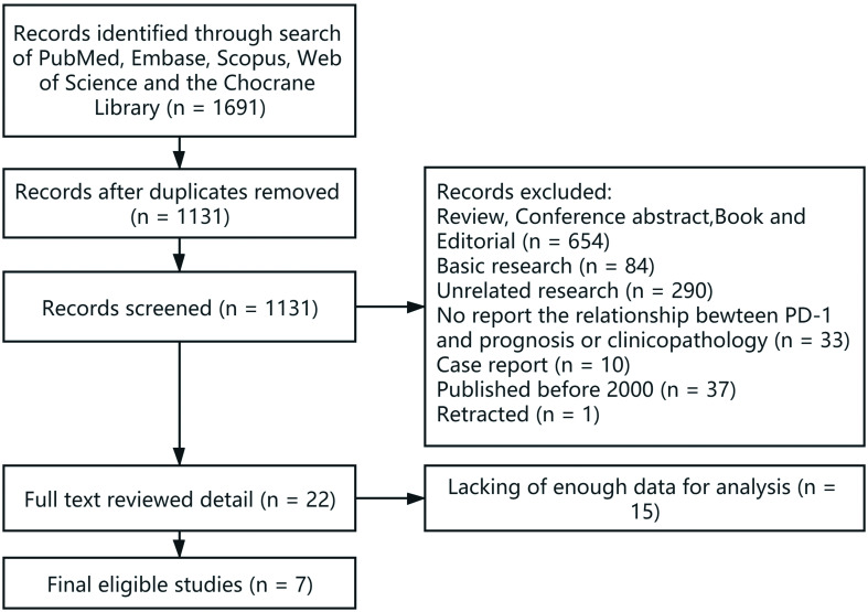
In the present study, we identified 1,691 articles with the initial searching strategy. Of these studies, we excluded 560 duplicates and deleted another 1,109 records after screening titles or abstracts. After thoroughly reviewing the full texts of 22 potentially eligible articles, 7 trials meeting the inclusion criteria were included in the final analysis. Figure 1 demonstrates the detailed selection process.
Fig. 1. Flow chart of the study’s identification of papers for meta-analysis.
Characteristics of the included studies
Table 1 lists the principal features of included studies. All studies were published in the last 20 years. The number of patients was 658 in total, ranging from 40 to 171. Two were cross-sectional studies and the rest were prospective studies. The total seven included studies were all implemented in China.
Table 1. Characteristics of studies included in the meta-analysis.
| References | Country | Case number | Method | Cut-off | Outcome | Follow-up time |
|---|---|---|---|---|---|---|
| Hsu et al.21 | Taiwan | 45 | Flow cytometry | Median percentage: 34.6% in CD3+ cells | Clinical characteristics | – |
| Shi et al.22 | China | 56 | Flow cytometry Immunohistochemistry | Median percentage: 16.31% in PMBC, 23.55% in NIL, 42.17% in TIL | Stage, DFS | 36 months |
| Zeng at el.23 | China | 141 | Flow cytometric | Median percentage in CD8+ cells: 6.55% in stage A, 10.37% in stage B, 18.34% in stage C | Clinical characteristics, OS, PFS | Median of 23 months (6–36 months) |
| Li et al.24 | China | 171 | Immunohistochemistry | IHC score median: 5.06 in tumor, 3.07 in tumor adjacent tissues | Clinical characteristics | – |
| Li et al.25 | China | 83 | ELISA | 10 ng/mL | OS, surgery | Median of 36 (1–77) months |
| Liu et al.26 | China | 40 | Flow cytometry | Median percentage: 7.05% in PMBC, 36.57% in NIL, 30.01% in TIL | DFS | 30 months |
| Liu et al.27 | China | 122 | Flow cytometry staining | Median percentage: 12.8% in CD8+ cells | Clinical characteristics, OS, PFS | Median of 59 weeks (32–84 weeks) |
NIL, nontumor infiltrating lymphocytes; TIL, Tumor infiltrating lymphocytes; PBMC, peripheral blood monocyte cell; PFS, progression-free survival.
Among the seven studies, five reported the correlation between survival and PD-1 expression, and some of them also explored the correlation between clinicopathological features and PD-1 expression, while two only examined the latter connection. Some did not directly report HRs and 95% CIs; hence, we calculated these statistics by adopting Kaplan-Meier curves. Heterogeneity existed in the method for detecting PD-1, the cut-off points of high PD-1 and the criteria of other clinical characteristics. We evaluated the study quality using the NOS. The score ranged from 6 to 8 (Table 2), suggesting that the methodology of the studies was relatively reliable.
Table 2. NOS quality assessment of the enrolled studies.
| References | Selection |
Comparability of cohorts on the basis of the design or analysis (study adjusts for ag, se) | Outcome |
Total | |||||
|---|---|---|---|---|---|---|---|---|---|
| Representativeness of the exposed cohort | Selection of the non-exposed cohort | Ascertainment of exposure | Demonstration that outcome of interest was not present at start of study | Assessment of outcome | Was follow-up long enough for outcome to occur | Adequacy of follow up of cohorts | |||
| Hsu et al.21 | – | * | * | – | ** | * | * | * | 7 |
| Shi et al.22 | * | * | * | – | ** | * | * | * | 8 |
| Zeng at el.23 | – | * | * | – | ** | * | * | * | 7 |
| Li et al.24 | * | * | * | – | ** | * | * | * | 8 |
| Li et al.25 | * | – | * | – | * | * | * | * | 6 |
| Liu et al.26 | * | * | * | – | – | * | * | * | 6 |
| Liu et al.27 | * | * | * | – | ** | * | * | * | 8 |
Survival analysis
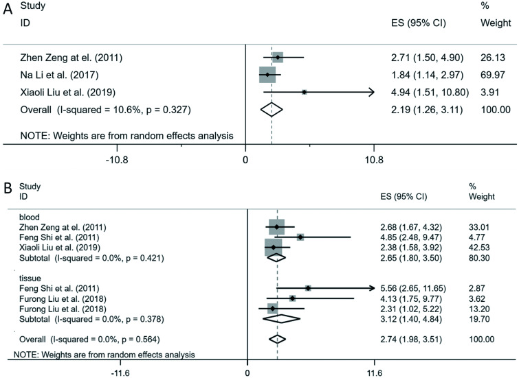
OS was measured in three of the seven included studies. A total of 314 patients from these three studies were evaluated to examine the correlation between PD-1 expression and OS. No significant heterogeneity existed among the included studies (χ2=2.24; p=0.327; I2=10.6%). Pooled results by random modeling revealed that a high level of circulating PD-1 was related to poor prognosis in term of shorter OS (HR=2.19, 95% CI: 1.26–3.12, p<0.001; Fig. 2A).
Fig. 2. Forest plot of studies evaluating the association between PD-1 expression and OS (A)/DFS (B) in patients with HBV-HCC.
Four of the seven included studies reported HR for DFS, including 423 patients. Figure 2B showed no heterogeneity in the included studies (χ2=3.90; p=0.564; I2=0%). Pooled results by random model revealed that high PD-1 predicted poorer DFS (HR=2.74, 95% CI: 1.76–3.73, p<0.001), independent of the sample resources. The subgroup analysis showed that circulating PD-1 could significantly predict the DFS (HR=2.65, 95% CI: 1.80–3.50, p<0.001; Fig. 2B), while the PD-1 expressed by tumor infiltrating lymphocytes (TILs) predicted better than the circulating PD-1 in the serum (HR=3.12, 95% CI: 1.40–4.84, p<0.001; Fig. 2B).
Correlation of PD-1 expression with clinical features
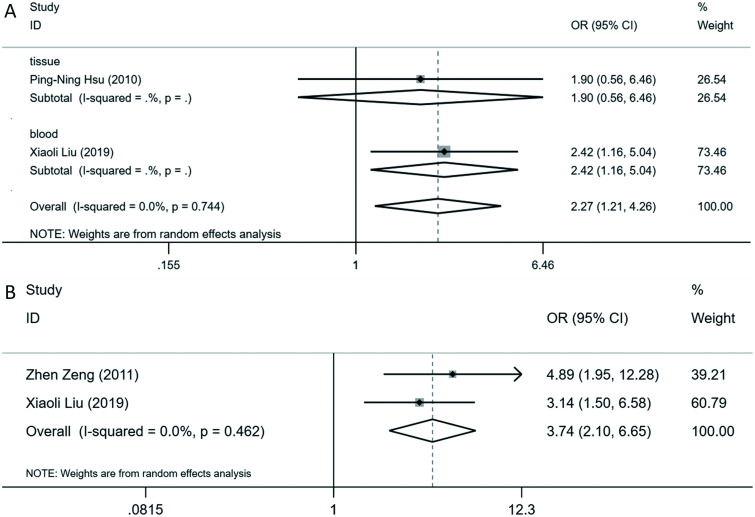
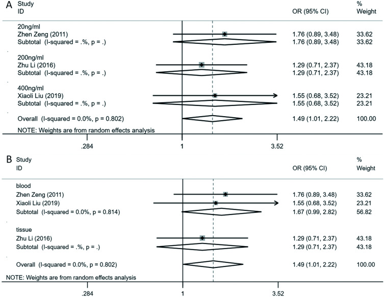
Table 3 illustrates the relationship between PD-1 overexpression and clinical parameters. A high level of PD-1 is associated with multiple tumors (OR=2.27, 95% CI: 1.21–4.26, p=0.011; Fig. 3A), advanced BCLC stages (OR=3.74, 95% CI: 2.10–6.65, p<0.001; Fig. 3B) and higher level of serum AFP (OR=1.50, 95% CI: 1.01–2.22, p=0.047; Fig. 4). However, the results of subgroup analysis showed no clinical significance. The other clinical parameters were not statistically associated with the PD-1 overexpression, including age, sex, tumor size, liver cirrhosis, portal vein invasion, vascular invasion, TNM stage and Child-Pugh score (Table 3).
Table 3. Relationship between high PD-1 and the clinicopathological features.
| Characteristic | Studies | Case number | Pooled OR (95% CI) | p | Heterogeneity |
Model | Publication bias Begg’s p | References | |
|---|---|---|---|---|---|---|---|---|---|
| I 2 | p | ||||||||
| Age | 3 | 434 | 1.148 (0.777–1.696) | 0.488 | 0% | 0.761 | Random | 0.296 | 23,24,27 |
| Sex (male/female) | 3 | 434 | 1.018 (0.568, 1.823) | 0.952 | 30% | 0.241 | Random | 1 | 23,24,27 |
| Number of tumors (multiple/solitary) | 2 | 167 | 2.268 (1.209, 4.257) | 0.011 | 0% | 0.744 | Random | 1 | 21,27 |
| Tumor size (>5 cm/≤5 cm) | 4 | 478 | 1.709 (0.420, 6.959) | 0.455 | 90.60% | <0.001 | Random | 0.734 | 21,23,24,27 |
| Liver cirrhosis (present/absent) | 2 | 167 | 1.522 (0.080–28.893) | 0.78 | 88.30% | 0.003 | Random | 1 | 21,27 |
| AFP (high/low) | 3 | 434 | 1.495 (1.005, 2.223) | 0.047 | 0% | 0.802 | Random | 1 | 23,24,27 |
| Vascular invasion (present/absent) | 3 | 308 | 0.535 (0.059–4.816) | 0.577 | 91.70% | <0.001 | Random | 1 | 21,23,27 |
| BCLC stage (C+D vs. A+B) | 2 | 263 | 3.738 (2.101, 6.651) | <0.001 | 0% | 0.462 | Random | 1 | 23,27 |
| TNM stage (3+4 vs. 1+2) | 3 | 272 | 2.116 (0.855, 5.237) | 0.105 | 60.50% | 0.08 | Random | 1 | 21,22,24 |
| HBV DNA level | 2 | 263 | 0.951 (0.553, 1.635) | 0.856 | 0% | 0.901 | Random | 1 | 23,27 |
| Child-Pugh score (B/A) | 3 | 357 | 1.704 (0.893–3.252) | 0.106 | 34.20% | 0.219 | Random | 1 | 21,23,24 |
Fig. 3. Forest plot of studies evaluating the association between PD-1 expression and tumor numbers (A)/BCLC stage (B) in patients with HBV-HCC.
Fig. 4. Forest plot of studies evaluating the association between PD-1 expression and AFP levels in patients with HBV-HCC.
A and B are two subgroup analyses respectively based on PD-1 resources and different AFP levels.
Publication bias and sensitivity analysis
We used Begg’s funnel plot to test potential publication bias. Sensitivity analysis was executed by sequentially omitting each trial one at a time. Supplementary Figure 1 shows the potential publication bias and sensitivity analysis results among the studies involved in the survival analysis. No apparent publication bias for analysis existed (Egger’s test: p=0.153 for OS and p=0.202 for DFS; Begg’s test: p=0.296 for OS and p=0.452 for DFS). The sensitivity analysis showed that no single trial remarkably altered the pooled results for OS and DFS, which indicated that our estimates were robust and reliable. Besides, there was no significant publication bias in the analysis of clinical features, either (Table 3). Sensitivity analysis demonstrated that deleting any single study did not remarkably affect the pooled ORs for the clinical parameters with significant differences (Supplementary Fig. 2).
Discussion
HCC, considered as a common highly aggressive tumor, has been disturbing the global public health system for its dismal prognosis. Elevated serum HBV DNA level serves as a reliable risk predictor independent of hepatitis B e antigen, serum alanine aminotransferase level, and liver cirrhosis.29 HBV-HCC has been the focus of research on HCC. Given the high prevalence of HBV in China, it could be profound to provide insight into the relationship between HBV-HCC and cutting-edge immunotherapy.
Since the first PD-1/PD-L1 inhibitors nivolumab and pembrolizumab were approved by the Food and Drug Administration (FDA) in 2014, this class has been rapidly developed and was approved for several solid tumors, such as melanoma and Hodgkin lymphoma.7 PD-1 is a negative regulator of T-cell activation by its mechanism of suppressing T-cell activity at different stages in the immune response when interacting with its two ligands, PD-L1 and PD-L2. When engaged by ligands, PD-1 inhibits kinase signaling pathways through phosphatase activity, rather than T-cell activation as in the physiological condition.30,31 PD-1/PD-L1 overexpression has been noticed in various solid tumors, and several studies have concluded that the overexpression of PD-1/PD-L1 plays an important role in regulating the T-cell-mediated antitumor response, leading to poor prognosis.32–34 Although the earliest and most widely recognized predictive biomarker was PD-L1, with several assays approved by FDA, it has not been proven as the definitive biomarker. In the TME with high density of CD8+ tumor infiltrating lymphocytes, PD-1, PD-L1/PD-L2 and cytotoxic T-lymphocyte antigen 4 (i.e. CTLA-4) might predict the prognosis and response to PD-1/PD-L1 blockade as well.35 In our subgroup analysis, PD-1 expressed by TILs predicted DFS better than the circulating PD-1 in the serum. This may be because the PD-1 expressed by TILs can interact with PD-L1 secreted by tumor cells, leading to immune escape of tumor cells. PD-1 is secreted into serum after being expressed in cells. The PD-1 in TILs has higher predictive efficiency and it is recommended to routinely detect PD-1 in TILs.
There have been several reviews and meta-analyses indicating the relationship between the high level of PD-L1 and worse prognosis in patients with HCC.36–38 However, two meta-analyses published last year found high expression of PD-1 predicted a better prognosis of HCC patients.39,40 We had reviewed some literature suggesting that high level of PD-1 is associated with a poor prognosis in HBV-HCC patients, and we decided to conduct a meta-analysis to explore the relationship between the PD-1 expression and prognosis in HBV-HCC. This meta-analysis, presented herein and based on seven studies with 658 patients, showed that the high expression of PD-1 statistically indicated a poor OS and DFS, whether the sample originated from blood or tumors. As for the clinicopathological parameters, our findings suggested that overexpression of PD-1 was significantly associated with multiple tumors, higher level of AFP and advanced BCLC stages of HCC. Chronic liver diseases with longstanding inflammation often induce T cell exhaustion and the appearance of regulatory T cells (i.e. Tregs). PD-1 is an important molecule in the pathway of immune escape of tumors. The increased expression can lead to immune escape of the tumor, leading to an increase of AFP and advanced BCLC stages. A tumor secreting AFP in large amounts is a prognostic sign of larger focus or multifocality, extrahepatic spread, and poor survival. Critelli et al.41 found that fast-growing HCC has poor differentiation and more angiogenesis, characterized by an immunosuppressed microenvironment under local up-regulation of PD-1 and PD-L1, along with elevated AFP, TGF-β, and IL-8. PD-1 may have a predictive value in AFP-nonsecreting HCC.
Among the included studies, two showed that high PD-1 expression was associated with larger tumor size, one showed that high PD-1 expression was associated with smaller tumor size and the results of two others showed that the PD-1 expression and tumor size were not statistically correlated (Supplementary Fig. 3). Two of included studies reported the relationship between PD-1 expression and TNM stage, which both showed that the PD-1 expression and TNM stage were not statistically correlated (Supplementary Fig. 4). Since there were studies that confirmed that the outcome of PD-1 inhibitor may be correlated with tumor size and TNM stage42,43 and two of the included studies indicated that high PD-1 expression was associated with larger tumor size, we speculated that the results may not be confirmative due to insufficient sample. It required further expansion of the sample to draw an accurate conclusion. The same was true for liver cirrhosis. Neither of the two articles that mentioned the relationship between cirrhosis and PD-1 expression showed the severity of cirrhosis. Most of the patients had low HBV DNA replication (<100 IU/mL). Liu et al.27 analyzed PD-1 level in peripheral blood mononuclear cells rather than TILs, which might not reflect the exact situation of immune infiltration in HCC tissue. Hsu et al.21 provided no information on antiviral therapy or details of cirrhosis. This could explain why such a vital factor of hepatitis B did not significantly correlate with PD-1 expression levels. The incidence of HCC increased with serum HBV DNA level in a dose-response relationship. Participants with persistent elevation of serum HBV DNA level during follow-up had the highest HCC risk.29 Regular antiviral treatment in some patients may not necessarily increase the expression of PD-1. Grouping HBV-HCC patients according to the amount of HBV replication to analyze the relationship between PD-1 and prognosis can define the predictive value of PD-1 in such patients more clearly.
The expression of PD-1 in T cells can be induced by up-regulated PD-L1 in tumor cells and by other molecules. Some studies have suggested that PD-L1-positive tumor cells have prominent immune cell infiltration in HCC, such as CD3+ TILs (representing overall T cells), CD8+ TILs (representing cytotoxic T cells), and tumor-associated macrophages (i.e. TAMs).27,37,44,45 This result may support the possibility of a role for an adaptive immune resistance mechanism. Some other research studies have pointed out that PD-1 expression is related to T cell exhaustion. Blocking the PD-1 pathway could reverse this phenotype, to restore anti-tumor immunity.46–48 In HBV-HCC, the reactivation of oncofetal gene SALL4 by HBV counteracts miR-200c in PD-L1-induced T cell exhaustion. Overexpression of miR-200c antagonizes HBV-mediated PD-L1 expression by targeting the 3′-UTR of the CD274 gene (encoding PD-L1) directly, which reverses antiviral CD8+ T cell exhaustion.49 Through analysis, we also found that the positive rate of PD-1 was relatively higher than that of PD-L1 in HCC tissue. In view of its high positive ratio, the diversity of detection methods and the stability of the results on tumor prognosis, we believe that PD-1 can be a marker to predict HBV-HCC prognosis. A meta-analysis in patients with pretreated advanced non-small-cell lung cancer indicated a slight benefit from anti-PD-1 compared to that from anti-PD-L1 inhibitors.50 An indirect comparison in advanced squamous non-small-cell lung cancer showed that, for PD-L1 low/negative patients, pembrolizumab had superior OS (HR=0.43, range: 0.24–0.76; p<0.01/HR=0.74, range: 0.40–1.38; p=0.35) and better progression-free survival (HR=0.80, range: 0.51–1.26; p=0.33/HR=0.46, range: 0.28–0.75; p<0.01) than atezolizumab. There has been no study comparing the efficacy between PD-1 inhibitors and PD-L1 inhibitors in HCC patients thus far. More clinical trials are required to determine which target is better for HBV-HCC, PD-1, or PD-L1. In our analysis, PD-1 had shown good predictive efficacy.
Han et al.44 pointed out that PD-1 expression in tumors was statistically related to the level in serum. The higher expression of PD-1 in tumors, the higher concentration of PD-1 in serum. The results of our analysis support this view. The PD-1 expression levels in tumor and in serum were consistent in predicting prognosis and clinical parameters. However, due to different detection methods and grouping levels, the relationship between PD-1 and survival may be inaccurate. Considering this point, though the heterogeneity was low, we still carried out the subgroup analysis to assure whether both of the detection methods could predict the prognosis.
To our knowledge, our study is the first meta-analysis focused on the prognostic value of PD-1 expression in HBV-HCC patients specifically. The results are different from those in all HCC patients regardless of HBV.
However, there are several limitations inherent to our study’s design. First, for some studies, we used the Engauge Digitizer to calculate HRs, which may cause bias. Second, the number of studies was insufficient. There were only three studies that investigated the correlation between PD-1 expression and OS and four that focused on the correlation between PD-1 and DFS. All studies originated from China, which may have limited the data extrapolation. Third, the grouping criteria of PD-1 level and other parameters of included studies varied. Different standards may increase bias. Finally, though our analysis upon prognosis in HBV-HCC showed the opposite result from the study in HCC, the relationship of PD-1 expression was not associated with HBV infection. The mechanism between PD-1 and HBV needs further study.
Conclusions
Our meta-analyses revealed that PD-1 expression was significantly correlated with shorter OS, DFS, higher level of AFP, multiple tumors and advanced BCLC stage of HBV-HCC. Based on the included studies, we found that PD-1 expressed in tumors and blood could reflect the immune status of patients, thereby increasing the reliability of the results. We can preliminarily assume that HBV enhances the expression of PD-1 through certain mechanisms and leads to poor prognosis of HBV-HCC patients. The prognostic role of PD-1 in HBV-HCC and the mechanism of how HBV mediated PD-1 expression still demand further investigation.
Supporting information
Abbreviations
- AFP
alpha fetoprotein
- BCLC
Barcelona Clinic Liver Cancer
- CI
confidence interval
- CTLA-4
cytotoxic T-lymphocyte antigen 4
- DFS
disease-free survival
- FDA
Food and Drug Administration
- HBV-HCC
hepatitis B virus-related hepatocellular carcinoma
- HCC
hepatocellular carcinoma
- HRs
hazard ratios
- HBV
hepatitis B virus
- mAbs
monoclonal antibodies
- NOS
Newcastle Ottawa quality assessment scale
- ORs
odds ratios
- OS
overall survival
- PD-1/PD-L1
programmed cell death 1/programmed death ligand 1
- TAM
tumor-associated macrophage
- TIL
tumor infiltrating lymphocyte
- NIL
nontumor infiltrating lymphocytes
- TME
tumor microenvironment
- TNM
tumor-node-metastasis
- Treg
regulatory T cell
Data sharing statement
All data are available upon request.
References
- 1.Bray F, Ferlay J, Soerjomataram I, Siegel RL, Torre LA, Jemal A. Global cancer statistics 2018: GLOBOCAN estimates of incidence and mortality worldwide for 36 cancers in 185 countries. CA Cancer J Clin. 2018;68(6):394–424. doi: 10.3322/caac.21492. [DOI] [PubMed] [Google Scholar]
- 2.Singal AG, Lampertico P, Nahon P. Epidemiology and surveillance for hepatocellular carcinoma: New trends. J Hepatol. 2020;72(2):250–261. doi: 10.1016/j.jhep.2019.08.025. [DOI] [PMC free article] [PubMed] [Google Scholar]
- 3.Chan SL, Wong VW, Qin S, Chan HL. Infection and cancer: The case of hepatitis B. J Clin Oncol. 2016;34(1):83–90. doi: 10.1200/JCO.2015.61.5724. [DOI] [PubMed] [Google Scholar]
- 4.Llovet JM, De Baere T, Kulik L, Haber PK, Greten TF, Meyer T, et al. Locoregional therapies in the era of molecular and immune treatments for hepatocellular carcinoma. Nat Rev Gastroenterol Hepatol. 2021;18:293–313. doi: 10.1038/s41575-020-00395-0. [DOI] [PubMed] [Google Scholar]
- 5.Roth GS, Decaens T. Liver immunotolerance and hepatocellular carcinoma: Patho-physiological mechanisms and therapeutic perspectives. Eur J Cancer. 2017;87:101–112. doi: 10.1016/j.ejca.2017.10.010. [DOI] [PubMed] [Google Scholar]
- 6.Constantinidou A, Alifieris C, Trafalis DT. Targeting programmed cell death -1 (PD-1) and ligand (PD-L1): A new era in cancer active immunotherapy. Pharmacol Ther. 2019;194:84–106. doi: 10.1016/j.pharmthera.2018.09.008. [DOI] [PubMed] [Google Scholar]
- 7.Gong J, Chehrazi-Raffle A, Reddi S, Salgia R. Development of PD-1 and PD-L1 inhibitors as a form of cancer immunotherapy: a comprehensive review of registration trials and future considerations. J Immunother Cancer. 2018;6(1):8. doi: 10.1186/s40425-018-0316-z. [DOI] [PMC free article] [PubMed] [Google Scholar]
- 8.Keir ME, Butte MJ, Freeman GJ, Sharpe AH. PD-1 and its ligands in tolerance and immunity. Annu Rev Immunol. 2008;26:677–704. doi: 10.1146/annurev.immunol.26.021607.090331. [DOI] [PMC free article] [PubMed] [Google Scholar]
- 9.Sharpe AH, Pauken KE. The diverse functions of the PD1 inhibitory pathway. Nat Rev Immunol. 2018;18(3):153–167. doi: 10.1038/nri.2017.108. [DOI] [PubMed] [Google Scholar]
- 10.Wang X, Sun Q, Liu Q, Wang C, Yao R, Wang Y. CTC immune escape mediated by PD-L1. Med Hypotheses. 2016;93:138–139. doi: 10.1016/j.mehy.2016.05.022. [DOI] [PubMed] [Google Scholar]
- 11.Wolchok JD, Chan TA. Cancer: Antitumour immunity gets a boost. Nature. 2014;515(7528):496–498. doi: 10.1038/515496a. [DOI] [PMC free article] [PubMed] [Google Scholar]
- 12.Rizvi NA, Hellmann MD, Brahmer JR, Juergens RA, Borghaei H, Gettinger S, et al. Nivolumab in combination with platinum-based doublet chemotherapy for first-line treatment of advanced non-small-cell lung cancer. J Clin Oncol. 2016;34(25):2969–2979. doi: 10.1200/JCO.2016.66.9861. [DOI] [PMC free article] [PubMed] [Google Scholar]
- 13.Hui R, Garon EB, Goldman JW, Leighl NB, Hellmann MD, Patnaik A, et al. Pembrolizumab as first-line therapy for patients with PD-L1-positive advanced non-small cell lung cancer: a phase 1 trial. Ann Oncol. 2017;28(4):874–881. doi: 10.1093/annonc/mdx008. [DOI] [PMC free article] [PubMed] [Google Scholar]
- 14.Koh YW, Jeon YK, Yoon DH, Suh C, Huh J. Programmed death 1 expression in the peritumoral microenvironment is associated with a poorer prognosis in classical Hodgkin lymphoma. Tumour Biol. 2016;37(6):7507–7514. doi: 10.1007/s13277-015-4622-5. [DOI] [PubMed] [Google Scholar]
- 15.Böger C, Behrens HM, Mathiak M, Krüger S, Kalthoff H, Röcken C. PD-L1 is an independent prognostic predictor in gastric cancer of Western patients. Oncotarget. 2016;7(17):24269–24283. doi: 10.18632/oncotarget.8169. [DOI] [PMC free article] [PubMed] [Google Scholar]
- 16.Xia H, Shen J, Hu F, Chen S, Huang H, Xu Y, et al. PD-L1 over-expression is associated with a poor prognosis in Asian non-small cell lung cancer patients. Clin Chim Acta. 2017;469:191–194. doi: 10.1016/j.cca.2017.02.005. [DOI] [PubMed] [Google Scholar]
- 17.Trojan J, Sarrazin C. Complete response of hepatocellular carcinoma in a patient with end-stage liver disease treated with nivolumab: Whishful thinking or possible? Am J Gastroenterol. 2016;111(8):1208–1209. doi: 10.1038/ajg.2016.214. [DOI] [PubMed] [Google Scholar]
- 18.Lim CJ, Lee YH, Pan L, Lai L, Chua C, Wasser M, et al. Multidimensional analyses reveal distinct immune microenvironment in hepatitis B virus-related hepatocellular carcinoma. Gut. 2019;68(5):916–927. doi: 10.1136/gutjnl-2018-316510. [DOI] [PubMed] [Google Scholar]
- 19.Sun C, Sun H, Zhang C, Tian Z. NK cell receptor imbalance and NK cell dysfunction in HBV infection and hepatocellular carcinoma. Cell Mol Immunol. 2015;12(3):292–302. doi: 10.1038/cmi.2014.91. [DOI] [PMC free article] [PubMed] [Google Scholar]
- 20.Koh S, Tan AT, Li L, Bertoletti A. Targeted therapy of hepatitis B virus-related hepatocellular carcinoma: Present and future. Diseases. 2016;4(1):10. doi: 10.3390/diseases4010010. [DOI] [PMC free article] [PubMed] [Google Scholar]
- 21.Hsu PN, Yang TC, Kao JT, Cheng KS, Lee YJ, Wang YM, et al. Increased PD-1 and decreased CD28 expression in chronic hepatitis B patients with advanced hepatocellular carcinoma. Liver Int. 2010;30(9):1379–1386. doi: 10.1111/j.1478-3231.2010.02323.x. [DOI] [PubMed] [Google Scholar]
- 22.Shi F, Shi M, Zeng Z, Qi RZ, Liu ZW, Zhang JY, et al. PD-1 and PD-L1 upregulation promotes CD8(+) T-cell apoptosis and postoperative recurrence in hepatocellular carcinoma patients. Int J Cancer. 2011;128(4):887–896. doi: 10.1002/ijc.25397. [DOI] [PubMed] [Google Scholar]
- 23.Zeng Z, Shi F, Zhou L, Zhang MN, Chen Y, Chang XJ, et al. Upregulation of circulating PD-L1/PD-1 is associated with poor post-cryoablation prognosis in patients with HBV-related hepatocellular carcinoma. PLoS One. 2011;6(9):e23621. doi: 10.1371/journal.pone.0023621. [DOI] [PMC free article] [PubMed] [Google Scholar]
- 24.Li Z, Li N, Li F, Zhou Z, Sang J, Chen Y, et al. Immune checkpoint proteins PD-1 and TIM-3 are both highly expressed in liver tissues and correlate with their gene polymorphisms in patients with HBV-related hepatocellular carcinoma. Medicine (Baltimore) 2016;95(52):e5749. doi: 10.1097/MD.0000000000005749. [DOI] [PMC free article] [PubMed] [Google Scholar]
- 25.Li N, Zhou Z, Li F, Sang J, Han Q, Lv Y, et al. Circulating soluble programmed death-1 levels may differentiate immune-tolerant phase from other phases and hepatocellular carcinoma from other clinical diseases in chronic hepatitis B virus infection. Oncotarget. 2017;8(28):46020–46033. doi: 10.18632/oncotarget.17546. [DOI] [PMC free article] [PubMed] [Google Scholar]
- 26.Liu F, Zeng G, Zhou S, He X, Sun N, Zhu X, et al. Blocking Tim-3 or/and PD-1 reverses dysfunction of tumor-infiltrating lymphocytes in HBV-related hepatocellular carcinoma. Bull Cancer. 2018;105(5):493–501. doi: 10.1016/j.bulcan.2018.01.018. [DOI] [PubMed] [Google Scholar]
- 27.Liu X, Li M, Wang X, Dang Z, Jiang Y, Wang X, et al. PD-1+ TIGIT+ CD8+ T cells are associated with pathogenesis and progression of patients with hepatitis B virus-related hepatocellular carcinoma. Cancer Immunol Immunother. 2019;68(12):2041–2054. doi: 10.1007/s00262-019-02426-5. [DOI] [PMC free article] [PubMed] [Google Scholar]
- 28.Wells G, Shea B, O’Connell D, Robertson J, Peterson J, Welch V, et al. The newcastle-ottawa scale (NOS) for assessing the quality of nonrandomised studies in meta-analyses. Available from: http://www3.med.unipmn.it/dispense_ebm/2009-2010/Corso%20Perfezionamento%20EBM_Faggiano/NOS_oxford.pdf.
- 29.Chen CJ, Yang HI, Su J, Jen CL, You SL, Lu SN, et al. Risk of hepatocellular carcinoma across a biological gradient of serum hepatitis B virus DNA level. JAMA. 2006;295(1):65–73. doi: 10.1001/jama.295.1.65. [DOI] [PubMed] [Google Scholar]
- 30.Ishida Y, Agata Y, Shibahara K, Honjo T. Induced expression of PD-1, a novel member of the immunoglobulin gene superfamily, upon programmed cell death. EMBO J. 1992;11(11):3887–3895. doi: 10.1002/j.1460-2075.1992.tb05481.x. [DOI] [PMC free article] [PubMed] [Google Scholar]
- 31.Freeman GJ, Long AJ, Iwai Y, Bourque K, Chernova T, Nishimura H, et al. Engagement of the PD-1 immunoinhibitory receptor by a novel B7 family member leads to negative regulation of lymphocyte activation. J Exp Med. 2000;192(7):1027–1034. doi: 10.1084/jem.192.7.1027. [DOI] [PMC free article] [PubMed] [Google Scholar]
- 32.Fusi A, Festino L, Botti G, Masucci G, Melero I, Lorigan P, et al. PD-L1 expression as a potential predictive biomarker. Lancet Oncol. 2015;16(13):1285–1287. doi: 10.1016/S1470-2045(15)00307-1. [DOI] [PubMed] [Google Scholar]
- 33.Chatterjee J, Dai W, Aziz NHA, Teo PY, Wahba J, Phelps DL, et al. Clinical use of programmed cell death-1 and its ligand expression as discriminatory and predictive markers in ovarian cancer. Clin Cancer Res. 2017;23(13):3453–3460. doi: 10.1158/1078-0432.CCR-16-2366. [DOI] [PubMed] [Google Scholar]
- 34.Tsutsumi S, Saeki H, Nakashima Y, Ito S, Oki E, Morita M, et al. Programmed death-ligand 1 expression at tumor invasive front is associated with epithelial-mesenchymal transition and poor prognosis in esophageal squamous cell carcinoma. Cancer Sci. 2017;108(6):1119–1127. doi: 10.1111/cas.13237. [DOI] [PMC free article] [PubMed] [Google Scholar]
- 35.Taube JM, Klein A, Brahmer JR, Xu H, Pan X, Kim JH, et al. Association of PD-1, PD-1 ligands, and other features of the tumor immune microenvironment with response to anti-PD-1 therapy. Clin Cancer Res. 2014;20(19):5064–5074. doi: 10.1158/1078-0432.CCR-13-3271. [DOI] [PMC free article] [PubMed] [Google Scholar]
- 36.Li JH, Ma WJ, Wang GG, Jiang X, Chen X, Wu L, et al. Clinicopathologic significance and prognostic value of programmed cell death ligand 1 (PD-L1) in patients with hepatocellular carcinoma: A meta-analysis. Front Immunol. 2018;9:2077. doi: 10.3389/fimmu.2018.02077. [DOI] [PMC free article] [PubMed] [Google Scholar]
- 37.Liao H, Chen W, Dai Y, Richardson JJ, Guo J, Yuan K, et al. Expression of programmed cell death-ligands in hepatocellular carcinoma: Correlation with immune microenvironment and survival outcomes. Front Oncol. 2019;9:883. doi: 10.3389/fonc.2019.00883. [DOI] [PMC free article] [PubMed] [Google Scholar]
- 38.Wang Q, Liu F, Liu L. Prognostic significance of PD-L1 in solid tumor: An updated meta-analysis. Medicine (Baltimore) 2017;96(18):e6369. doi: 10.1097/MD.0000000000006369. [DOI] [PMC free article] [PubMed] [Google Scholar]
- 39.Yang J, Zhang W, Zhang Z, Song F, Ding M, Zhao X, et al. Clinicopathological and prognostic roles of the expression levels of the programmed cell death-1 gene in patients with hepatocellular carcinoma: A systematic review and meta-analysis. Genet Test Mol Biomarkers. 2020;24(10):641–648. doi: 10.1089/gtmb.2020.0063. [DOI] [PubMed] [Google Scholar]
- 40.Li XS, Li JW, Li H, Jiang T. Prognostic value of programmed cell death ligand 1 (PD-L1) for hepatocellular carcinoma: a meta-analysis. Biosci Rep. 2020;40(4):BSR20200459. doi: 10.1042/BSR20200459. [DOI] [PMC free article] [PubMed] [Google Scholar]
- 41.Critelli R, Milosa F, Faillaci F, Condello R, Turola E, Marzi L, et al. Microenvironment inflammatory infiltrate drives growth speed and outcome of hepatocellular carcinoma: a prospective clinical study. Cell Death Dis. 2017;8(8):e3017. doi: 10.1038/cddis.2017.395. [DOI] [PMC free article] [PubMed] [Google Scholar]
- 42.Shi X, Yu PC, Lei BW, Li CW, Zhang Y, Tan LC, et al. Association between programmed death-ligand 1 expression and clinicopathological characteristics, structural recurrence, and biochemical recurrence/persistent disease in medullary thyroid carcinoma. Thyroid. 2019;29(9):1269–1278. doi: 10.1089/thy.2019.0079. [DOI] [PubMed] [Google Scholar]
- 43.Li Z, Li B, Peng D, Xing H, Wang G, Li P, et al. Expression and clinical significance of PD-1 in hepatocellular carcinoma tissues detected by a novel mouse anti-human PD-1 monoclonal antibody. Int J Oncol. 2018;52(6):2079–2092. doi: 10.3892/ijo.2018.4358. [DOI] [PMC free article] [PubMed] [Google Scholar]
- 44.Han X, Gu YK, Li SL, Chen H, Chen MS, Cai QQ, et al. Pre-treatment serum levels of soluble programmed cell death-ligand 1 predict prognosis in patients with hepatitis B-related hepatocellular carcinoma. J Cancer Res Clin Oncol. 2019;145(2):303–312. doi: 10.1007/s00432-018-2758-6. [DOI] [PMC free article] [PubMed] [Google Scholar]
- 45.Gabrielson A, Wu Y, Wang H, Jiang J, Kallakury B, Gatalica Z, et al. Intratumoral CD3 and CD8 T-cell densities associated with relapse-free survival in HCC. Cancer Immunol Res. 2016;4(5):419–430. doi: 10.1158/2326-6066.CIR-15-0110. [DOI] [PMC free article] [PubMed] [Google Scholar]
- 46.Sakuishi K, Apetoh L, Sullivan JM, Blazar BR, Kuchroo VK, Anderson AC. Targeting Tim-3 and PD-1 pathways to reverse T cell exhaustion and restore anti-tumor immunity. J Exp Med. 2010;207(10):2187–2194. doi: 10.1084/jem.20100643. [DOI] [PMC free article] [PubMed] [Google Scholar]
- 47.Liu J, Zhang S, Hu Y, Yang Z, Li J, Liu X, et al. Targeting PD-1 and tim-3 pathways to reverse CD8 T-Cell exhaustion and enhance ex vivo t-cell responses to autologous dendritic/tumor vaccines. J Immunother. 2016;39(4):171–180. doi: 10.1097/CJI.0000000000000122. [DOI] [PubMed] [Google Scholar]
- 48.Liu J, Liu Y, Meng L, Liu K, Ji B. Targeting the PD-L1/DNMT1 axis in acquired resistance to sorafenib in human hepatocellular carcinoma. Oncol Rep. 2017;38(2):899–907. doi: 10.3892/or.2017.5722. [DOI] [PMC free article] [PubMed] [Google Scholar]
- 49.Sun C, Lan P, Han Q, Huang M, Zhang Z, Xu G, et al. Oncofetal gene SALL4 reactivation by hepatitis B virus counteracts miR-200c in PD-L1-induced T cell exhaustion. Nat Commun. 2018;9(1):1241. doi: 10.1038/s41467-018-03584-3. [DOI] [PMC free article] [PubMed] [Google Scholar]
- 50.Tartarone A, Roviello G, Lerose R, Roudi R, Aieta M, Zoppoli P. Anti-PD-1 versus anti-PD-L1 therapy in patients with pretreated advanced non-small-cell lung cancer: a meta-analysis. Future Oncol. 2019;15(20):2423–2433. doi: 10.2217/fon-2018-0868. [DOI] [PubMed] [Google Scholar]
Associated Data
This section collects any data citations, data availability statements, or supplementary materials included in this article.


