


Abstract
Purpose
Gonadotropin-resistant ovary syndrome (GROS) is a rare endocrine disorder that causes hypergonadotropic hypogonadism, amenorrhea, and infertility. This study reports live birth in two women with GROS who underwent fertility treatment with in vitro maturation (IVM).
Methods
Both patients had primary infertility, amenorrhea (primary and secondary), typical secondary sexual characters, elevated gonadotropin levels, normal ovarian reserve, normal chromosomal characteristics, and previous nonresponsiveness gonadotropin stimulations. One patient had polymorphism of the follicle-stimulating hormone receptor, which is a predictor of poor ovarian response. Given unresponsiveness to exogenous gonadotropin stimulations, IVM with human chorionic gonadotropin priming (hCG-IVM) was performed in both patients. All transferrable embryos were vitrified.
Results
Both patients achieved pregnancy after their first frozen embryos transfer, and each delivered a healthy baby boy.
Conclusions
These results suggest that IVM should be a first-line therapeutic option for patients with GROS.
Keywords: In vitro maturation, Gonadotropin-resistant ovary syndrome, Live birth, Infertility
Background
In vitro maturation (IVM) is an assisted reproductive technique (ART) in which immature oocytes are retrieved from small antral follicles (diameter 2–10 mm) in the ovaries with mild or no ovarian stimulation, then subsequently matured in culture in vitro. After this maturation process, the remainder of the treatment procedure is consistent with that used in conventional intracytoplasmic sperm injection (ICSI) [1–3]. Therefore, IVM is a gentler approach to assisted reproductive technologies (ART) because complications such as ovarian hyperstimulation syndrome (OHSS) can be avoided [1]. No cases of OHSS after IVM have been reported in the literature to date.
Recent studies have demonstrated that IVM is safe for both women and their babies [4, 5]. In addition, a randomized controlled trial showed that IVM was not less effective than in vitro fertilization (IVF)/ICSI in women with high antral follicle count (AFC) [6]. Currently, IVM treatment has been advocated in clinical practice in the settings of polycystic ovary syndrome, fertility preservation, inadequate response, oocyte maturation defects, and gonadotropin resistance ovary syndrome (GROS) [1].
GROS is a rare endocrine syndrome characterized by hypergonadotropic anovulation (WHO group 3) and consequent infertility. Women with GROS experience amenorrhea (primary or secondary) despite having an intact uterus and vagina, elevated levels of follicle-stimulating hormone (FSH) and luteinizing hormone (LH), normal ovarian reserve, no concomitant autoimmune disease, and an inappropriate response to ovarian stimulation [7, 8]. The etiology of GROS remains unclear. However, both genetics and immunology might play a role. Several mutations with loss of function and single-nucleotide morphisms of the FSH receptor (FSHR) have been described to date [9–21]. The lack of response to ovarian stimulation in patients with GROS means that conventional IVF is not possible. Therefore, oocyte donation was previously considered the only option for ART in these patients. However, IVM is increasingly being recognized as a potential alternative [11, 22], which would provide the opportunity for a woman with GROS to achieve fertilization of her own oocytes. Several live births after use of IVM in GROS have been reported [7, 15, 18, 22, 23]. This paper reports successful live births with healthy babies after IVM in two women with GROS.
Methods
Karyotyping and FSHR polymorphism analysis
Karyotyping was performed by G-banding at 500–550 band level. A 2-mL sample of peripheral blood was taken on the day of oocyte pick-up. Lymphocytes were cultured with phytohemagglutinin, and cells were harvested according to cytogenetic standard procedures of hypotonic treatment and methanol/acetic acid fixation (3:1). Giemsa stain was used in this technique. FSHR polymorphism analysis was conducted in patient 2. Genomic DNA was extracted from peripheral leucocytes using the DNeasy blood and tissue extraction kit (Qiagen; catalog no.69504) then genotyping of single-nucleotide polymorphisms (SNPs) was performed using predesigned TaqMan SNP assays (Life Technologies) for FSHR polymorphisms, including c.2039A>G (p.Asn680Ser, assay C_2676874_10), c.919A>G (p.Thr307Ala, assay C_2676873_30), -29G >A (C_426553_10), and -211 G>T (C_27829553_10).
Hormonal testing
Hormonal tests performed included LH (Roche Cobas e411, sensitivity: 25.8 mIU/mL, coefficient of variability: 5.8%), FSH (Roche Cobas e411, sensitivity: 25.0 mIU/mL, coefficient of variability: 5.8%), thyroid-stimulating hormone (TSH, Beckman Access, sensitivity: 2.7 ng/dL, coefficient of variability: 5.6%), free thyroxine (fT4, Beckman Access, sensitivity: 2.7 ng/dL, coefficient of variability: 5.6%), and prolactin (Roche Cobas e411, sensitivity: 38.0 μg/L, coefficient of variability: 5.5%).
Oocyte retrieval
Oocyte retrieval was performed 36 h after human chorionic gonadotropin (hCG) administration, using a double-needle system, with a 17-G external needle and a 19-G internal needle (Kitazato®, Japan). The pressure during the collection was about 100–120 mmHg. After aspiration, follicular fluid was transferred to the lab, where immature oocytes were identified under a stereomicroscope using the sliding technique; subsequently, all fluid was poured into a strainer for a final check for any missed oocytes.
IVM, ICSI, embryo culture, and vitrification
Oocyte maturation checks were performed to separate “in vivo” mature oocytes from immature ones. “In vivo” mature oocytes underwent ICSI on the same day. Immature oocytes were placed in pre-maturation medium for 2 h, then transferred to maturation medium (Origio®, Denmark) and cultured for 20 h, after which mature oocytes underwent ICSI. For the remaining oocytes, maturation was re-checked after another 4 h, with ICSI performed in any additional mature oocytes. A fertilization check was performed under an inverted microscope at 16–18 h after insemination. Embryos were cultured in Global full LP (LifeGlobal®, Canada) at 37 °C, 6% carbon dioxide, and 5% oxygen. Embryo evaluation was performed at 44 ± 2 or 68 ± 1 h after fertilization using the Istanbul consensus. All potential embryos were vitrified (Cryotech®, Japan) on day 2 or day 3 after ICSI. Day-2 embryos had to have at least 2 cells, no multinucleation, and fragmentation of ≤ 25%, while day-3 embryos had to have at least 6 cells, no multinucleation, and fragmentation of ≤ 25%. Combined oral contraceptive pills were given to induce menstruation.
Endometrial preparation for embryo transfer and pregnancy follow-up
Oral estradiol (Valiera 2 mg; Laboratorios Recalcine, Chile) 8 mg/day was started from days 2–3 of menstruation. When endometrial thickness reached at least 8 mm, vaginal micronized progesterone (Cyclogest 400 mg; Accord-UK Limited, UK) 800 mg/day was initiated. Transfer of thawed embryos was performed 2–3 days after starting progesterone. A beta-hCG blood test for pregnancy was performed 2 weeks after embryo transfer, with a level > 5 mIU/mL defined as a positive pregnancy test. Luteal phase support with oral estradiol (Valiera 2 mg; Laboratorios Recalcine, Chile) 8 mg/day and vaginal progesterone (Cyclogest 400 mg; Accord-UK Limited, UK) 800 mg/day were given until the seventh week of gestation.
Results
Case description
The two patients were treated at IVFMD, My Duc Hospital, Ho Chi Minh City, Vietnam, between 2019 and 2020, and were aged 27 years when they came to our clinic. They both had amenorrhea, with menstruation induced by oral contraceptive pills. The first patient had primary amenorrhea, while the second had normal puberty and menstruation until the age of 22 years. Both patients had a history of primary infertility. Physical examinations found normal mature secondary sexual characteristics and no hyperandrogenic signs. No significant medical or surgical history was reported, and there was no remarkable family history of inheritable conditions.
Both patients had undergone two previous unsuccessful attempts at ovarian stimulation, one each at another hospital and one each at our hospital. In patient 1, the first stimulation cycle was cancelled after 3 days of FSH injections with a total dose of 900 IU due to inadequate response; the second did not result in growth of any follicles after 7 days of injections of FSH+hMG with a total dose of 3000 IU and was converted to hCG-IVM. In patient 2, the first stimulation cycle resulted in three immature oocytes retrieved but no embryos were produced after 10 days of FSH+hMG injections with a total dose of 3900 IU, and the second unsuccessful cycle was then converted to hCG-IVM after 7 days of FSH+hMG injections with a total dose of 3000 IU.
Fertility investigations
Comprehensive evaluations were performed in both patients, including pelvic ultrasound scan, hormonal and ovarian reserve testing, and karyotype analysis. The ultrasound scan was normal in patient 1, but patient 2 showed a hypoplastic uterus with polycystic ovarian (PCO) morphology. Both patients had normal thyroid function (TSH and fT4), high levels of FSH and LH, and elevated anti-Mullerian hormone levels (Table 1). Patient 1 had a normal AFC on ultrasound, while patient 2 had PCO morphology in both ovaries (Table 1). The results of FSHR polymorphism testing in patient 2 showed the homozygous threonine FSHR polymorphism (alanine was replaced by threonine at position 307: Ala307Thr (rs6165) (Table 1).
Table 1.
Patients’ baseline and laboratory characteristics
| Characteristics | Patient 1 | Patient 2 |
|---|---|---|
| Body mass index, kg/m2 | 20 | 22 |
| Follicle-stimulating hormone, IU/mL | 46.53 | 91.84 |
| Luteinizing hormone, IU/mL | 48.15 | 35.2 |
| Anti-Müllerian hormone, ng/mL | 2.22 | 5.53 |
| Antral follicle count, n | 14 | PCO-like ovaries |
| Prolactin, ng/m | 9.83 | 2.93 |
| Thyroid-stimulating hormone, μIU/mL | 1.488 | 1.222 |
| Free thyroxine, ng/dL | 1.04 | 0.97 |
| Karyotypes | Normal | 46XX, 22pstk+ |
| Follicle-stimulating hormone receptor polymorphisms | - | Ala307Thr (rs6165) |
Oocytes retrieval and IVM treatment
On the second day of the induced cycle, both patients came to the clinic for infertility treatment, where they were provided with full details of their options and potential treatments. IVM was recommended but patients requested to have ovarian stimulation first, and if this was unsuccessful, they agreed to undergo IVM.
The first patient was treated with recombinant FSH (rFSH) starting at 300 IU/day. Four days later, ultrasound scan showed that the largest follicle had a diameter of 4 mm, and the rest had a diameter of 2 mm. The gonadotropin dosage was increased to 600 IU/day (300 IU/day of rFSH and 300 IU/day of human menopausal gonadotropin [hMG]), but no change in follicle size was detected on ultrasound 3 days later. The endometrium was thin (3 mm) and the estradiol level was low (42 pg/mL).
Patient 2 was treated with hMG 300 IU/day, with an ultrasound scan 4 days later which showed small follicles (diameter 2–4 mm) and endometrial thickness of 5 mm. Increasing the gonadotropin dosage to 600 IU/day (rFSH 300 IU/day and hMG 300 IU/day) for 3 days had no effect, and the estradiol level was extremely low (< 5 ng/mL).
Both patients agreed to undergo IVM and were given a single dose of hCG 10,000 IU; oocyte retrieval was scheduled for 36 h later. Mature embryos were obtained from both patients and vitrified (Table 2).
Table 2.
IVM cycle outcomes
| Characteristics | Patient 1 | Patient 2 |
|---|---|---|
| Number of oocytes: | 7 | 13 |
| Metaphase II | 0 | 4 |
| Metaphase I | 2 | 4 |
| Germinal vesicle | 5 | 5 |
| Number of matured oocytes after “in vitro” maturation culture | 2 | 3 |
| Number of oocytes fertilized | 2 | 5 |
| Number of embryos | Two day-2 | Three day-3 and two day-2 |
| Number of embryos from “in vivo” matured oocytes | 0 | Three day-3 |
| Number of embryos from matured oocytes after in vitro maturation culture | Two day-2 | Two day-2 |
| Number of embryos transferred | Two day-2 (grades I and II)* | Two day-3 (2 grade II)* |
| Number of embryos frozen | 0 | One day-3 (grade II)* and two day-2 (grade II)* |
| Endometrial thickness (mm) | 9.5 | 11 |
| Clinical pregnancy, n | 1 | 1 |
| Live birth, n | 1 | 1 |
| Infant birth statistics: | ||
| Weight, g | 3100 | 3310 |
| Length, cm | 48 | 45 |
| APGAR scores at 1 and 5 min | 8 and 9 | 8 and 9 |
*According to Alpha consensus on embryo assessment
Embryo transfer and pregnancy monitoring
After embryo vitrification, patients were given oral contraceptives to induce menstruation and returned to the clinic on the second day of the menstrual cycle. Two embryos (Fig. 1) were implanted in each patient (Table 2). Both pregnancies progressed normally and resulted in delivery of a healthy male infant to each patient by cesarean section (Table 2). Patient 2 has three additional frozen embryos for future use.
Fig. 1.
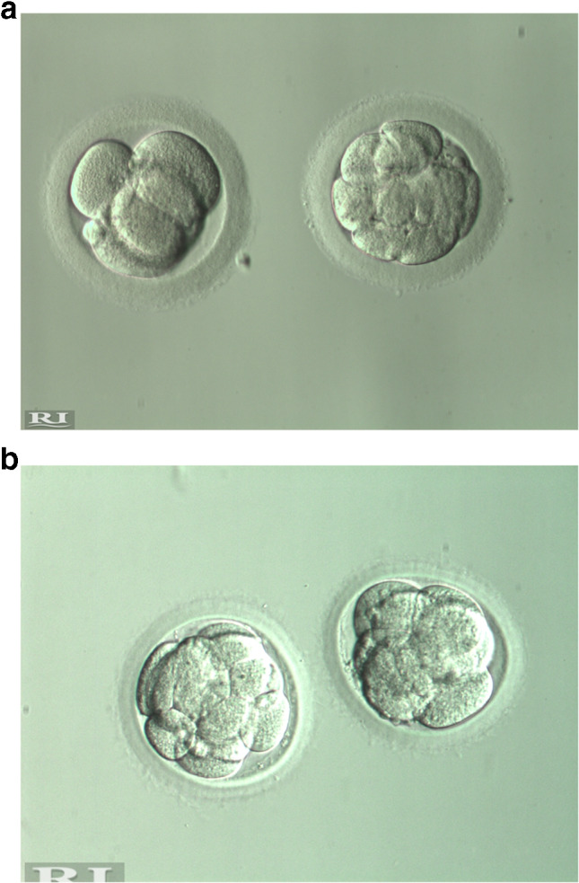
Embryos for transfer: A Two day-2 embryos from patient 1; B Two day-3 embryos from patient 2
Discussion
Although GROS was first reported more than 50 years ago [8], there have not been many live births after IVM in these patients [15, 18, 22, 23]. This paper described two successful live births in patients with GROS who had experienced failed IVF cycles due to unresponsiveness to ovarian stimulation with FSH/hMG.
The main features of GROS include endogenous hypergonadotropinemia, a normal number of ovarian follicles, and lack of response to stimulation with exogenous human gonadotropins. A high level of FSH and normal level of AMH are the hormonal characteristics of GROS. AMH represents the number of primordial follicles up to small antral follicles of < 8 mm. FSH is controlled by the estradiol level of the developing follicle. Normally, the developing follicle secretes an increasing level of estradiol; in turn, estradiol exerts negative feedback on the pituitary causing a reduction in FSH level. In GROS, the follicle does not respond to gonadotropin, there is no growth of the follicle and no associated increase in estradiol level, hence no negative feedback of estradiol on the pituitary, and therefore FSH continues to increase. In GROS, the number of follicles is normal, but the follicles do not respond to gonadotropins. Therefore, AMH level is normal but FSH level is high. Patients with GROS experience a number of complications, including primary/secondary amenorrhea and infertility, but fail ART due to an inadequate ovarian response to exogenous FSH [24–26]. To accurately diagnose GROS, a comprehensive evaluation should be performed in patients presenting with amenorrhea, including FSH, LH, and AMH levels, an ultrasound scan to evaluate AFC and FSHR genotyping/phenotyping. Currently, levels of inhibin B and AMH, and the AFC, are thought to provide the best indication of ovarian reserve in patients with GROS. Levels of inhibin B in currently reported cases have varied. In the first GROS case with live birth, reported by Grynberg et al. [15], the inhibin B level was very low, while it was normal in another case [18], but not reported in another [22]. Due to the strong correlation between inhibin B level and the diameter of antral follicles, differences in follicle sizes might influence the inhibin B level. Meanwhile, intrafollicular AMH levels slowly increase in parallel with increasing follicle diameter [27]. Therefore, in patients with GROS, AFC and AMH are highly correlated and sufficiently reflect oocyte quantity. In the current literature, patients both have normal or elevated AMH and AFC levels [18, 22, 23], and ovarian reserves in our patients are similar in previously reported cases.
The etiology of GROS is still unknown. Previous papers reported no unusual surgical, autoimmune disease, or genetic predisposition (i.e., family histories, karyotypes, gonadotropin receptors, and post-receptor defects) [28, 29]. Therefore, it was hypothesized that abnormal interaction between FSH and its receptor might play a role [28, 29]. Certainly, a variety of FSHR mutations have been described in the literature [9–21]. FSHR polymorphism testing was performed in patient 2, who showed heterozygosity for the threonine variant located at FSHR rs6165 (p. A307Thr). Interestingly, others have also demonstrated that the FSHR (rs6165) was associated with a lower yield of oocytes retrieved and a higher risk for inadequate response to ovarian stimulation [24]. Moreover, the FSHR (rs6165) genotype also influences the number of embryos produced [30]. The molecular mechanism for the effects of genotypes and ovarian response could be the change from a polar (T) to a nonpolar, hydrophobic (A) amino acid, thereby removing a potential O-linked glycosylation site [24], and this might be involved in the hormone-binding ability of FSHR and FSH-mediated signal transduction during ovarian stimulation [31]. Thus, as recently discussed [11], FSHR genetic testing should be used to help determine suitability of individual GROS patients for IVM.
In our report, there was a potential role for the injection of hCG 10,000 IU in the maturation of the oocytes without IVM culture. The “in vivo” matured oocytes were harvested in both cases (two in patient 1 and four in patient 2). The presence of “in vivo” matured oocytes was not reported in previous papers regardless of hCG usage [15, 18, 22]. The hCG priming approach used in our patients is currently the most widely used of all possible IVM regimens. One of the disadvantages of hCG IVM is that oocytes are at varying maturity levels at oocyte pick-up [31]. In our report, the first patient had two “in vivo” mature oocytes (metaphase [M] II) that did not develop to cleavage stage, while there were four such oocytes in the second patient. It is interesting to know that there could be the opportunity for oocytes from patients with GROS to be matured “in vivo” despite the small follicle sizes. Of these oocytes in our report, three embryos were successfully cultured to day 3, and patient 2 had transfer of embryos cultured from “in vivo” mature oocytes. However, given that the standard IVM should be done without FSH and hCG priming, a clinical approach for IVM in which no FSH and hCG are given before oocyte retrieval should be considered [1]. Recently, Kornilov et al. described immature oocyte retrieval for IVM culture without FSH and hCG [23]. The patient was scheduled for oocyte retrieval on the eighth day of oral contraceptive-induced menstruation, and fifteen immature oocytes were retrieved, resulting in three blastocysts; two of these were euploidy after preimplantation genetic testing for aneuploidy and a single embryo transfer was performed resulting in the birth of a healthy baby [23].
Over the last 5 years, a non-hCG IVM system has been developed. This uses oocyte prematuration (or pre-IVM) culture systems to prevent spontaneous in vitro maturation processes and maintain cumulus-oocyte gap junctional communication [32, 33]. Such methodology has been termed capacitation IVM (CAPA-IVM) [34]. A recent randomized controlled trial showed that CAPA-IVM was not less effective than IVF/ICSI in women with high AFC [6]. This non-hCG IVM system has the potential to become the standard IVM protocol in clinical settings.
In clinical practice, patients with GROS are often considered to have a poor ovarian response or chronic anovulation. For poor ovarian response, new POSEIDON criteria have recently been established to individualize the approach to poor responders [35]. Poor responders are stratified into four groups based on age, ovarian reserve, and previous ovarian response. Those with adequate ovarian reserve but low response to stimulation are classified into groups 1 and 2. Poor responses in these patients are due to low FSH sensitivity and GROS [35]. In addition, for patients with anovulation WHO group III, a precise differential diagnosis should clearly distinguish between patients with primary ovarian insufficiency and those with GROS [22]. For those diagnosed with GROS, IVM may offer a new potential treatment alternative to oocyte donation.
Although IVM could be a first-line treatment options for patients with GROS, there are some barriers to its widespread implementation. These include inappropriate concerns regarding effectiveness and safety, lack of uniform protocols, few centers with the appropriate expertise, and lack of appropriate training [34, 36]. Therefore, specific actions are needed to provide incentives for the broad uptake of IVM worldwide. First, there is a need for a standard IVM protocol. A variety of clinical and laboratory approaches are currently used for IVM, including such as conventional, hCG- and non-hCG-primed, and FSH- and non-FSH primed. Secondly, specific training should be organized for specialists. For example, activities should include training on the technology needed for physicians to aspirate unexpanded cumulus-oocyte complexes from antral follicles, and training courses for embryologists on handling of unexpanded cumulus-oocyte complexes on and embryo maturation culture skills. Thirdly, more studies on IVM-related issues should be conducted, such as those relating to the health of babies born with the use of this technique. Finally, it is essential to improve community recognition for IVM as a safe, affordable, convenient, and authentic ART technique [1, 34].
The outcomes in our two patients emphasize the potential role of IVM in infertility treatment for patients with GROS. However, this report was limited by the lack of information on the FSHR variant of the first patient. This is essential to expand knowledge on the role of FSHR variants in ovarian response and to confirm the consistency of FSHR (rs6165) genotypes in patients with GROS.
Conclusion
The details of these cases show that a “gentle” ART option exists for women with GROS. In order to maximize the success of this approach, a comprehensive evaluation is required, including FSHR genotyping. There is also a need for training to facilitate small follicle OPU and detect unexpanded cumulus-oocyte complexes, then to apply the IVM culture technique. This would allow personalized treatment for women with GROS and improve fertility outcomes in these challenging patients.
Author contribution
HLL, TTNL, and VTTT designed the study and monitored data collection. Planning for the first draft of the manuscript was undertaken by HLL. The first draft of the paper was written by HLL and VNAH. The final draft of the paper was written by HLL and LNV. All authors were involved in the decision to publish the paper and in critical revisions of the manuscript. HLL acts as overall guarantor and accepts full responsibility.
Declarations
Conflict of interest
LNV has received speaker and conference fees from Merck; and grant, speaker, and conference fees from Merck Sharpe & Dohme and Ferring. TMH has received speaker fees from Merck, Merck Sharp & Dohme, and Ferring. All other authors declare no competing interests.
Footnotes
Publisher’s note
Springer Nature remains neutral with regard to jurisdictional claims in published maps and institutional affiliations.
References
- 1.De Vos M, Grynberg M, Ho TM, Yuan Y, Albertini DF, Gilchrist RB. Perspectives on the development and future of oocyte IVM in clinical practice. J Assist Reprod Genet. 2021;38:1265–1280. doi: 10.1007/s10815-021-02263-5. [DOI] [PMC free article] [PubMed] [Google Scholar]
- 2.Edwards RG. Maturation in vitro of human ovarian oöcytes. Lancet. 1965;2:926–929. doi: 10.1016/s0140-6736(65)92903-x. [DOI] [PubMed] [Google Scholar]
- 3.Edwards RG, Bavister BD, Steptoe PC. Early stages of fertilization in vitro of human oocytes matured in vitro. Nature. 1969;221:632–635. doi: 10.1038/221632a0. [DOI] [PubMed] [Google Scholar]
- 4.Ho VNA, Braam SC, Pham TD, Mol BW, Vuong LN. The effectiveness and safety of in vitro maturation of oocytes versus in vitro fertilization in women with a high antral follicle count. Hum Reprod. 2019;34:1055–1064. doi: 10.1093/humrep/dez060. [DOI] [PubMed] [Google Scholar]
- 5.Vuong LN, Le AH, Ho VNA, Pham TD, Sanchez F, Romero S, et al. Live births after oocyte in vitro maturation with a prematuration step in women with polycystic ovary syndrome. J Assist Reprod Genet. 2020;37:347–357. doi: 10.1007/s10815-019-01677-6. [DOI] [PMC free article] [PubMed] [Google Scholar]
- 6.Vuong LN, Ho VNA, Ho TM, Dang VQ, Phung TH, Giang NH, et al. In-vitro maturation of oocytes versus conventional IVF in women with infertility and a high antral follicle count: a randomized non-inferiority controlled trial. Hum Reprod. 2020;35:2537–2547. doi: 10.1093/humrep/deaa240. [DOI] [PubMed] [Google Scholar]
- 7.Galvão A, Segers I, Smitz J, Tournaye H, De Vos M. In vitro maturation (IVM) of oocytes in patients with resistant ovary syndrome and in patients with repeated deficient oocyte maturation. J Assist Reprod Genet. 2018;35:2161–2171. doi: 10.1007/s10815-018-1317-z. [DOI] [PMC free article] [PubMed] [Google Scholar]
- 8.Jones GS, De Moraes-Ruehsen M. A new syndrome of amenorrhae in association with hypergonadotropism and apparently normal ovarian follicular apparatus. Am J Obstet Gynecol. 1969;104:597–600. doi: 10.1016/s0002-9378(16)34255-7. [DOI] [PubMed] [Google Scholar]
- 9.Allen LA, Achermann JC, Pakarinen P, Kotlar TJ, Huhtaniemi IT, Jameson JL, et al. A novel loss of function mutation in exon 10 of the FSH receptor gene causing hypergonadotrophic hypogonadism: clinical and molecular characteristics. Hum Reprod. 2003;18:251–256. doi: 10.1093/humrep/deg046. [DOI] [PubMed] [Google Scholar]
- 10.Beau I, Touraine P, Meduri G, Gougeon A, Desroches A, Matuchansky C, et al. A novel phenotype related to partial loss of function mutations of the follicle stimulating hormone receptor. J Clin Invest. 1998;102:1352–1359. doi: 10.1172/jci3795. [DOI] [PMC free article] [PubMed] [Google Scholar]
- 11.Benammar A, Fanchin R, Filali-Baba M, Vialard F, Fossard C, Vandame J, et al. Utilization of in vitro maturation in cases with a FSH receptor mutation. J Assist Reprod Genet. 2021;38:1311–1321. doi: 10.1007/s10815-021-02249-3. [DOI] [PMC free article] [PubMed] [Google Scholar]
- 12.Bramble MS, Goldstein EH, Lipson A, Ngun T, Eskin A, Gosschalk JE, et al. A novel follicle-stimulating hormone receptor mutation causing primary ovarian failure: a fertility application of whole exome sequencing. Hum Reprod. 2016;31:905–914. doi: 10.1093/humrep/dew025. [DOI] [PMC free article] [PubMed] [Google Scholar]
- 13.Doherty E, Pakarinen P, Tiitinen A, Kiilavuori A, Huhtaniemi I, Forrest S, et al. A Novel mutation in the FSH receptor inhibiting signal transduction and causing primary ovarian failure. J Clin Endocrinol Metab. 2002;87:1151–1155. doi: 10.1210/jcem.87.3.8319. [DOI] [PubMed] [Google Scholar]
- 14.Gromoll J, Simoni M, Nordhoff V, Behre HM, De Geyter C, Nieschlag E. Functional and clinical consequences of mutations in the FSH receptor. Mol Cell Endocrinol. 1996;125:177–182. doi: 10.1016/s0303-7207(96)03949-4. [DOI] [PubMed] [Google Scholar]
- 15.Grynberg M, Peltoketo H, Christin-Maître S, Poulain M, Bouchard P, Fanchin R. First birth achieved after in vitro maturation of oocytes from a woman endowed with multiple antral follicles unresponsive to follicle-stimulating hormone. J Clin Endocrinol Metab. 2013;98:4493–4498. doi: 10.1210/jc.2013-1967. [DOI] [PubMed] [Google Scholar]
- 16.Katari S, Wood-Trageser MA, Jiang H, Kalynchuk E, Muzumdar R, Yatsenko SA, et al. Novel inactivating mutation of the fsh receptor in two siblings of indian origin with premature ovarian failure. J Clin Endocrinol Metab. 2015;100:2154–2157. doi: 10.1210/jc.2015-1401. [DOI] [PMC free article] [PubMed] [Google Scholar]
- 17.Kuechler A, Hauffa BP, Köninger A, Kleinau G, Albrecht B, Horsthemke B, et al. An unbalanced translocation unmasks a recessive mutation in the follicle-stimulating hormone receptor (FSHR) gene and causes FSH resistance. Eur J Hum Genet. 2010;18:656–661. doi: 10.1038/ejhg.2009.244. [DOI] [PMC free article] [PubMed] [Google Scholar]
- 18.Li Y, Pan P, Yuan P, Qiu Q, Yang D. Successful live birth in a woman with resistant ovary syndrome following in vitro maturation of oocytes. J Ovarian Res. 2016;9:54. doi: 10.1186/s13048-016-0263-6. [DOI] [PMC free article] [PubMed] [Google Scholar]
- 19.Meduri GM, Vu Hai MT, Jolivet A, Takemori S, Kominami S, Driancourt MA, et al. Comparison of cellular distribution of LH receptors and steroidogenic enzymes in the porcine ovary. J Endocrinol. 1996;148(3):435–446. doi: 10.1677/joe.0.1480435. [DOI] [PubMed] [Google Scholar]
- 20.Nakamura Y, Maekawa R, Yamagata Y, Tamura I, Sugino N. A novel mutation in exon8 of the follicle-stimulating hormone receptor in a woman with primary amenorrhea. Gynecol Endocrinol. 2008;24:708–712. doi: 10.1080/09513590802454927. [DOI] [PubMed] [Google Scholar]
- 21.Touraine P, Beau I, Gougeon A, Meduri G, Desroches A, Pichard C, et al. New natural inactivating mutations of the follicle-stimulating hormone receptor: correlations between receptor function and phenotype. Mol Endocrinol. 1999;13:1844–1854. doi: 10.1210/mend.13.11.0370. [DOI] [PubMed] [Google Scholar]
- 22.Flageole C, Toufaily C, Bernard DJ, Ates S, Blais V, Chénier S, et al. Successful in vitro maturation of oocytes in a woman with gonadotropin-resistant ovary syndrome associated with a novel combination of FSH receptor gene variants: a case report. J Assist Reprod Genet. 2019;36:425–432. doi: 10.1007/s10815-018-1394-z. [DOI] [PMC free article] [PubMed] [Google Scholar]
- 23.Kornilov NV, Pavlova MN, Yakovlev PP. The live birth in a woman with resistant ovary syndrome after in vitro oocyte maturation and preimplantation genetic testing for aneuploidy. J Assist Reprod Genet. 2021;38:1303–1309. doi: 10.1007/s10815-021-02085-5. [DOI] [PMC free article] [PubMed] [Google Scholar]
- 24.Alviggi C, Conforti A, Santi D, Esteves SC, Andersen CY, Humaidan P, et al. Clinical relevance of genetic variants of gonadotrophins and their receptors in controlled ovarian stimulation: a systematic review and meta-analysis. Hum Reprod Update. 2018;24:599–614. doi: 10.1093/humupd/dmy019. [DOI] [PubMed] [Google Scholar]
- 25.Latronico AC, Arnhold IJP. Gonadotropin resistance. In: Maghnie M, Loche S, Cappa M, Ghizzoni L, Lorini R, editors. Endocrine development. Basel: S. Karger AG; 2013. [Google Scholar]
- 26.Talbert LM, Raj MH, Hammond MG, Greer T. Endocrine and immunologic studies in a patient with resistant ovary syndrome. Fertil Steril. 1984;42:741–744. doi: 10.1016/s0015-0282(16)48200-2. [DOI] [PubMed] [Google Scholar]
- 27.Andersen CY, Schmidt KT, Kristensen SG, Rosendahl M, Byskov AG, Ernst E. Concentrations of AMH and inhibin-B in relation to follicular diameter in normal human small antral follicles. Hum Reprod. 2010;25:1282–1287. doi: 10.1093/humrep/deq019. [DOI] [PubMed] [Google Scholar]
- 28.Conway GS, Conway E, Walker C, Hoppner W, Gromoll J, Simoni M. Mutation screening and isoform prevalence of the follicle stimulating hormone receptor gene in women with premature ovarian failure, resistant ovary syndrome and polycystic ovary syndrome. Clin Endocrinol. 1999;51:97–99. doi: 10.1046/j.1365-2265.1999.00745.x. [DOI] [PubMed] [Google Scholar]
- 29.Haller-Kikkatalo K, Salumets A, Uibo R. Review on autoimmune reactions in female infertility: antibodies to follicle stimulating hormone. Clin Dev Immunol. 2012;2012:762541. doi: 10.1155/2012/762541. [DOI] [PMC free article] [PubMed] [Google Scholar]
- 30.Trevisan CM, Peluso C, Cordts EB, de Oliveira R, Christofolini DM, Barbosa CP, et al. Ala307Thr and Asn680Ser polymorphisms of FSHR gene in human reproduction outcomes. Cell Physiol Biochem. 2014;34:1527–1535. doi: 10.1159/000366356. [DOI] [PubMed] [Google Scholar]
- 31.Agrawal G, Dighe RR. Critical involvement of the hinge region of the follicle-stimulating hormone receptor in the activation of the receptor. J Biol Chem. 2009;284:2636–2647. doi: 10.1074/jbc.M808199200. [DOI] [PubMed] [Google Scholar]
- 32.Sánchez F, Lolicato F, Romero S, De Vos M, Van Ranst H, Verheyen G, et al. An improved IVM method for cumulus-oocyte complexes from small follicles in polycystic ovary syndrome patients enhances oocyte competence and embryo yield. Hum Reprod. 2017;32:2056–2068. doi: 10.1093/humrep/dex262. [DOI] [PubMed] [Google Scholar]
- 33.Son WY, Tan SL. Laboratory and embryological aspects of hCG-primed in vitro maturation cycles for patients with polycystic ovaries. Hum Reprod Update. 2010;16:675–689. doi: 10.1093/humupd/dmq014. [DOI] [PubMed] [Google Scholar]
- 34.Smitz JE, Thompson JG, Gilchrist RB. The promise of in vitro maturation in assisted reproduction and fertility preservation. Semin Reprod Med. 2011;29:24–37. doi: 10.1055/s-0030-1268701. [DOI] [PubMed] [Google Scholar]
- 35.Esteves SC, Alviggi C, Humaidan P, Fischer R, Andersen CY, Conforti A, et al. The POSEIDON Criteria and Its Measure of Success Through the Eyes of Clinicians and Embryologists. Front Endocrinol (Lausanne) 2019;10:814. doi: 10.3389/fendo.2019.00814. [DOI] [PMC free article] [PubMed] [Google Scholar]
- 36.Vuong LN, Ho TM, Gilchrist RB, Smitz J. The Place of In Vitro Maturation in Assisted Reproductive Technology. Fertil Reprod. 2019;01:11–15. doi: 10.1142/s2661318219300022. [DOI] [Google Scholar]