Summary:
Lip fillers have a great impact on the facial aesthetic industry, where several techniques have been proposed for lip beautification in terms of both the results and delivering a safe injection procedure. The study aimed to report a personal experience with a new lip filler technique, via inserting a microcannula through three entry points, resembling an inverted Mercedes Benz sign. Ten female patients between 22 and 29 years of age had a lip filler treatment with a cross-linked hyaluronic acid injected using a microcannula through two entry points at both Glogau-Klein points of the upper lip and one entry point at the midline of the lower lip. The filler product was deposited in both retrograde and aliquots fashion in the superficial muscular plane. All patients reported a high degree of satisfaction with the results of the procedure, with slight swelling and bruising transiently present in some of the patients. Unlike the conventional cannula technique, this new technique offers artistry in accentuating the cupid’s bow and redrawing the lips.
Takeaways
Question: Can microcannula be used to reshape the lips and to accentuate the cupid bow and lip tubercles?
Findings: The proposed technique provides satisfactory results in terms of reshaping the lips and providing a safe injection.
Meaning: The three point technique provides a safe and artistic approach for lip filler injections.
INTRODUCTION
Many people consider pouty and flipped lips to be a sign of a youthful and attractive look.1 That is why many techniques have been advocated for lip augmentation, which may be surgical or nonsurgical. Using hyaluronic acid filler to enhance the lip volume is one of the most requested services in our daily facial aesthetic practice.2,3 The term beautification refers to beautifying the lips with respect to the normal lip proportions, which ideally should be 1:1.6. However, this ratio may change in African and Asian patients.4
Attractive lip shapes are in the eye of their beholder: older women seek rejuvenation to treat the signs of aging by achieving a more youthful and hydrated lip.5 Nowadays millennial patients request what is called “Russian lips” (in other words, the tenting technique), where the filler is placed in a vertical struts injection along the whole lip, violating both lip borders (the vermillion line and the wet/dry border) with a possible risk of vascular occlusion and/or filler spread into the skin side of the lip (ergotrid area), creating a plateau appearance (or mustache appearance), as described by Harris.6 This technique aims at achieving a flat profile with an increase in the vermillion height where the ratio is almost 50/50 upper to lower lip. In the latter technique, the filler is placed with a needle.
Several complications associated with lip fillers have been reported by many clinicians, such as ischemia, tissue necrosis, and visual loss.7–9 Detailed knowledge of anatomy, lip zones, rheological properties of the filler, safe injection techniques, and the use of a microcannula10 and/or duplex ultrasound11,12 may minimize the risk of lip filler complications. The technique described here is my innovative modified approach to achieve a lip eversion, increased vermillion height, and redrawing the cupid’s bow to give a pouty appearance with the use of a largely borne cannula, which carries a lower risk of vascular occlusion and complications, especially those associated with the original Russian lip technique.
MATERIALS AND METHODS
The present study included a sample of 10 White women aged 22–29 years who desired to have a lip filler treatment. Both medical and dental history were taken for all patients, as well as the history of any previous injectables and/or COVID-19 vaccine. Written informed consent was obtained for all patients explaining the full details regarding the procedure along with the side effects and possible complications. Full assessments were performed for the mouth at rest and dynamics, any asymmetry, skin quality, lip ratio, and shape. Less than a syringe of hyaluronic acid filler (Belotero Volume, Merz Company) was used for all the patients, and all injections were performed using a microcannula (SoftFil Paris, France).
All patients were anesthetized using Scandonest plain 3% anesthesia (Mepivacaine Hydrochloride 3% without vasoconstrictor). The anesthesia was injected at five points extra orally, two points at 1 cm from the alar base of the nose bilaterally, one point at the subnasale, and two points opposite to the mental foramen bilaterally. This was followed by the application of betadine on the lips and perioral area.
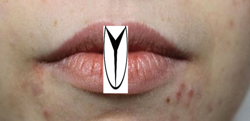
Two vertical lines were drawn from the ala of the nose down to the inferior border of the chin in addition to another line at the midline of both the upper and lower lip, the two vertical lines from the ala outlines, the borders of the injection process where the filler product must be confined within this area, as shown in Supplemental Video 1 (See Video 1 [online], which displays the outline for the injection technique followed by injection of the upper lip.). Three entry points were chosen for the whole injection procedure resembling an inverted triangle or an inverted Mercedes Benz sign, the upper lip was injected through two entry points that were chosen at each Glogau-Klein (GK) point (the ski slope shape of the lip in the profile as you move from the skin above the lip down onto the pink vermillion). The lower lip, on the other hand, was injected through one entry point right at the midline (Fig. 1). Filler injection for the upper lip was performed in the following fashion: a pilot needle was used to create an opening right at the tip of cupid’s bow (GK point) on the right side of the upper lip, as shown in Supplemental Video 2 (See Video 2 [online], which shows the injection technique for the upper lip.). After creating the opening, a softfil microcannula (22 G 50 mm) was used to enter through the opening in a vertical fashion until it reached slightly behind the wet/dry border. The injection was administered in the form of aliquots (0.1 mm) at this area followed by retrograde injection, leaving a microdroplet at the GK point upon exit. The same maneuver was performed many times until achieving the desired pouty appearance in addition to redirecting the cannula from the same entry point to fill in the sides of the lip. The same steps were applied to the left side of the upper lip. Aliquots were mainly placed in the upper lip tubercles.
Fig. 1.
The three entry points resemble an inverted Mercedes Benz shape.
Video 1. This video demonstrates the outline for the injection technique followed by injection of the upper lip.
Video 2. This video demonstrates the injection technique for the upper lip.
On the other hand, injection of the lower lip was performed in the following manner: a pilot needle was used to create an opening right at the midline of the lower lip at the vermillion border, as shown in Supplemental Video 3 (See Video 3 [online], which shows the injection technique for the lower lip.). Softfil microcannula (22 G 50 mm) was used to enter through this opening, followed by redirecting the cannula to both sides of the lip to volumize the lip in a retrograde injection fashion; aliquots were also placed at the lower lip tubercles.
Video 3. This video demonstrates the injection technique for the lower lip.
All injections for the upper and lower lip were performed in a superficial plane as judged by visualization of the shadow of the tip of the cannula through the lip before injection. No injections were performed on either lip border (external and internal), where the internal border is the wet/dry border, and the external border is the vermillion line. For those patients who required definition for the philtrum lines, injections were performed using the same cannula from the same entry point at the upper lip GK point.
RESULTS
Ten patients aged between 22 and 29 years were treated. Of the total patients, 80% reported mild pain at the time of anesthesia injection especially at the subnasale point, with complete comfort after the anesthetic effect started before performing the filler injection. In total, 20% reported comfort through the entire procedure. Some degree of erythema, edema, and bruising was observed in 70% of the patients, which settled down after a couple of days except for the bruising, which lasted up to 1 week in some of the patients. The bruising was only observed at the point of entry at the GK point in the upper lip, where it would be visible on only one side of the lip (the left side or the right side). There were no cases of vascular occlusion or any adverse events as injections were administered with a large-borne softfil cannula and in a safe plane, avoiding the junction of the wet/dry border. There were no cases of filler spread into the ergotrid area, creating a mustache appearance, because no injections were performed at the vermillion border. At the end of the study, 100% of the patients reported a high degree of satisfaction based on a verbal survey and agreed on repeating the whole procedure over again. They also stated that they would recommend the procedure to a friend or relative.
DISCUSSION
Unlike needles, a microcannula provides less risk of vascular occlusion according to anatomical evidence-based research because needles can easily penetrate a blood vessel during injection, where cannulas with blunt ends carry a lower risk.13 However, both small borne cannulas (27 G) and using too much force during the injection may violate a blood vessel with subsequent intraluminal filler placement, causing a vascular occlusion.14
It is of no doubt that using cannulas in lip filler injections provides a degree of safety, as proven in other research.15 However, the conventional technique does not provide much artistry in reshaping the lips; therefore, most clinicians tend to use the needle alone or with a cannula to fulfill this purpose. In my proposed technique, reshaping and defining the cupid’s bow along with accentuating the lip tubercles can be achieved, which offers a degree of artistry.
Moreover, a large number of patients request the Russian lips, where the technique is performed using a needle. The proposed technique could also be a safer alternative due to the use of a large-borne softfil cannula (22 G) with minimal force, in addition to avoiding the junction of the wet/dry border where most of the vessels lie; thus, less risk of vascular occlusion tends to occur. Softfil cannula is different from any other type of cannula in that the internal diameter of the cannula exceeds the outer one; thus the extrusion force is kept to minimal offering a high degree of precision with the injection. In addition to that, the incidence of vessel cannulation is very minimal. In my technique, no injections were performed at the vermillion border, and this eliminates the risk of filler spread, which creates a mustache appearance, as seen in the original Russian technique, which violates the external lip border.6 The zones of injection depend on the need for lip contouring and augmentation, where there are different zones in the lips, as reported by Jacono.16 In this study, all injections were contained within the medial zone of the lip to preserve the natural tapering of the lips as they go on laterally. However, if there is a deficiency in the lateral part of the lips or the patient requests a lateral lip pout, peristomal injections could be applied by extending the injection process beyond the vertical lines drawn from the ala, as proposed in this technique.
CONCLUSIONS
This is a new modified technique for filler injection, unlike the conventional cannula procedure, and this technique may provide a satisfactory result especially for those demanding what is called Russian lips. Further studies with a larger sample size are needed to provide sufficient data about the proposed technique, in addition to considering the use of a cannula with a shorter length than the one used in this technique for better control during the injection procedure.
Footnotes
Published online 14 December 2021.
Disclosure: The author has no financial interest to declare in relation to the content of this article.
Related Digital Media are available in the full-text version of the article on www.PRSGlobalOpen.com.
REFERENCES
- 1.Wollina U, Goldman A. Lip enhancement and mouth corner lift with fillers and botulinum toxin A. Dermatol Ther. 2020;33:e14231. [DOI] [PubMed] [Google Scholar]
- 2.Nikolis A, Bertucci V, Solish N, et al. An objective, quantitative assessment of flexible hyaluronic acid fillers in lip and perioral enhancement. Dermatol Surg. 2021;47:e168–e173. [DOI] [PMC free article] [PubMed] [Google Scholar]
- 3.Votto SS, Read-Fuller A, Reddy L. Lip augmentation. Oral Maxillofac Surg Clin North Am. 2021;33:185–195. [DOI] [PubMed] [Google Scholar]
- 4.Heidekrueger PI, Juran S, Szpalski C, et al. The current preferred female lip ratio. J Craniomaxillofac Surg. 2017;45:655–660. [DOI] [PubMed] [Google Scholar]
- 5.Wollina U. Perioral rejuvenation: Restoration of attractiveness in aging females by minimally invasive procedures. Clin Interv Aging. 2013;8:1149–1155. [DOI] [PMC free article] [PubMed] [Google Scholar]
- 6.Harris S. The Harris classification of filler spread. Prime Journal. 2020 [Google Scholar]
- 7.Gupta A, Miller PJ. Management of lip complications. Facial Plast Surg Clin North Am. 2019;27:565–570. [DOI] [PubMed] [Google Scholar]
- 8.Haneke E. Managing complications of fillers: Rare and not-so-rare. J Cutan Aesthet Surg. 2015;8:198–210. [DOI] [PMC free article] [PubMed] [Google Scholar]
- 9.San Miguel Moragas J, Reddy RR, Hernández Alfaro F, et al. Systematic review of “filling” procedures for lip augmentation regarding types of material, outcomes and complications. J Craniomaxillofac Surg. 2015;43:883–906. [DOI] [PubMed] [Google Scholar]
- 10.Blandford AD, Hwang CJ, Young J, et al. Microanatomical location of hyaluronic acid gel following injection of the upper lip vermillion border: comparison of needle and microcannula injection technique. Ophthalmic Plast Reconstr Surg. 2018;34:296–299. [DOI] [PMC free article] [PubMed] [Google Scholar]
- 11.Mlosek RK, Słoboda K, Malinowska S. High frequency ultrasound imaging as a “potential” way of evaluation modality in side effects of lip augmentation—case report. J Cosmet Laser Ther. 2019;21:203–205. [DOI] [PubMed] [Google Scholar]
- 12.Urdiales-Gálvez F, De Cabo-Francés FM, Bové I. Ultrasound patterns of different dermal filler materials used in aesthetics. J Cosmet Dermatol. 2021;20:1541–1548. [DOI] [PMC free article] [PubMed] [Google Scholar]
- 13.Fulton J, Caperton C, Weinkle S, et al. Filler injections with the blunt-tip microcannula. J Drugs Dermatol. 2012;11:1098–1103. [PubMed] [Google Scholar]
- 14.Pavicic T, Webb KL, Frank K, et al. Arterial wall penetration forces in needles versus cannulas. Plast Reconstr Surg. 2019;143:504e–512e. [DOI] [PubMed] [Google Scholar]
- 15.Chopra R, Graivier M, Fabi S, et al. A multi-center, open-label, prospective study of cannula injection of small-particle hyaluronic acid plus lidocaine (SPHAL) for lip augmentation. J Drugs Dermatol. 2018;17:10–16. [PubMed] [Google Scholar]
- 16.Jacono AA. A new classification of lip zones to customize injectable lip augmentation. Arch Facial Plast Surg. 2008;10:25–9. [DOI] [PubMed] [Google Scholar]


