Abstract
Advances in genome-scale metabolic models (GEMs) and computational drug discovery have caused the identification of drug targets at the system-level and inhibitors to combat bacterial infection and drug resistance. Here we report a structural systems pharmacology framework that integrates the GEM and structure-based virtual screening (SBVS) method to identify drugs effective for Escherichia coli infection. The most complete genome-scale metabolic reconstruction integrated with protein structures (GEM-PRO) of E. coli, iML1515_GP, and FDA-approved drugs have been used. FBA was performed to predict drug targets in silico. The 195 essential genes were predicted in the rich medium. The subsystems in which a significant number of these genes are involved are cofactor, lipopolysaccharide (LPS) biosynthesis that are necessary for cell growth. Therefore, some proteins encoded by these genes are responsible for the biosynthesis and transport of LPS which is the first line of defense against threats. So, these proteins can be potential drug targets. The enzymes with experimental structure and cognate ligands were selected as final drug targets for performing the SBVS method. Finally, we have suggested those drugs that have good interaction with the selected proteins as drug repositioning cases. Also, the suggested molecules could be promising lead compounds. This framework may be helpful to fill the gap between genomics and drug discovery. Results show this framework suggests novel antibacterials that can be subjected to experimental testing soon and it can be suitable for other pathogens.
1. Introduction
The experimental drug discovery process is expensive, resource-intensive, and time-consuming. Computational drug discovery approaches facilitate the identification and evaluation of potential drug molecules. Therefore, these methods can be an effective plan to accelerate drug development and reduce costs. Such methods are essential in the early stage of drug discovery [1,2]. Furthermore, the drug resistance of pathogens in humans is a critical emerging issue nowadays. Therefore, finding new drug targets, and consequently, new anti-infective agents are necessary. On the other hand, due to the complexity of infectious diseases, effective therapeutic strategies are required. Therefore, identifying multiple druggable targets, e.g., by systems biology approaches, is preferred to those approaches which find single-targets.
In some of the previous studies, conserved genes in pathogens obtained by comparative genomic analyses were assumed as drug targets [3]. However, genome-scale metabolic models (GEMs) can provide more biological information, and analyzing the metabolic networks as a system-oriented approach will accelerate the process of finding essential drug targets [4,5]. SBVS is a computational approach that searches a set of ligands to discover potential active molecules for a protein. There is no need for physically existing molecules and this is an advantage of virtual screening methods [6].
In this work, we present a structural systems pharmacology framework to identify drug-target [4] interactions by coupling analyzing a genome-scale metabolic model integrated with protein structures (GEM-PRO) and a SBVS method. This framework uses the advantages of both approaches and is valuable for the drug discovery field. This strategy is able to identify drug targets on the system-level aspect and then drugs for their inhibition simultaneously. Here, we have focused on the gram-negative bacterium Escherichia coli K-12 MG1655 as a case. Intestinal pathogenic E. coli (IPEC) causes intestinal infection, including diarrhea or dysentery. Enteropathogenic E. coli (EPEC) is a subgroup of IPEC and E. coli K-12 is a well-known model for EPEC strains [7]. E. coli K-12 is the most completely characterized organism and a laboratory strain.
We used GEM-PRO of E. coli for extraction of essential genes for the growth as druggable targets, and then, we identified potential modulators of the targets via a SBVS method. We applied our computational strategy for doing drug repurposing against E. coli which can accelerate drug discovery efforts. We anticipate this framework can be applied for other bacterial pathogens with validated GEM to inhibit their caused infection.
2. Material and methods
2.1. Genome-scale metabolic network model
Genome-scale metabolic models (GEMs) are fundamental and widely-trusted tools in systems biology to study metabolism in silico. The GEMs are shown to be useful for data interpretation and physiological predictions [8,9]. We used the genome-scale metabolic reconstruction iML1515 and iML1428-iso (a context-specific version) of E. coli strain K-12 substrain MG1655 (Taxonomy ID: 83333) integrated with proteins (GEM-PRO) [10]. These reconstructions have the most comprehensive information to date for E. coli metabolism. The former model, iML1515, includes related protein structures and integrates systems and structural biology. The context-specific model iML1428-iso considers only dominant isozymes of iML1515 reactions, that is, the isozymes with higher expression in glucose M9 medium. This model is more accurate in predicting gene knockout. Therefore, iML1428-iso was used in subsequent analysis. The model iML1428-iso contains 1429 genes, 2712 reactions, 1877 metabolites. We checked the model validation with MEMOTE (Metabolic Model Tests), a standardized testing suite for GEMs [11]. To obtain the growth rate and perform the subsequent metabolic simulations, a rich medium was considered, to simulate the human body conditions. The components of the defined medium are listed in Supplementary S1 Table.
2.2. Identification of potential drug targets
For constraint-based modeling of metabolic fluxes in GEMs, we used COBRApy v 0.16.0 [12], with ‘glpk’ as the linear programming solver [13]. To predict the essentiality of metabolic genes, single gene deletion simulations were done using flux balance analysis (FBA) by considering the gene-protein-reaction (GPR) relationships. For each gene, the flux of its corresponding reaction was constrained to zero, and then, FBA was used to study its effect on biomass production rate [14]. The wild-type biomass reaction in the model (i.e., BIOMASS_Ec_iML1428_WT_75p37M) was set as the objective function of FBA. The rich medium was considered for the simulations by adding some of the exchange reactions to the model using the lower bound set equal to -0.1 mmol/gDW/h. A gene is considered essential for the cell if its knock out decreases the growth rate to less than five percent of its maximum value.
2.3. Subsystems and GO terms of essential genes
The subsystems associated with the essential genes were identified [10]. Additionally, the essential genes were enriched with gene ontology (GO) terms from the UniProt Knowledgebase (UniProtKB) [15]. GO terms describe the biological role of genes from three different aspects, namely Biological Process (BP), Molecular Function (MF), and Cellular Component (CC) [16]. CC terms determine which genes are associated with the cell membrane. The relevance of the essential genes and their encoded products (i.e., the potential drug targets) to fight against the bacterium is investigated by analyzing their subsystems and associated GO terms.
2.4. Exclusion of identified essential genes with human homologs
To choose potential drug targets from the list of essential genes of E. coli, those genes that have at least a human homolog are excluded from the list. To achieve this goal, we used the PathoSystems Resource Integration Center (PATRIC) (https://www.patricbrc.org) [17]. We used the information about human homologs for the E. coli K12 MG1655 obtained by BLASTP in the PATRIC database.
2.5. Identifying 3D structures and their co-crystallized ligands for the essential gene product
For linking the metabolic network to the 3D structures of its proteins, we utilized ssbio. The ssbio package provides a framework to work with structural information of proteins in genome-scale network reconstructions [18]. Representative structures were mapped to each identified essential gene. They were selected based on QC/QA criteria such as resolution, number of mutations, and completeness [10]. UniProt IDs are obtained from the UniProt metadata and mapped to each gene. The chemical compound (cognate ligands) information was obtained from the Ligand Expo database [19] and Protein Data Bank (PDB) metadata of the GEM-PRO. We mapped information of the bound ligands to the essential genes using this extracted data. Ligand Expo database provides chemical and structural information about small molecules within the structures of the PDB. The chains of protein structures with bound ligands needed to run the next step SBVS method are detected using the PDB metadata.
After excluding the essential genes that have human homologs, some further filters were applied to select only the most informative protein-ligand complexes. Briefly, the genes with no experimental structure were excluded. Among the essential genes, 117 (62%) of them have experimentally resolved structures. Using information from Ligand Expo database that has 123871 pairs of protein-ligands, the experimental structures with no co-crystallized ligands were also removed, as the bound ligand is needed to describe a protein binding pocket for performing the SBVS in the next step. So, we considered 103 genes whose protein structures have bound ligands (54.35%). Additionally, the essential genes that have structures with only a metal ion as the bound ligand were excluded. Moreover, the semi-manually curated BioLiP database was used to remove biologically irrelevant ligand-protein interactions, which are related to those molecules that are added merely for obtaining protein crystals. After removing proteins with irrelevant bound ligands, 70 essential genes remained. The protein products of some of these 70 genes have more than one bound ligand, and hence, 92 protein-ligand pairs were considered for performing PLPS2.
Those structures that could pass the above filters were used in the SBVS step. We downloaded the structure files of these shortlisted proteins from the RCSB Protein Data Bank (PDB) website.
2.6. FDA-approved drugs
The data set used for doing SBVS is the 3D structure of FDA-approved drugs downloaded from the ZINC15 library [20]. ZINC15 is a free database of commercially-available, ready-to-dock, and 3D compounds for virtual screening. Open Babel, an open-source chemistry toolkit, was applied to find and remove potentially redundant molecules from the data set [21]. Finally, the data set of 1404 MOL2 files was used in the SBVS step.
2.7. Structure-based virtual screening to rank FDA-approved drugs against drug targets
We generated multiple conformations (maximum of 50 conformers) for each molecule using the Confab [22] option of Open Babel to consider the molecule flexibility. Confab needs a 3D structure of a molecule as the input file and generates diverse low-energy conformers. A default RMSD cutoff of 0.5 Å was set in this step.
Structure-based virtual screening was performed with FDA-approved drugs for identified drug targets of E. coli using PL-PatchSurfer2 (PLPS2) [23]. Further inspection was done to possibly select the most potential and pathogen-specific compounds that could inhibit more than one drug target at once. These compounds are proposed to be used in polypharmacology cases. To apply PLPS2, a protein structure file (PDB) with a co-crystallized ligand bound to it for identification of a binding pocket, and a set of small molecule files (MOL2) are needed. PLPS2 finds complementarities on surfaces between binding pockets and conformers of molecules. First, after detecting the binding pocket, the separation of the bound ligand from its target is automatically done for all targets. The molecular surface of the targets and the conformations of molecules are created by the Adaptive Poisson-Boltzmann Solver (APBS) software package [24]. The input file for APBS is prepared by PDB2PQR software, via converting the PDB file to PQR format by assigning atom charge and radius information [25]. After that, the generated surfaces are ‘sliced’ into overlapping local patches to assess the local matching of the target pocket and the molecule conformation. For the surfaces of the patches, four features, namely shape, electrostatic potential (calculated using APBS), atom-based hydrophobicity (calculated using XLOGP3 method) [26], and hydrogen-bond acceptor/donor are represented with three-dimensional Zernike descriptors (3DZDs) [27]. 3DZD is a vector representation of a mathematical 3D function in Euclidean space, and it is invariant to rotation. SSIC files are generated with the information of patches for targets and ligands. The number of patches, the coordinates of the center of patches, and 3DZDs of four features are in the SSIC files. Then, to extract compatible patch pairs, a comparison between patches of a binding pocket and a molecule conformation is performed using the Auction algorithm. Then, identified complementaries are estimated using a score. The score ranks ligands against each drug target. To calculate the score for each molecule, the Boltzmann-Weighted Score (BS) has been used [28,29]. To sort molecules, BS uses a weighted average of scores of all molecule conformers. The performance of PLPS2 has been examined with four data sets. This SBVS approach works faster than the other available common methods, including AutoDock Vina, DOCK6, and ROCS. It has been shown that the surface patch representation [30] enhances tolerance to conformation changes of targets, and this is an advantage of PLPS2.
This approach ranks FDA-approved drugs against each identified essential target. Therefore, the best-ranked molecules were obtained for each target. Besides, the best-ranked molecules that have good interaction with more than one target are proposed as potential polypharmacology cases. The polypharmacology opportunities were determined based on three different strategies. In the first method, the drugs in the only top (first) rank of all targets were checked. Also, the top five ranks and 1-percentile were considered in the second and third methods, respectively. In the two latter methods, we checked molecules in other top ranks to prevent the loss of the possibly effective molecules. In the 1-percentile approach, we divide the distance between the best and the worst BS values into 100 equal parts for each target, and then, we take the ligands (drugs) that are in the one percent of this distance (their scores are better than the 1-percentile). Then, 30 ligands that have been filtered for more proteins were selected.
Also, agglomerative hierarchical clustering dendrograms are shown on the heat maps for both targets and drug molecules via seaborn [31] which is a Python data visualization library based on matplotlib [32]. The individual data points are as one cluster and in each iteration combines using a bottom-up approach. The method used for calculating the distance between the newly formed clusters is “average” and the metric to compute the distance between m points is “Euclidean distance” (2-norm). Score values of the final selected ligands are normalized between 0 and 1 for each essential target. Then, rescaling of the scores is done with a linear function according to the following formula based on each row (each target):
| (1) |
Where xi is the BS value of the molecule, max (x) is the maximum of BS among molecules for each target, and min (x) is the minimum of BS among molecules for each target. Therefore, the best ligand for each target obtains the highest normalized score. Performing PLPS2 and creating all input files needed for different steps were carried out automatically using Python programming.
2.8. ATC-code of the selected drugs
We inspected the characteristics of the final shortlisted molecules which were predicted to stop the growth of the bacterium in the DrugBank [33]. DrugBank is a free database with drug information, their mechanisms, interactions, and targets. Anatomical Therapeutic Chemical (ATC) code of selected top ligands was checked from the World Health Organization (WHO) Guidelines 2020. The drug’s ATC Classification System classifies the active ingredients of drugs in a hierarchy with five different levels. We investigated whether our shortlisted drugs are anti-infectives.
3. Results and discussion
3.1. Identification of essential metabolic genes as potential drug targets
We used iML1428, the context-specific genome-scale metabolic network of E. coli K-12 integrated with proteins (GEM-PRO), to determine the maximum growth rates in minimal and rich media. Then, we identified essential genes for the growth of the bacterium. We simulated growth on a rich medium to simulate the human body condition [14,34]. The rich medium assumption was applied by opening the flux of exchange reactions of those metabolites that exist in the yeast extract [35]. The availability of nutrients has a major impact on metabolic fluxes.
We used flux variability analysis (FVA) for identifying blocked reactions. From the list of all 2712 metabolic reactions in iML1428-iso, there are 260 universally blocked reactions (9.58%), which cannot carry any nonzero flux while all model boundaries are unconstrained. Also, we found 968 (35.69%) and 895 (33.00%) blocked reactions in minimal and rich media, respectively.
The wild-type biomass reaction (BIOMASS_Ec_iML1428_WT_75p37M) was set as the objective function. The ultimate goal of this study is to find drugs that can prevent bacterial growth, and therefore, biomass objective function [36] is appropriate for predicting the potential drug targets [37,38]. The growth was zero in the minimal medium (glc lb = -10 mmol/gDW/h) for the organism. To find the reason, we validated the model by MEMOTE. According to MEMOTE report, when the model is simulated on the provided minimal medium, one precursor (adenosylcobalamin [’adocbl_c’]) of biomass reaction cannot be produced. This metabolite is one of the biologically active forms of vitamin B12. To solve this problem, we set the flux lower bound of adenosylcobalamin (EX_adocbl_e) exchange reaction to -0.1 mmol/gDW/h. Finally, the optimization succeeded and the aerobic growth rates were 0.880 1/h and 1.065 1/h by FBA in the minimal and rich media, respectively.
In the next step, to identify the essential genes for growth, single-gene knockout simulations were done using the FBA method in a rich medium. Firstly, each gene of the model is knocked out, and the maximum flux value through the objective reaction is calculated by FBA. The flux values smaller than 10−8 are considered zero, as they are presumably originated from computational numerical errors. Then, we selected those genes whose knocking out results in decreased growth or no growth phenotype. More precisely, those genes whose knockout make the growth rate to decrease to <5% of the maximum growth rate (i.e., <0.053 1/h) were chosen. Finally, we identified 195 essential metabolic genes for growth in the simulated rich medium using FBA method, which comprises 10.7% of genes in the network. The products of these genes were considered potential targets for drug discovery. The list of essential genes and their UniProt IDs (i.e., the potential drug targets) is presented in Supplementary S1 File.
3.2. Subsystems and GO terms of the essential genes
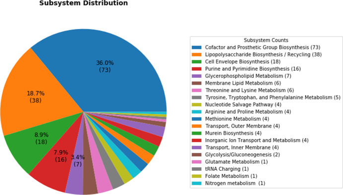
We investigated the subsystems/pathways of identified essential genes. The majority of these genes were found to be involved in the ‘Cofactor and Prosthetic Group Biosynthesis’ (73 genes), ‘Lipopolysaccharide Biosynthesis/Recycling’ (38 genes), ‘Cell Envelope Biosynthesis’ (18 genes), and ‘Purine and Pyrimidine Biosynthesis’ (16 genes) pathways. These subsystems are obviously important for bacterial growth.
As the most identified essential genes are in the “Cofactor and Prosthetic Group Biosynthesis” subsystem, we have provided their description and BP GO terms in Supplementary S2 Table. Cofactors have an important role in metabolism. Therefore, genes involved in the biosynthesis process of cofactors could be potential drug targets. According to Supplementary S2 Table, "NAD salvage" and "de novo NAD biosynthetic" are the biological processes of some identified essential genes. NAD cofactor is needed in some biological processes of prokaryotes like redox balance and energy metabolism [39]. On the other hand, some enzymes use NAD as a substrate in processes like DNA repair and degrade it [40]. Therefore, NAD biosynthetic process is required and could provide drug targets to fight against bacteria [39]. As well, genes involved in the coenzyme A and FAD biosynthetic process could be antibacterial targets [41]. Also, vitamins that are organic cofactors, their biosynthetic process are potential drug targets. For example, the biosynthetic pathway of folic acid is a useful target for sulfonamide antibiotics. Besides, the active form of thiamin (thiamin diphosphate) is a vital cofactor for organisms and is necessary for the activity of branched-chain amino acid metabolic enzymes [42,43]. As we can see in Supplementary S2 Table, some of the predicted essential genes are in the biosynthetic process of cobalamin (vitamin B12), thiamine (vitamin B1), riboflavin (vitamin B2), folic acid (vitamin B9), and menaquinone (vitamin K2).
Cell-membrane-related enzymes could be good options for better drug accessibility to stop or slow down the growth of pathogens [44]. Membrane-related essential genes and their membrane-related CC GO terms are shown in Table 1.
Table 1. Membrane-related essential genes.
| Essential genes | UniProt IDs | Gene description | Membrane-related CC GO term IDs | GO Terms |
|---|---|---|---|---|
| b4262 | P0ADC6 | lipopolysaccharide transport system protein LptG | GO:0005886 | Plasma membrane |
| GO:0043190 | ATP-binding cassette (ABC) transporter complex | |||
| GO:0016021 | Integral component of membrane | |||
| GO:1990351 | Transporter complex | |||
| b3201 | P0A9V1 | lipopolysaccharide transport system ATP binding protein LptB | GO:0043190 | ATP-binding cassette (ABC) transporter complex |
| GO:0005886 | Plasma membrane | |||
| GO:1990351 | Transporter complex | |||
| b3843 | P0AAB4 | 3-octaprenyl-4-hydroxybenzoate decarboxylase | GO:0005886 | Plasma membrane |
| b4177 | P0A7D4 | adenylosuccinate synthetase | GO:0016020 | Membrane |
| b0182 | P10441 | lipid A disaccharide synthase | GO:0019897 | Extrinsic component of plasma membrane |
| b3619 | P67910 | ADP-L-glycero-D-mannoheptose 6-epimerase | GO:0016020 | Membrane |
| b0586 | P11454 | apo-serine activating enzyme | GO:0005886 | Plasma membrane |
| b0594 | P10378 | 2,3-dihydroxybenzoate-AMP ligase | GO:0016020 | Membrane |
| b0635 | P0AD65 | peptidoglycan DD-transpeptidase MrdA | GO:0005887 | Integral component of plasma membrane |
| b0158 | P37028 | vitamin B12 ABC transporter periplasmic binding protein | GO:0030288 | Outer membrane-bounded periplasmic space |
| b2476 | P0A7D7 | phosphoribosylaminoimidazole-succinocarboxamide synthase | GO:0016020 | Membrane |
| b3966 | P06129 | cobalamin outer membrane transporter | GO:0016021 | Integral component of membrane |
| GO:0031230 | Intrinsic component of cell outer membrane | |||
| GO:0046930 | Pore complex |
On the other hand, analysis of the biological processes in which these genes are involved showed that ‘cell wall organization’, ‘lipopolysaccharide biosynthesis’, ‘lipopolysaccharide transport’, and ‘peptidoglycan biosynthetic process’ are the enriched GO terms. The rigid cell wall of Gram-negative bacteria is protection against osmotic lysis. Furthermore, the cell surface of the bacteria composed of LPS, known as endotoxin, provides the first line of protection against antibiotics and other harmful agents [45,46]. Besides, LPS is synthesized in the inner membrane of the cell and is transported to the outer membrane by transporter targets. LPS doesn’t allow antibiotics to enter the cell by creating a barrier and makes bacteria resistant to many antibiotics [47,48]. Therefore, the products of these genes related to cell wall and biosynthesis and transportation of LPS have a high level of importance for bacterial growth and survival [49,50]. Bacterial cell wall compounds are good potential drug target opportunities for killing bacteria or overcoming drug resistance [45,46,51,52]. Fig 1 provides a complete overview of the subsystem distribution of 195 essential genes.
Fig 1. Subsystem distribution of 195 essential genes associated with their frequency.
3.3. Exclusion of identified essential genes having human homologs
To avoid any probable interference with normal human functions, in the next step, those essential genes that have a human homolog were excluded from the initial list of potential genes [14]. According to the PATRIC platform, there are 54 human homologs for the complete reference genome of E. coli K-12 MG1655. We obtained UniProt IDs of these genes using their PATRIC IDs and the Retrieve/ID mapping tool of the UniProt website (https://www.uniprot.org/uploadlists/). Then, the set of these 54 human homologs was compared to the set of the 195 essential genes of E. coli, to find the common genes. Four essential genes with human homologs are listed in Table 2.
Table 2. Essential genes with human homologs.
| Locus tag | UniProt ID | Gene symbol | Human homolog | EC number |
|---|---|---|---|---|
| b2530 | P0A6B7 | iscS | NFS1 (cysteine desulfurase) | 2.8.1.7 |
| b2827 | P15640 | thyA | thyA (Thymidylate synthase) | 2.1.1.45 |
| b4005 | P15640 | purD | GART (Phosphoribosylamine-glycine ligase) | 6.3.4.13 |
| b2942 | P0A817 | metK | MAT2A (methionine adenosyltransferase 2A) | 2.5.1.6 |
3.4. Identifying structures and their co-crystallized ligands for each essential gene product
We utilized E. coli GEM-PRO (iML1515_GP) to obtain representative structures of the 191 essential genes using ssbio for performing SBVS. Information about co-crystallized ligands is obtained from the Ligand Expo database and mapped to the essential genes. For all drug targets, the chains with bound ligands are identified from the metadata incorporated in iML1515_GP, using ssbio. To perform the SBVS, a ligand-binding pocket is needed and the mentioned filters for the selection of the most informative protein-ligand complexes were applied. At last, 70 essential genes remained. The structures that can succeed in these filtrations are used in the SBVS step. The protein products of some of these 70 essential genes have more than one bound ligand, and hence, 92 protein-ligand pairs are considered for performing PLPS2. Finally, we downloaded the PDB structure of these shortlisted proteins. The complete list of the essential genes and their related information are presented in Supplementary S2 File.
3.5. Structure-based virtual screening
We identified essential targets in the previous steps. We ranked the ligands (FDA-approved drugs) against each target by running PLPS2 as an SBVS method. PLPS2 generates a molecular surface for the proteins and molecule conformations using APBS. To investigate the level of matching between a binding pocket and a molecule conformation, generated surfaces are divided into multiple patches. Shape, electrostatic potential, atom-based hydrophobicity, and hydrogen-bonding are represented for the patches with 3DZDs [30]. SSIC files with information about patches are generated for each protein and its ligands. After that, detected compatible pairs of patches are ranked using BS. The ranked ligands against each identified target associated with their score values are shown in Supplementary S3 File. Each row of the matrix is related to ranked ligands for each essential target. As we used FDA-approved drugs, the best-ranked molecules could be opportunities for drug repositioning with predicted antibacterial indication.
In addition to the above results, polypharmacology (multi-target) cases considering all selected drug targets were obtained using three frequency-based methods. Based on the first method that considers only the drugs in the top rank, those drugs that interact with more than 3 essential targets are listed in Table 3.
Table 3. Polypharmacology molecules selected based on the top rank of targets.
| Molecule | Number of targets | Targets |
|---|---|---|
| ZINC000085537014 | 5 | 1WXI_AMP, 1Q7B_NAP, 5M29_CBY, 3KQJ_UD1, 1EQ2_NAP |
| ZINC000003914596 | 3 | LI52_CTP, 5EJ8_TD6, 3ETH_ATP |
| ZINC000003941829 | 3 | 1GSA_ADP, 1GSA_GSH, 5ETP_5RZ |
| ZINC000085432544 | 3 | 2O1S_TDP, 1HV9_UD1, 5M1D_4LU |
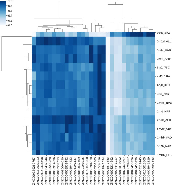
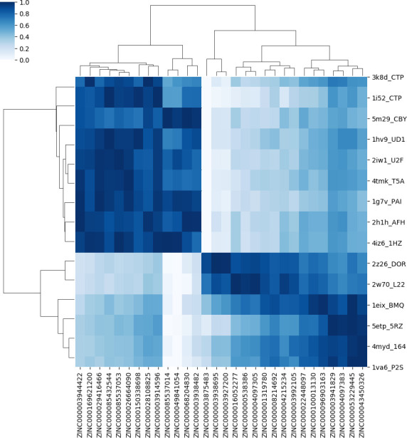
The percentile-based approach is more suitable in cases where the scores of ranked molecules for each essential target are very close to each other and causes the loss of fewer good molecules. Figs 2 and 3 show the heat maps of selected ligands from the top five ranks and 1-percentile methods, respectively. The selected ligands are polypharmacology cases that can stop the growth of the pathogen using the inhibition of multiple drug targets.
Fig 2. The heat map of selected ligands from the top five ranks of essential targets.

Fig 3. The heat map of selected ligands from the 1-percentile method.

The colorbar of the heatmaps ranges from white (assigned to the lowest normalized score values) to dark blue (assigned to the highest normalized score values) for each target. Thus, for each drug target, by moving row-wise on the heat map, one can find the best interacting ligand as the one with the darkest blue shade on that row.
The target dendrogram shows the hierarchical clustering of 92 protein-ligand pairs. This clustering is based on the similarity of the surface patches (which, in turn, is translated to similarity of the score rankings) of the selected drugs that are proposed as potentially active compounds for the pathogen. Enzymes are in the same cluster if they are similar in the binding ordering of these drugs. According to the ligand dendrogram, selected ligands are in the same cluster if their score values to the essential enzymes are similar. The labels displayed on the vertical axis of heat maps represent the top 15 targets to which the selected molecules have the strongest interactions. According to Fig 2, the cluster of 5etp is different from the cluster of the other 14 targets. Also, according to Fig 3, the cluster of 3K8D, LI52, 5M29, 1HV9, 2IW1, 4TMK, 1G7V, 2H1H, and 4IZ6 is different from the cluster of 2Z26, 2W70, 1EIX, 5ETP, 4MYD, and 1VA6 considering the selected ligand.
3.6. Investigation of ATC-code and safety of the selected drugs
We have presented drug IDs and ATC-codes of selected molecules (drugs) in Table 4. The first level of ATC-code is the anatomical main group and contains one letter. The second level shows the therapeutic subgroup and is two digits. The third level is the therapeutic/pharmacological subgroup and has one letter. The fourth level indicates the chemical/therapeutic/pharmacological subgroup and is one letter. The fifth level is the chemical substance and has two digits. According to WHO Guidelines 2020, anti-infectives are classified in J, A01AB, A02BD, A07A, D01, D06, D07C, D09AA, D10AF, G01, R02AB, and S01/S02/S03 groups [53]. Therefore, among the drugs in Table 4, Rifaximin (DB01220), Doxycycline (DB00254), Tobramycin (DB00684), Rifampicin (DB01045), Grazoprevir (DB11575), Tetracycline (DB00759), Minocycline (DB01017), Retapamulin (DB01256) are in the category of anti-infectives that have been suggested by our pipeline. It should be noted that, although the result is obtained based on integrating validated GEM with the validated SBVS method, they need to be tested experimentally. Also, we have extracted the ChEMBL IDs of the selected drugs from the UniChem database. Then, we have obtained the human genes (targets) related to these drugs via ChEMBL API (https://www.ebi.ac.uk/chembl) [54]. The ten drugs that have no known human targets are listed in Table 4. Among the selected drugs, Grazoprevir and Retapamulin are anti-infectives, and also they have no human targets. Also, there is a record of Retapamulin [55,56] in the treatment of bacterial infections in DrugBank. These results could be a sign of validation of our work. The compounds without any known human targets could be better options for experimental tests against E. coli infection.
Table 4. IDs and ATC-codes of the selected molecules.
| ZINC ID | Drug Name | DrugBank ID | ATC-code | ChEMBL ID | Known Human Targets? | method |
|---|---|---|---|---|---|---|
| ZINC000003944422 | Ritonavir | DB00503 | J05AE03 | CHEMBL163 | 2,3 | |
| ZINC000169621200 | Rifaximin | DB01220 | A07AA11/ D06AX11 | CHEMBL1617 | 2,3 | |
| ZINC000029416466 | Saquinavir | DB01232 | J05AE01 | CHEMBL296480 | No | 3 |
| ZINC000085432544 | Vinblastine | DB00570 | L01CA01 | CHEMBL159 | 1,2,3 | |
| ZINC000085537053 | Docetaxel | DB01248 | L01CD02 | CHEMBL92 | 3 | |
| ZINC000026664090 | Saquinavir | DB01232 | J05AE01 | CHEMBL296480 | No | 2,3 |
| ZINC000150338698 | Capreomycin | DB00314 | J04AB30 | 2,3 | ||
| ZINC000028108825 | Gadofosveset trisodium | DB06705 | V08CA11 | CHEMBL1615469 | No | 2,3 |
| ZINC000003914596 | Saquinavir | DB01232 | J05AE01 | CHEMBL114 | 1,2,3 | |
| ZINC000085537014 | Cobicistat | DB09065 | J05AR09/J05AR15/J05AR14/J05AR22 /J05AR18 /V03AX03 | CHEMBL2095208 | 1,2,3 | |
| ZINC000049841054 | Carfilzomib | DB08889 | L01XX45 | CHEMBL451887 | 2,3 | |
| ZINC000068204830 | Daclatasvir | J05AP58/J05AP07 | CHEMBL2023898 | 2,3 | ||
| ZINC000003938482 | Posaconazole | J02AC04 | CHEMBL1397 | 2,3 | ||
| ZINC000003875483 | Oxymorphone | N02AA11 (WHO) | CHEMBL963 | 2,3 | ||
| ZINC000003938695 | Norgestimate | DB00957 | G03AA11 | CHEMBL1200934 | 3 | |
| ZINC000003927200 | Drospirenone | DB01395 | G03AC10/G03FA17/G03AA12 | CHEMBL1509 | 3 | |
| ZINC000016052277 | Doxycycline | DB00254 | J01AA02/A01AB22/J01AA20 | CHEMBL1433 | 3 | |
| ZINC000000538386 | Sufentanil | DB00708 | N01AH03 | CHEMBL658 | 3 | |
| ZINC000004097305 | Flunisolide | DB00180 | R01AD04/R03BA03 | CHEMBL1512 | 3 | |
| ZINC000001319780 | Buprenorphine | DB00921 | N07BC01/N07BC51/N02AE01 | CHEMBL560511 | 3 | |
| ZINC000008214692 | Tobramycin | DB00684 | S01AA12/J01GB01 | CHEMBL1747 | 2,3 | |
| ZINC000004215234 | Cefditoren | DB01066 | J01DD16 | CHEMBL1743 | No | 3 |
| ZINC000003992105 | Fluticasone furoate | DB08906 | R03AL08/R01AD12/R03AK10/R03BA09 | CHEMBL1676 | 3 | |
| ZINC000022448097 | Gadoxetic acid | DB08884 | V08CA10 | CHEMBL2110606 | No | 2,3 |
| ZINC000100013130 | Midostaurin | DB06595 | L01XE39 | CHEMBL608533 | 3 | |
| ZINC000096903163 | Diacetyl benzoyl lathyrol | DB11260 | CHEMBL552128 | No | 3 | |
| ZINC000003941829 | Fosamprenavir | DB01319 | J05AE07 | CHEMBL1664 | 1,2,3 | |
| ZINC000004097383 | Pancuronium | DB01337 | M03AC01 | CHEMBL185073 | 2,3 | |
| ZINC000053229445 | Rocuronium | DB00728 | M03AC09 | CHEMBL1201244 | 2,3 | |
| ZINC000043450326 | Omacetaxine mepesuccinate | DB04865 | L01XX40 | 2,3 | ||
| ZINC000169289767 | Trypan blue | DB09158 | CHEMBL1089641 | 2 | ||
| ZINC000169621223 | Rifampicin | DB01045 | J04AM02/J04AB02/J04AM06 /J04AM05/J04AM07 | CHEMBL374478 | 2 | |
| ZINC000085555528 | Vinblastine | DB00570 | L01CA01 | CHEMBL22969 | 2 | |
| ZINC000008101127 | Indocyanine green | DB09374 | CHEMBL1615807 | No | 2 | |
| ZINC000049637509 | Isavuconazonium | DB06636 | J02AC05 | CHEMBL1183349 | No | 2 |
| ZINC000095551509 | Grazoprevir | DB11575 | J05AP11/J05AP54 | CHEMBL2063090 | No | 2 |
| ZINC000084441937 | Tetracycline | DB00759 | S02AA08/A02BD02/A01AB13/J01AA07 /S01AA09/J01AA20 /J01RA08/S03AA02 /D06AA04 /A02BD08 | CHEMBL1440 | 2 | |
| ZINC000014879992 | Minocycline | DB01017 | J01AA08/J01AA20 /A01AB23/ | CHEMBL1434 | 2 | |
| ZINC000019796087 | Nicardipine | DB00622 | C08CA04 | CHEMBL1598680 | 2 | |
| ZINC000043450324 | Omacetaxine mepesuccinate | DB04865 | L01XX40 | CHEMBL175858 | 2 | |
| ZINC000100013500 | Retapamulin | DB01256 | D06AX13 | CHEMBL1658 | No | 2 |
4. Conclusion
The discovery of novel antibacterial agents is necessary due to the rapid worldwide emergence of antibiotic resistance. GEMs are representative models of organisms at the metabolism level and they are good frameworks for the investigation of bacterial phenotypes. In this study, we have developed a structural systems pharmacology framework based on analyzing a metabolic network and a SBVS approach. The coupling of these two methods was done to achieve better results. As an example of its application, we have represented that this framework works well for E. coli and we could find anti-infective molecules for it. This can also be a general pipeline for the development of novel antibacterials for other bacterial pathogens that have GEM. Here, we have performed constraint-based flux analysis (FBA) on the most complete E. coli GEM-PRO for the rational and system-level identification of essential genes whose knocking out causes the growth of the pathogen to stop. 195 genes that are essential for the survival of the pathogen are identified and high-priority proteins related to these genes are detected as potential drug targets to carry out SBVS. These targets are the most promising candidates due to the availability of experimental structure in the PDB database and having cognate biologically relevant ligands. The SBVS method was performed with FDA-approved drugs for these targets by PLPS2. It evaluates interactions between a protein and a small molecule, based on molecular surfaces with shape, electrostatic potential, hydrophobicity, and hydrogen bonding features. Finally, we have identified new potential inhibitors among available FDA-approved drugs to stop the growth of the pathogen. Working with available drugs instead of other small molecules is an advantage because of the fast gaining drug resistance of pathogens. Therefore, the quicker discovery of new safe drugs is urgent. Here, we have predicted a new therapeutic indication (antibacterial) for reported drugs. Consequently, these drugs can be proposed as drug repositioning opportunities. It will be valuable if the proposed anti-infective drugs will be shifted to in vitro and in vivo experiments soon for validation of the results.
Supporting information
(DOCX)
(DOCX)
(XLSX)
(XLSX)
(XLSX)
(TIF)
Data Availability
All relevant data are within the paper and its Supporting Information files.
Funding Statement
The author(s) received no specific funding for this work.
References
- 1.Bajorath, J., Computer-aided drug discovery [version 1; referees: 3 approved]. 2015.
- 2.Kennedy D.M.F., Computational and Structural Approaches to Drug Discovery, in: Stroud R.M., Finer-Moore J. (eds.). The Royal Society of Chemistry, Cambridge, UK (2008). xvii + 382 pp., £89–00, ISBN: 0-85404-365-1. International Journal of Biological Macromolecules, 2008. 42(5): p. 491–492. [Google Scholar]
- 3.Payne D.J., et al., Drugs for bad bugs: confronting the challenges of antibacterial discovery. Nature reviews Drug discovery, 2007. 6(1): p. 29–40. doi: 10.1038/nrd2201 [DOI] [PubMed] [Google Scholar]
- 4.Kim H.U., Sohn S.B., and Lee S.Y., Metabolic network modeling and simulation for drug targeting and discovery. Biotechnology Journal, 2012. 7(3): p. 330–342. doi: 10.1002/biot.201100159 [DOI] [PubMed] [Google Scholar]
- 5.Ma H. and Goryanin I., Human metabolic network reconstruction and its impact on drug discovery and development. Drug discovery today, 2008. 13(9–10): p. 402–408. doi: 10.1016/j.drudis.2008.02.002 [DOI] [PubMed] [Google Scholar]
- 6.Lionta E., et al., Structure-based virtual screening for drug discovery: principles, applications and recent advances. Current topics in medicinal chemistry, 2014. 14(16): p. 1923–1938. doi: 10.2174/1568026614666140929124445 [DOI] [PMC free article] [PubMed] [Google Scholar]
- 7.Chang R.L., et al., Antibacterial mechanisms identified through structural systems pharmacology. BMC systems biology, 2013. 7(1): p. 102. doi: 10.1186/1752-0509-7-102 [DOI] [PMC free article] [PubMed] [Google Scholar]
- 8.Gu C., et al., Current status and applications of genome-scale metabolic models. Genome biology, 2019. 20(1): p. 1–18. doi: 10.1186/s13059-018-1612-0 [DOI] [PMC free article] [PubMed] [Google Scholar]
- 9.McCloskey D., Palsson B.Ø., and Feist A.M., Basic and applied uses of genome-scale metabolic network reconstructions of Escherichia coli. Molecular systems biology, 2013. 9(1): p. 661. doi: 10.1038/msb.2013.18 [DOI] [PMC free article] [PubMed] [Google Scholar]
- 10.Monk J.M., et al., i ML1515, a knowledgebase that computes Escherichia coli traits. Nature biotechnology, 2017. 35(10): p. 904–908. doi: 10.1038/nbt.3956 [DOI] [PMC free article] [PubMed] [Google Scholar]
- 11.Lieven C., et al., MEMOTE for standardized genome-scale metabolic model testing. Nature biotechnology, 2020. 38(3): p. 272–276. doi: 10.1038/s41587-020-0446-y [DOI] [PMC free article] [PubMed] [Google Scholar]
- 12.Ebrahim A., et al., COBRApy: constraints-based reconstruction and analysis for python. BMC systems biology, 2013. 7(1): p. 1–6. doi: 10.1186/1752-0509-7-74 [DOI] [PMC free article] [PubMed] [Google Scholar]
- 13.Makhorin, A., GLPK (GNU linear programming kit). http://www.gnu.org/s/glpk/glpk.html, 2008.
- 14.Perumal D., et al., Targeting multiple targets in Pseudomonas aeruginosa PAO1 using flux balance analysis of a reconstructed genome-scale metabolic network. Journal of drug targeting, 2011. 19(1): p. 1–13. doi: 10.3109/10611861003649753 [DOI] [PubMed] [Google Scholar]
- 15.UniProt: The universal protein knowledgebase in 2021. Nucleic Acids Research, 2021. 49(D1): p. D480–D489. doi: 10.1093/nar/gkaa1100 [DOI] [PMC free article] [PubMed] [Google Scholar]
- 16.Consortium G.O., The gene ontology (GO) project in 2006. Nucleic acids research, 2006. 34(suppl_1): p. D322–D326. doi: 10.1093/nar/gkj021 [DOI] [PMC free article] [PubMed] [Google Scholar]
- 17.Davis J.J., et al., The PATRIC Bioinformatics Resource Center: expanding data and analysis capabilities. Nucleic acids research, 2020. 48(D1): p. D606–D612. doi: 10.1093/nar/gkz943 [DOI] [PMC free article] [PubMed] [Google Scholar]
- 18.Mih N., et al., ssbio: a Python framework for structural systems biology. Bioinformatics, 2018. 34(12): p. 2155–2157. doi: 10.1093/bioinformatics/bty077 [DOI] [PMC free article] [PubMed] [Google Scholar]
- 19.Feng Z., et al., Ligand Depot: a data warehouse for ligands bound to macromolecules. Bioinformatics, 2004. 20(13): p. 2153–2155. doi: 10.1093/bioinformatics/bth214 [DOI] [PubMed] [Google Scholar]
- 20.Sterling T. and Irwin J.J., ZINC 15–ligand discovery for everyone. Journal of chemical information and modeling, 2015. 55(11): p. 2324–2337. doi: 10.1021/acs.jcim.5b00559 [DOI] [PMC free article] [PubMed] [Google Scholar]
- 21.O’Boyle N.M., et al., Open Babel: An open chemical toolbox. Journal of cheminformatics, 2011. 3(1): p. 1–14. doi: 10.1186/1758-2946-3-1 [DOI] [PMC free article] [PubMed] [Google Scholar]
- 22.O’Boyle N.M., Vandermeersch T., and Hutchison G.R., Confab–generation of diverse low energy conformers. Journal of Cheminformatics, 2011. 3(1): p. 1–1. doi: 10.1186/1758-2946-3-1 [DOI] [PMC free article] [PubMed] [Google Scholar]
- 23.Shin W.-H. and Kihara D., Virtual ligand screening using PL-PatchSurfer2, a molecular surface-based protein–ligand docking method, in Computational Drug Discovery and Design. 2018, Springer. p. 105–121. [DOI] [PubMed] [Google Scholar]
- 24.Baker N.A., et al., Electrostatics of nanosystems: application to microtubules and the ribosome. Proceedings of the National Academy of Sciences, 2001. 98(18): p. 10037–10041. doi: 10.1073/pnas.181342398 [DOI] [PMC free article] [PubMed] [Google Scholar]
- 25.Unni S., et al., Web servers and services for electrostatics calculations with APBS and PDB2PQR. Journal of computational chemistry, 2011. 32(7): p. 1488–1491. doi: 10.1002/jcc.21720 [DOI] [PMC free article] [PubMed] [Google Scholar]
- 26.Cheng T., et al., Computation of octanol− water partition coefficients by guiding an additive model with knowledge. Journal of chemical information and modeling, 2007. 47(6): p. 2140–2148. doi: 10.1021/ci700257y [DOI] [PubMed] [Google Scholar]
- 27.Novotni, M. and R. Klein. 3D Zernike descriptors for content based shape retrieval. in Proceedings of the eighth ACM symposium on Solid modeling and applications. 2003.
- 28.Paulsen J.L. and Anderson A.C., Scoring ensembles of docked protein: ligand interactions for virtual lead optimization. Journal of chemical information and modeling, 2009. 49(12): p. 2813–2819. doi: 10.1021/ci9003078 [DOI] [PMC free article] [PubMed] [Google Scholar]
- 29.Vosmeer C.R., et al., Towards automated binding affinity prediction using an iterative linear interaction energy approach. International journal of molecular sciences, 2014. 15(1): p. 798–816. doi: 10.3390/ijms15010798 [DOI] [PMC free article] [PubMed] [Google Scholar]
- 30.Shin W.-H., et al., PL-PatchSurfer2: improved local surface matching-based virtual screening method that is tolerant to target and ligand structure variation. Journal of chemical information and modeling, 2016. 56(9): p. 1676–1691. doi: 10.1021/acs.jcim.6b00163 [DOI] [PMC free article] [PubMed] [Google Scholar]
- 31.Waskom M., et al., Seaborn: statistical data visualization. Astrophysics Source Code Library, 2020: p. ascl: 2012.015. [Google Scholar]
- 32.Hunter J.D., Matplotlib: A 2D graphics environment. IEEE Annals of the History of Computing, 2007. 9(03): p. 90–95. [Google Scholar]
- 33.Wishart D.S., et al., DrugBank 5.0: a major update to the DrugBank database for 2018. Nucleic acids research, 2018. 46(D1): p. D1074–D1082. doi: 10.1093/nar/gkx1037 [DOI] [PMC free article] [PubMed] [Google Scholar]
- 34.Papp B., Pál C., and Hurst L.D., Metabolic network analysis of the causes and evolution of enzyme dispensability in yeast. Nature, 2004. 429(6992): p. 661–664. doi: 10.1038/nature02636 [DOI] [PubMed] [Google Scholar]
- 35.Ogoshi R.C., et al., Acidifying and yeast extract in diets for adults cats. Animal Science Journal, 2014. 85(5): p. 555–561. doi: 10.1111/asj.12166 [DOI] [PubMed] [Google Scholar]
- 36.Bintener T., Pacheco M.P., and Sauter T., Towards the routine use of in silico screenings for drug discovery using metabolic modelling. Biochemical Society Transactions, 2020. 48(3): p. 955–969. doi: 10.1042/BST20190867 [DOI] [PMC free article] [PubMed] [Google Scholar]
- 37.Kitano H., A robustness-based approach to systems-oriented drug design. Nature reviews Drug discovery, 2007. 6(3): p. 202–210. doi: 10.1038/nrd2195 [DOI] [PubMed] [Google Scholar]
- 38.Kim H.U., et al., Integrative genome-scale metabolic analysis of Vibrio vulnificus for drug targeting and discovery. Molecular systems biology, 2011. 7(1): p. 460. doi: 10.1038/msb.2010.115 [DOI] [PMC free article] [PubMed] [Google Scholar]
- 39.Boshoff H.I., et al., Biosynthesis and recycling of nicotinamide cofactors in Mycobacterium tuberculosis: an essential role for NAD in nonreplicating bacilli. Journal of Biological Chemistry, 2008. 283(28): p. 19329–19341. doi: 10.1074/jbc.M800694200 [DOI] [PMC free article] [PubMed] [Google Scholar]
- 40.Rodionova I.A., et al., Metabolic and bactericidal effects of targeted suppression of NadD and NadE enzymes in mycobacteria. MBio, 2014. 5(1): p. e00747–13. doi: 10.1128/mBio.00747-13 [DOI] [PMC free article] [PubMed] [Google Scholar]
- 41.Gerdes S.Y., et al., From genetic footprinting to antimicrobial drug targets: examples in cofactor biosynthetic pathways. 2002, Am Soc Microbiol. doi: 10.1128/JB.184.16.4555-4572.2002 [DOI] [PMC free article] [PubMed] [Google Scholar]
- 42.Du Q., Wang H., and Xie J., Thiamin (vitamin B1) biosynthesis and regulation: a rich source of antimicrobial drug targets? International journal of biological sciences, 2011. 7(1): p. 41. doi: 10.7150/ijbs.7.41 [DOI] [PMC free article] [PubMed] [Google Scholar]
- 43.Mobegi F.M., et al., From microbial gene essentiality to novel antimicrobial drug targets. BMC genomics, 2014. 15(1): p. 1–11. doi: 10.1186/1471-2164-15-958 [DOI] [PMC free article] [PubMed] [Google Scholar]
- 44.Naclerio G.A. and Sintim H.O., Multiple ways to kill bacteria via inhibiting novel cell wall or membrane targets. Future Medicinal Chemistry, 2020. 12(13): p. 1253–1279. doi: 10.4155/fmc-2020-0046 [DOI] [PubMed] [Google Scholar]
- 45.Thomanek N., et al., Intricate crosstalk between lipopolysaccharide, phospholipid and fatty acid metabolism in Escherichia coli modulates proteolysis of LpxC. Frontiers in microbiology, 2019. 9: p. 3285. doi: 10.3389/fmicb.2018.03285 [DOI] [PMC free article] [PubMed] [Google Scholar]
- 46.Bertani B. and Ruiz N., Function and biogenesis of lipopolysaccharides. EcoSal Plus, 2018. 8(1). doi: 10.1128/ecosalplus.ESP-0001-2018 [DOI] [PMC free article] [PubMed] [Google Scholar]
- 47.Zhang G., Meredith T.C., and Kahne D., On the essentiality of lipopolysaccharide to Gram-negative bacteria. Current opinion in microbiology, 2013. 16(6): p. 779–785. doi: 10.1016/j.mib.2013.09.007 [DOI] [PMC free article] [PubMed] [Google Scholar]
- 48.Simpson B.W., et al., Lipopolysaccharide transport to the cell surface: biosynthesis and extraction from the inner membrane. Philosophical Transactions of the Royal Society B: Biological Sciences, 2015. 370(1679): p. 20150029. doi: 10.1098/rstb.2015.0029 [DOI] [PMC free article] [PubMed] [Google Scholar]
- 49.Dhar S., et al., Cell-wall recycling and synthesis in Escherichia coli and Pseudomonas aeruginosa–their role in the development of resistance. Journal of Medical Microbiology, 2018. 67(1): p. 1–21. doi: 10.1099/jmm.0.000636 [DOI] [PubMed] [Google Scholar]
- 50.Sompairac N., et al., Metabolic and signalling network maps integration: application to cross-talk studies and omics data analysis in cancer. BMC bioinformatics, 2019. 20(4): p. 140. doi: 10.1186/s12859-019-2682-z [DOI] [PMC free article] [PubMed] [Google Scholar]
- 51.Schuerholz T., et al., Bacterial cell wall compounds as promising targets of antimicrobial agents II. Immunological and clinical aspects. Current drug targets, 2012. 13(9): p. 1131–1137. doi: 10.2174/138945012802002438 [DOI] [PubMed] [Google Scholar]
- 52.Wang X. and Quinn P.J., Lipopolysaccharide: biosynthetic pathway and structure modification. Progress in lipid research, 2010. 49(2): p. 97–107. doi: 10.1016/j.plipres.2009.06.002 [DOI] [PubMed] [Google Scholar]
- 53.WHO Collaborating Centre for Drug Statistics Methodology, Guidelines for ATC classification and DDD assignment 2020. Oslo, 2020.
- 54.Gaulton A., et al., The ChEMBL database in 2017. Nucleic acids research, 2017. 45(D1): p. D945–D954. doi: 10.1093/nar/gkw1074 [DOI] [PMC free article] [PubMed] [Google Scholar]
- 55.Daum R.S., Kar S., and Kirkpatrick P., Retapamulin. 2007, Nature Publishing Group. [Google Scholar]
- 56.Dhingra D., Parakh A., and Ramachandran S., Retapamulin: a newer topical antibiotic. Journal of postgraduate medicine, 2013. 59(2): p. 127. doi: 10.4103/0022-3859.113842 [DOI] [PubMed] [Google Scholar]
Associated Data
This section collects any data citations, data availability statements, or supplementary materials included in this article.
Supplementary Materials
(DOCX)
(DOCX)
(XLSX)
(XLSX)
(XLSX)
(TIF)
Data Availability Statement
All relevant data are within the paper and its Supporting Information files.


