



Abstract
A relationship between environmental exposure to air pollution and cognitive impairment and neurological disorders has been described. Previous literature has focused on the direct effects of the air pollution components on neuronal and glial cells, as well as on involvement of oxidative stress and neuroinflammation on microglia and astrocyte reactivity. However, other mechanisms involved in the air pollution effects on central nervous system (CNS) toxicity can be playing critical roles. Increasingly, extracellular vesicle’s (EVs) mediated intercellular communication is being recognized as impacting the development of cognitive impairment and neurological disorders like Alzheimer’s disease and others. Here we describe the available evidence about toxic air pollutants and its components on brain, an involvement of brain cells specific and EVs types (based in the origin or in the size of EVs) in the initiation, exacerbation, and propagation of the neurotoxic effects (inflammation, neurodegeneration, and accumulation of neurotoxic proteins) induced by air pollution in the CNS. Additionally, we discuss the identification and isolation of neural-derived EVs from human plasma, the most common markers for neural-derived EVs, and their potential for use as diagnostic or therapeutic molecules for air pollution-related cognitive impairment and neurodegeneration.
Keywords: Extracellular Vesicles, Air pollution, Neurodegeneration, Cognitive Impairment, PM2.5
Ambient Air Pollution: Generalities
Ambient air pollution is a significant contributor to the burden of disease worldwide (GDB, 2019), and has been linked to the development of cognitive impairment and neurodegenerative diseases, such as cognitive aging, Parkinson’s disease (PD), and Alzheimer’s Disease (AD), and related dementia’s (Calderon-Garciduenas et al., 2018; Woodward et al., 2017a). Impaired cognition and dementia are leading causes of loss of independence in daily activities (McGuire et al., 2006), hospitalization (Chodosh et al., 2004), and mortality among older individuals (Bassuk et al., 2000; James et al., 2014). According to the World Health Organization (WHO) ambient air pollution, the by-product of emissions from industry, power generation, transportation, and domestic burning, is responsible for an estimated 4.2 million deaths annually. Most of the world population currently resides in regions where ambient air pollution levels surpass the WHO’s health-based air-quality guidelines (Turner et al., 2020). Cognitive impairment and other neurogenerative diseases exert a substantial burden on patients’ lives, affecting not only their skills but also their family roles, emotionality, self-confidence, as well as the lives of those close to them, with an increasing dependence and burden (Frank et al., 2006).
Ambient air pollution is mixture of several components like gases, metals, and particulate matter (PM) (Table 1). Evidence from both human and animal studies have shown that ambient air pollution targets the brain. Therefore, ambient air pollution is now recognized as having a neurotoxic effect on the central nervous system (Costa et al., 2017; Esmaeil Mousavi et al., 2017; Krauskopf et al., 2018; Levesque et al., 2011; Woodward et al., 2017a; Woodward et al., 2017b).
Table 1.
Air pollution is a mixture of several primary and secondary toxicants and can come from natural or human-derived activities. It contains organic (e.g., volatile organic components) or inorganic compounds; metals (e.g., lead [Pb], nickel [Ni], chromium [Cr], aluminum [Al], and iron [Fe]), gases (e.g., carbon monoxide [CO], sulfur oxides [SO], sulfur dioxide [SO2], nitrogen dioxide [NO2], and nitrogen oxide [NO]), ground-level ozone (O3), coarse and fine particulate matter [aerodynamic diameter ≤10 and < 2.5 μm (PM10, PM2.d ultrafine particulate matter [aerodynamic diameter ≤ 0.1 μm (PM0.1)] (Shang and Sun, 2018).
| Ambient Air Pollution | Sources | Components |
|---|---|---|
| Traffic-Related Air Pollution (TRAP) | Diesel Exhaust Motor Vehicle Exhaust Organic and Inorganic |
Gases: Ozone (O3), Carbon monoxide (CO), Sulfur oxide (SO), Sulfur Dioxide (SO2), Nitric oxides (NOx) Metals*: lead (Pb), Nickel (Ni), Chromium (Cr), Aluminum (Al), Iron (Fe) Liquids*: (droplets), polycyclic aromatic hydrocarbons, hapones, and steranes |
| Particulate Air Pollution | Incomplete combustion (burning fossil fuels and biomass). Industrial (power plants, oil refineries, factories, etc.). Natural (volcanoes, wildfires, wind-blown dust) |
Black carbon and other particulate matter: coarse (PM10, 10 μm-2.5 um), fine (PM2.5, <2.5 μm), ultrafine (PM0.1, <0.1 μm) |
May be particle-bound
A genetic component to the development of neurodegenerative diseases and AD exists (e.g., autosomal dominant mutations affect Aβ processing), although the majority of cases of AD occur sporadically and have a complex etiology related to aging (Pihlstrøm et al., 2018). However, in considering genetic risk factors for neurodegeneration, it is important to note that several genes linked to the development of neurodegenerative disease are also involved in the generation of oxidative stress via mitochondrial dysfunction or antioxidant factors (Neal and Richardson, 2018). Apart from genetic factors, PM can also induce mitochondrial dysfunction and trigger oxidative stress (Daiber et al., 2020). Taken together, genetic polymorphisms in antioxidant enzymes or mitochondrial protective factors make the brain susceptible to reactive oxygen species, such that exposure to environmental toxicants like pesticides, metals, and air pollution can exacerbate oxidative stress in the brain (Milani et al., 2020; Neal and Richardson, 2018).
Indeed, oxidative stress, chronic inflammation, and glial activation, are conventionally accepted factors in mechanisms of action for ambient air pollution-related neurotoxicity (Costa et al., 2017). These factors have been demonstrated experimentally to cause neuronal damage and oligodendrocyte dysfunction, which present as white-matter abnormalities and loss of cognitive function (Liddelow et al., 2017; Tsutsumi et al., 2019). They’ve also been found to be involved with the development of several neurological disorders and neurologic diseases such as autism spectrum disorder, schizophrenia, AD, PD, multiple sclerosis, and amyotrophic lateral sclerosis (Allen et al., 2017; Esmaeil Mousavi et al., 2017; Fiandaca et al., 2015; Wang and Hay, 2015).
The solid components of ambient air pollution, particularly PM2.5–0.1 (aerodynamic diameter < 2.5–0.1 μm), increase the risk for cognitive impairment and neurodegenerative disease. Human studies in children and adults have revealed brain volume loss in white matter, epithelial and endothelial damage contributing to blood-brain barrier (BBB) breakdown and inducing attention deficit or memory-loss in response to air pollution exposure (Calderon-Garciduenas et al., 2016; Chen et al., 2015).
Anthropogenic ambient air pollution is primary derived from traffic-related-air pollution (TRAP), resulting mostly from diesel fuel incomplete combustion (Costa et al., 2017). So, for clarity, this review will focus on the contributions of the PM components of ambient air pollution toxicity on the brain. Evidence suggests that dose, composition, and surface size of PM determine the potential to elicit low-grade, chronic inflammation, and oxidative damage (Valavanidis et al., 2008). Fine and ultrafine (nano-sized) particulate matter (PM2.5–0.1) increases the risk for cognitive impairment and neurodegenerative disease. PM2.5–0.1 is one of the most studied and considered one of the most hazardous air pollution components, due to its small size which allows PM2.5–0.1 to deposit deeper into the respiratory tract and enter the systemic circulation (Nemmar et al., 2002; Nemmar et al., 2001).
PM2.5–0.1 from TRAP is an agglomeration consisting of black carbon (BC), with nitrogen or sulfur gases, and metals such as Mg, Cu, Pb, Fe (Bai et al., 2018). The metal constituents of PM2.5–0.1 contribute to oxidative stress as they are catalyst for reactive oxygen species generation and cellular oxidant production (Pavanello et al., 2016). The soluble metal components of PM2.5–0.1 are reported to be responsible for EV- mediated neurotoxicity in mouse models (Chen et al., 2020). PM2.5–0.1 has been found to cause neurodevelopmental disorder and neurodegeneration in children and adults, respectively (Allen et al., 2017; Calderon-Garciduenas et al., 2018). Studies revealed brain volume loss in white matter, epithelial and endothelial damage contributing to BBB breakdown and inducing attention deficit or memory-loss in response to air pollution exposure (Calderon-Garciduenas et al., 2016; Chen et al., 2015).
Additionally, aging is a stochastic process which leads to a gradual decline in cellular, tissue and organ function, especially in brain. There is consistent evidence linking exposure to PM air pollution with poor age-related cognitive performance (Chen and Schwartz, 2009; Fougere et al., 2015; Power et al., 2011; Zeng et al., 2010) and accelerated cognitive decline (Weuve et al., 2012), such as AD and PD. Authors found that exposure to air pollution causes neuroinflammation and oxidative stress which damages several major classes of cellular molecules also found to be involved with aging and aged related diseases (Bondy, 2016; Migliore and Coppede, 2009). In particular, exposure to BC, a component of TRAP, surrogate for exposure to fossil fuel combustion, and fraction of the PM2.5, has been associated with poor cognition in older men (Power et al., 2011).
Moreover, TRAP also contains halogenated hydrocarbons and polycyclic aromatic hydrocarbons (PAHs), that are also linked to neurotoxicity (Li et al., 2020). Since ambient air pollution consists of a complex mixture of organic and inorganic matter, metals, and gases, a myriad of mechanisms may exist by which air pollution exerts its toxic effects on the brain, apart from age and genetic related factors.
For instance, mouse models have demonstrated that sub-chronic exposure to TRAP increases brain proteins related to the aforementioned neurologic diseases, including amyloid beta (Aβ), tau, and a-synuclein enrichment, particularly in the frontal and temporal lobe regions (Levesque et al., 2011).
However, there are no specific biomarkers to identify those at increased risk of age-related impaired cognition or neurodegeneration from air pollution exposure, which is a critical public health gap that hampers effective targeted prevention (Sperling et al., 2011). As such, it is imperative that both diagnostic and therapeutic intervention for air pollution related cognitive impairment and neurological disease be developed.
In this review, we examine the plausibility of EVs as mediators of the aforementioned toxic effect of ambient air pollution and the neurological response that leads to cognitive impairment and neurological disease. We propose that air pollution exerts a direct and indirect neurotoxic (neuroinflammation and neurodegeneration) effect on the central nervous system (CNS) by triggering EVs release through a mechanism involving activation of glial cells and altered autophagy function. Moreover, EVs may impact gene regulation via their miRNA cargo. An association between long-term PM2.5 air pollution and increased levels of EVs miRNAs was identified in serum, and in silico pathway analysis of PM2.5 associated EVs miRNAs identified pathways including oxidative stress and inflammation (Boda et al., 2020; Rodosthenous et al., 2016) indicating an epigenetic role of EVs in the pathogenesis of neurodegeneration from air pollution exposure.
We will also explore the available evidence concerning EVs as a form of “liquid biopsy” for determining air pollution exposure and, human cognitive impairment and neurogenerative diseases, in particular AD.
Overview of Extracellular Vesicles
Recently, biomolecular mechanisms involving the release of what was thought to be inconsequential cellular debris, but now knowns as EVs, have been explored as mediators of air pollution induced toxicity and pathogenesis in the brain (Rodosthenous et al., 2016). EVs are nano-sized (0.03–1μm) lipid bilayer membrane bound vesicles that are constitutively shed or released from virtually every cell of the body and engage in intercellular and inter-organ transfer of biological information (Pavanello et al., 2016; Stahl et al., 2019). EVs are actively released by all cell types into the extracellular space and systemic circulation. The EVs were detected in many biological fluids as in serum, plasma, cerebrospinal fluids, and others (D’Anca et al., 2019; Urabe et al., 2020). They shuttle bioactive molecular cargo, such as, RNAs, proteins, and metabolites, that control gene expression or otherwise modify phenotype of their target cells (Chen et al., 2014; Fruhbeis et al., 2013b; Kim et al., 2017). EVs uptake confers epigenetic or phenotypic changes in the recipient cell through the transfer of miRNA, chemokines and cytokines, metabolites, or other cellular components. Importantly, EVs have recently been recognized and studied for their potential ability to be harnessed for the development of novel noninvasive biomarkers for various human conditions (Urabe et al., 2020).
Though it is not known how EVs released from parent cells are targeted to recipient cells, cell-to-cell transfer of EVs may not be random. Small EVs (< 200 nm) in particular have been reported to recognize specific cellular populations (Basso and Bonetto, 2016), and to be targeted to specific cells, based in part, on their cell of origin (Kur et al., 2020). Furthermore, EVs function may vary by subtype; small EVs and medium EVs (< 200 nm) from the same cell may have opposing action. Under physiologically normal conditions small EVs participate in Aβ intracellular clearance, and also promote extracellular Aβ plaque formation (Falker et al., 2016). Conversely, medium EVs have been implicated in the conversion of aggregated Aβ into neurotoxic non-fibrillar soluble Aβ species, which are preferentially taken up by glial cells comparative to fibrils (Dinkins et al., 2014).
Classification
EVs were categorized based upon their biogenesis in two groups the exosomes such as endosome-origin and ectosomes plasma membrane-derived (microvesicles)(Akers et al., 2013; Doyle and Wang, 2019). Actually, is consider use of operational terms for EV subtypes that refer to a) physical characteristics of EVs, such as size or density; b) biochemical composition; or c) descriptions or cell of origin in the place of terms such as exosome and microvesicles (Thery et al., 2018; Wei et al., 2021).
EVs vary not only in size, but in shape and cargo, suggesting that functionality may be related to physical and biological characteristics. EVs facilitate intercellular signaling in the CNS that is beneficiary to neurons under physiological conditions (Janas et al., 2016). Brain EVs participate in neurotransmitter signaling, neuron-derived EVs have neurotrophic characteristics that aid in synaptic function, guard against neuronal damage by regulating synaptic vesicle cycle (Gao et al., 2020), and EVs miRNA cargo that may confer neuroprotection (Xu et al., 2019a). Under normal conditions EVs participate in cell maintenance by promoting development and differentiation, proliferation, and migration, along with myelination, synaptic plasticity and immunity (Wang et al., 2017a).
Cargo
Among the protein cargo present in EVs are, antigens that can elicit an immune response, pro-inflammatory cytokines that promote an inflammatory microenvironment, enzymes that play a role in autophagy and exosome regulation, and neurotoxic proteins and prion proteins (PrPc) that can initiate and propagate neurodegeneration. Exosome PrPc is present on the EVs membrane, where it serves as a receptor for oligomeric Aβ and specifically binds aggregated Aβ (Hartmann et al., 2017). The binding of Aβ species to PrPc might be neuroprotective, as it facilitates their clearance from the brain through transmission across the BBB and the acceleration of the conversion of insoluble Aβ to non-toxic soluble forms (Hartmann et al., 2017). However, it may also contribute to neurodegeneration, as it is known to cause synaptic dysfunction (Benilova et al., 2012).
EVs contain various RNA molecules, including protein-coding mRNA and non-coding RNAs, such as miRNAs, transfer RNAs, ribosomal RNAs, small nuclear RNAs, or circular RNAs. Relevant to neurodegeneration and damage linked to air pollution are EVs-miRNAs, which can cause alterations in cellular functions through gene suppression and can propagate the toxic effects of TRAP and generate changes that can initiate the development of neurodegenerative diseases like AD, PD, and Huntington’s disease through phenotype dysregulation. MiRNAs are evolutionary conserved small non-coding RNAs that regulate gene expression by targeting mRNA (Gouin et al., 2017) and mediate epigenetic changes by targeting critical enzymes involved in establishing epigenetic memory (Chuang and Jones, 2007). Physiologically, the sorting of miRNA into EVs may be a function of cell homeostasis to maintain appropriate miRNAs levels within cells. For example, the activation state of the parent cell determines the amount of endogenous miRNA targets, the level of which regulates where miRNA is translocated from the cytoplasm into multivesicular bodies for secretion in EVs (Squadrito et al., 2014). This indicates that exosome production and release is essential for the normal function of neuronal and glial cells. Disruption to this mechanism by direct or indirect effects of air pollution may lead to pathological changes such as cellular dysfunction and morphological aberrations in the brain.
EVs & Air Pollution
Most studies concerning the role of EVs in response to exposure to TRAP have been focused on the respiratory or vascular systems effects (Alkoussa et al., 2020). Meanwhile, information detailing the involvement of brain-derived EVs regarding air pollution-induced damage and neurodegeneration is scarce. However, findings from research about EVs and air pollution in other organ systems might be useful for extrapolating the EVs potential role in brain-related toxic responses to air pollution (Xu et al., 2019a). For instance, chronic PM2.5 exposure has been shown to stimulate the release of EVs from the pulmonary epithelium; affecting a regulatory mechanism involving alterations in EVs-miRNAs cargo composition (Xu et al., 2019a). Additionally, long-term exposure to PM2.5 was found to influence plasma EVs-miRNAs expression, revealing miRNAs correlated with the cardiovascular toxic effects: oxidative stress, inflammation, and coagulation activation (Pavanello et al., 2016).
Similar biological responses to PM exposure have been shown to occur in the BBB and brain, where oxidative stress, inflammation, and apoptosis of neuronal and glial cells have been found with continuous exposure to air pollution (Hajipour et al., 2020). Additionally, recent animal and human studies have shown that environmental exposures affect EV release (Bollati et al., 2015; Emmerechts et al., 2012). Interestingly, oxidative stress and inflammation have been found to stimulate EV release primarily from microglia and astrocytes, leading to neurodegeneration (Brites and Fernandes, 2015; Tsutsumi et al., 2019; Wang et al., 2017a).
Indeed, air pollution has been implicated in both AD pathogenesis (Fernandes et al., 2018) and EVs release (Martin et al., 2019) through the induction of inflammation, oxidative stress, and glial-cell activation. Therefore, it is plausible that an EVs associated mechanism or pathway mediates air pollution-induced CNS pathogenesis.
Particulate Matter Air Pollution & the Brain
Penetration
Previous studies have identified several factors that may increase susceptibility to air pollution effects on cardiovascular and respiratory disease (Clougherty and Kubzansky, 2010). These become important, as inhalation is the primary route by which PM2.5–0.1 enters the systemic circulation and directly or indirectly affect various organ systems, including the brain.
PM2.5–0.1 can enter the bloodstream via the respiratory system, and activate systemic pathways causing inflammation, oxidative stress, and coagulation; indirectly affecting the BBB (Shang et al., 2019).Systemic inflammation has been shown to induce the release of EVs from activated platelet and vascular endothelial cells, thereby, impairing endothelial function in several tissues including the brain (Kong et al., 2020).
PM2.5–.01 can directly enter the CNS via the olfactory bulb neurons, where it may bypass the lungs and travels up olfactory nerves to the brain. Some evidence suggests that the olfactory pathway could be the site of AD and PD initiation several years prior to the onset of symptoms (Calderon-Garciduenas et al., 2018). A study that evaluated the olfactory bulbs of adults chronically exposed to high levels of air pollution found AD and PD-related pathology to present in the form of elevated tau, Aβ, and Lewy bodies (Calderon-Garciduenas et al., 2018). Animal models have shown that within the olfactory bulb, PM2.5 can induce microglia activation and increased release of microglia-derived EVs (Chen et al., 2020); microglia activation also being implicated in neuronal disease. These results indicate that glial cell activation stimulates the release of EVs.
A summary of the air pollution potential entry into the central nervous system and its effect on glial cells and EVs production and isolation is shown in Figure 1.
Figure 1. Effect of Air Pollution on CNS Cells & EVs Release.
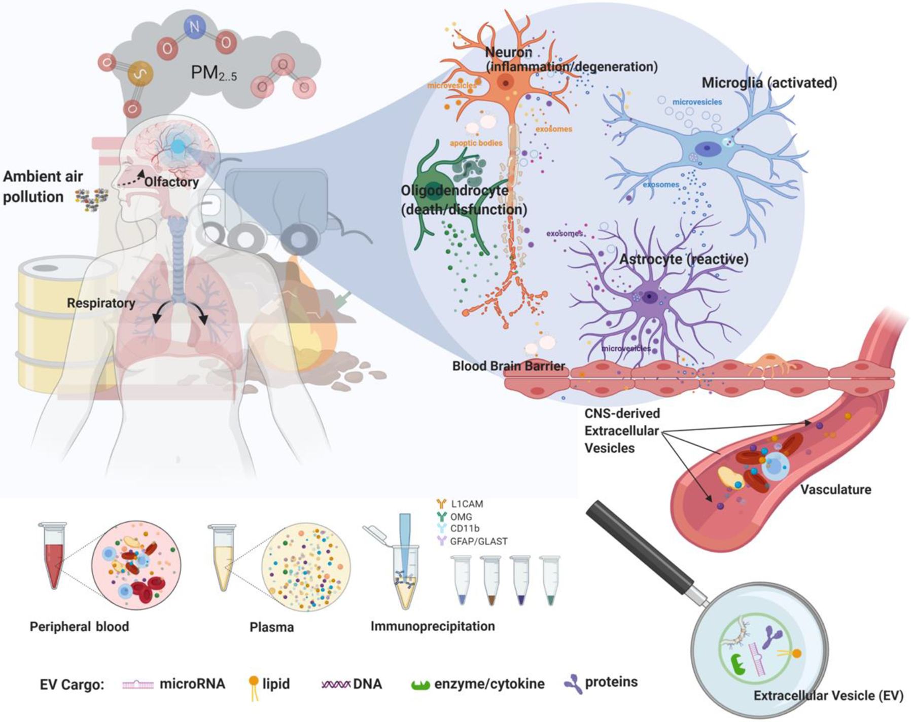
Air pollution triggers microglia activation and reactive astrocytes inducing neuronal inflammation/excitation and oligodendrocyte dysfunction. It results in the increased release of EVs from glial and neuronal cells. EVs can cross the BBB to enter the systemic circulation whereby, they may be isolated from blood/plasma and identified through centrifugation, filtering, or immunoprecipitation with cell-type specific antibodies for molecular analyses. PM2.5: Particulate matter with diameter < 2.5 μm. (Allen et al., 2017; Chen et al., 2020; Liddelow et al., 2017)
Propagation
It is not surprising that PM air pollution is a risk factor for neurological disorder as it is known to have deleterious effects on CNS endothelial cells and glial cells (Bai et al., 2018; Block and Calderon-Garciduenas, 2009; Murgoci et al., 2020). It is increasingly evidenced that air pollution induces microglia and astrocyte activation and plays a critical role in neurodegeneration (Woodward et al., 2017a; You et al., 2020). Changes to the white matter of the brain have also been found, which suggests oligodendrocyte dysfunction is an-other consequence of air pollution exposure (Bai et al., 2019; Calderon-Garciduenas et al., 2016; Li et al., 2020; Woodward et al., 2017a; Woodward et al., 2017b).
Glial Cell Activation
Microglia, astrocytes, oligodendrocytes, and ependymal cells are involved in the shuttling of immunomodulatory mediators and enzymes that participate in neurotransmission like TNF-α, IL1b, and glutaminase (Wang et al., 2017a; Woodward et al., 2017a). Particulate air pollution, specifically, diesel exhaust particles, were shown to increase TNF-α in vitro (Bai et al., 2018). PM2.5–0.1 triggers dysregulation of this system and results in microglia activation, reactive astrocytes, oligodendrocyte loss, BBB permeability, and increased release of EVs, the latter possibly being an attempt to regain niche balance (Calderon-Garciduenas et al., 2016; Wang et al., 2017a; Xiaoyu Chen and Fang Zhang, 2020). Continuous activation of these mechanisms may lead to loss of synaptic integrity, neuronal excitation, and decreased neuronal viability as dysfunctional glia release various pro-inflammatory cytokines and EVs-miRNAs (Fernandes et al., 2018). Diesel fuel exhaust not only caused neuroinflammation and oxidative stress, but stimulated autophagy and increased tau expression (Bai et al., 2018). Interestingly, EVs also participate in the formation and spread of neurotoxic protein species such as α-synuclein, tau, and Aβ in the extracellular space (Perez et al., 2019), and autophagy plays a role in the regulation of EVs.
EVs Production and Release
Autophagy is a cellular degradation process and recycling mechanism, also involved EVs processing, and degradation of tau and Aβ (Abdulrahman et al., 2018; Shang and Sun, 2018). Autophagy dysfunction has been described as being associated with neurodegeneration and AD (Bai et al., 2018). Since PM2.5–0.01 can activate autophagy, TRAP may conversely decrease the formation of EVs. This was evidenced by experiments showing that autophagy stimulation results in intracellular accumulation of neurotoxic proteins due to decreased EVs release, while autophagy inhibition increased the release of EVs and their cargo (Abdulrahman et al., 2018). Additionally, acute exposure to PM2.5–0.1 (ultrafine BC) was found to initiate neuroinflammation and phosphorylated tau (p-tau) accumulation (progressive development of AD-like features) in a neuronal cell line, which were related to changes in autophagy (Shang et al., 2019). Sheng found that inhibition of PM-induced autophagy in its initial stage aggravated p-tau accumulation and increased amyloidosis, but in the final stages of autophagy dysfunction alleviated accumulation, the progressive development of AD-like features in neuronal cell lines. They surmised that besides degradation, an unknown mechanism allows autophagy to transport Aβ and tau into the extracellular space. An implication that gives credence for the role of EVs in mediating air pollution induced neurodegenerative disorders and diseases.
The findings of Adulrahman, that EVs release ameliorated the toxic effect of PM2.5–0.1, suggest that EVs play an intricate counterbalancing role with autophagy in the homeostatic regulation of neurotoxic proteins, both with the goal of preventing the intracellular accumulation of toxic metabolites and proteins. Indeed, increasing evidence supports a role alongside autophagy for the selective secretion of neurotoxic proteins and RNAs by EVs (Baixauli et al., 2014). PM-induced autophagy and altered EVs release may interfere with the homeostatic communication between brain cells, as stimulation of autophagy inhibits EVs release, and conversely, the inhibition of autophagy increased the release of EVs (Abdulrahman et al., 2018).
EVs Mediated Seeding & Spread of Neurotoxic Proteins
EVs play a significant role in maintaining normal synaptic transmission and nerve re-generation but can also contribute to neurodegeneration and AD (Perez et al., 2019). One of the pathways by which tau degradation occurs is via endosome-lysosomal system, which involves the small GTPase Rab35 and the endosomal sorting complex required for transport (ESCRT) machinery, the same pathway by which EVs are produced (Vaz-Silva et al., 2018). EVs are reportedly involved in the propagation and extracellular aggregation of neurotoxic protein species, such as, tau and Aβ (Perez et al., 2019; Wang et al., 2017b). In vitro experimentation found tau protein was found to be released from and taken up by neurons via EVs, and that mediated trans-neuronal transfer of tau was promoted by depolarization and depended on synaptic connectivity (Wang et al., 2017b). Tau was found to be both hypo-phosphorylated and aggregate competent in neuron-derived EVs, meaning that exosome - associated tau is full length and not truncated (Guix et al., 2018; Wang et al., 2017b).
Additionally, EVs have been repeatedly shown to be involved in AD, as they contribute to the spread of Aβ by clustering around Aβ plaques and promote the conversion of fibrillary Aβ to more toxic soluble Aβ1–42, which is also linked to neuronal death (Joshi et al., 2014; Tsutsumi et al., 2019; Zheng et al., 2017).
Physiological & Pathological Roles of Glia-derived EVs on Neuronal Function
In the next paragraphs, we will highlight the interplay between air pollution, EVs, and neurotoxic proteins. As transfer of EVs to neurons was shown to be mediated by microglia, astrocytes, and oligodendrocytes (Brites and Fernandes, 2015), we will illustrate how EVs participate in neurotoxicity instigated by air pollution. A summary of the physiological and harmful effects of EVs on the central nervous system is shown in Figure 2.
Figure 2. Effects and functions of EVs in the central nervous system.
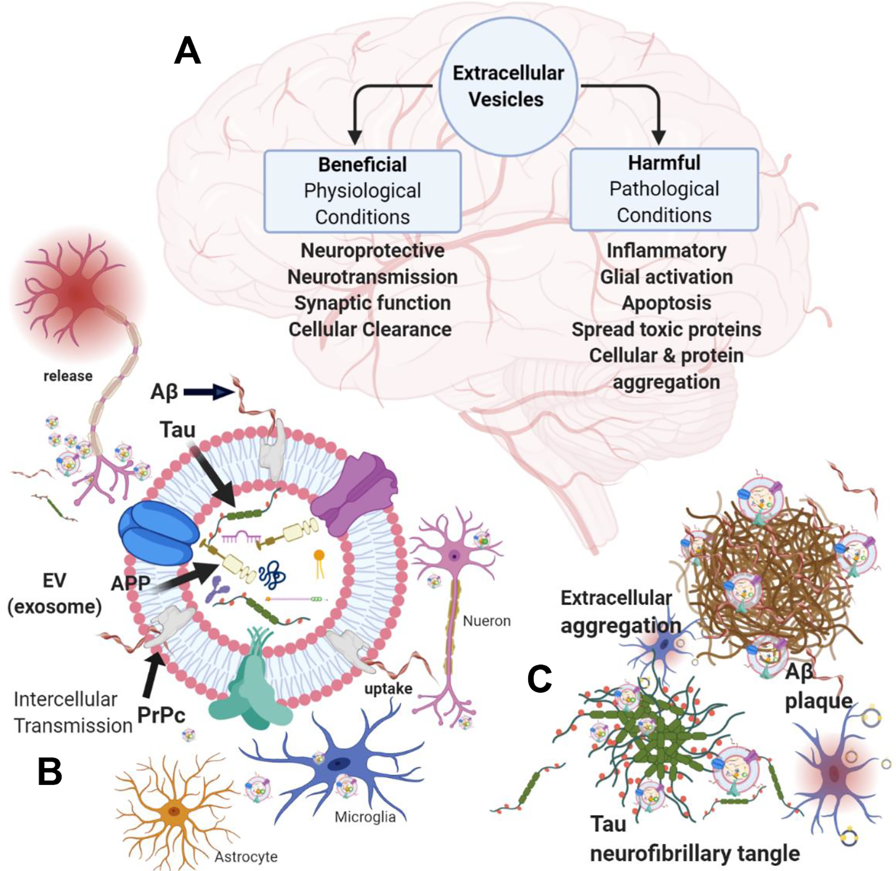
A: Under physiological conditions, EVs help maintain brain homeostasis and neuronal function. However, under pathological conditions, EVs may promote inflammation, neurodegeneration, and myelin loss and serve as conduits for the intercellular spread and aggregation of neurotoxic proteins. B: Neurons release EVs that can contain tau protein or carry Aβ fibrils anchored apically to membrane-bound PrPc. Microglia, astrocytes, and other neurons participate in the clearance and uptake of neuronal-derived EVs. C: EVs can seed the aggregation of extracellular free tau and Aβ, forming neurofibrillary tangles and plaques that activate microglia and promote microglial EV uptake.
EVs and Microglia
Physiological Function
Microglia are the resident immune cells of the brain, and along with their EVs participate in several brain functions. Microglia-EVs promote ceramide and sphingosine production in neurons, influencing synaptic activity and neuronal signaling (Antonucci et al., 2012). In addition to participating in appropriate neurotransmitter function, microglia-derived EVs also contribute to regulating the inflammatory microenvironment within the CNS by generating cytokines and functioning as immune mediators (Brites and Fernandes, 2015; Lemaire et al., 2020). Thereby, under normal physiological conditions, microglia-derived EVs participate in maintaining proper brain health and function (Lemaire et al., 2020).
Another role of microglia as phagocytes is scavenging and clearing debris from the brain (Bachiller et al., 2018). Microglia and their EVs facilitate clearance via EVs release and shuttling of exogenous EVs into lysosomal compartments for degradation or recycling into new EVs (Asai et al., 2015; Bachiller et al., 2018; Fernandes et al., 2018; Joshi et al., 2014), thereby regulating EVs presence in the brain. For example, EVs containing neurotoxic proteins released from neurons and astrocytes promote aggregation and fibrilization of Aβ and tau, respectively. Microglia-EVs associate with insoluble aggregated Aβ in the extracellular space and promote the formation of soluble Aβ forms that triggers clearance of Aβ by microglia uptake (Joshi et al., 2014). Microglial role in clearing neurotoxic proteins is also supported by reports stating that tau-containing EVs specifically target microglia for uptake (Wang et al., 2017b). Aβ and tau-associated EVs that are taken-up by microglia are then transported through the endocytic pathway to microglial lysosomes for degradation (Iqbal et al., 2010).
Neurotoxic Effect
Microglia have been described as primary targets of air pollution induced damage. PM2.5–0.1 can induce microglial cytotoxicity through oxidative stress, lipid peroxidation, inflammation, and activation, but also through the dysregulation of autophagy and apoptosis in microglial cells (Bai et al., 2019). PM from diesel fuel exhaust was determined to cause sustained microglial activation in mice. This activation resulted in hippocampal neuronal disorder. The p65 phosphorylation of NFκB, associated with microglial activation, leads to the release of proinflammatory cytokines and reactive oxygen species (ROS), resulting in decreased neuronal survival in vitro (Duffy et al., 2018; Li et al., 2020). BC and diesel-exhaust particles have also induced the ex-pression of Iba-1, a marker for microglia, and mediators of inflammation such as PGE2, TNF-α, and IL-6, suggesting that microglia are primary targets for air pollution, inducing inflammation in the brain (Bai et al., 2019). Activation of microglia has also been described when exposed to nanosized PM in vitro, suggesting a role in accelerated cognitive aging and a higher risk of dementia in humans (Woodward et al., 2017b).
The involvement of microglia activation in air-pollution associated cognitive impairment could be enormous. In the absence of microglia, diesel-exhaust PM did not result in neuronal death in vitro, but upon co-culture with microglia, neuronal cytotoxicity in-creased 2–3-fold (Roque et al., 2016). Additionally, inhibition of microglia-derived EV release abrogates excitotoxicity from excessive glutamate production by microglia exposed to PM2.5 (Xiaoyu Chen and Fang Zhang, 2020). Microglial activation results in the release of proinflammatory cytokines that not only contribute to chronic inflammation, but also activates sphingomyelinases in the neutral sphingomyelinase pathway that is also associated with exosome release. Indeed, EVs release is enhanced during inflammatory microglia activation from PM (Martin et al., 2019). The activation of sphingomyelinases mediated by the production of proinflammatory mediators from activated microglia maybe deleterious if prolonged, as it can increase the release of EVs and cause the enrichment of ceramide in EVs which may trigger apoptosis in neuronal cells (Dinkins et al., 2014). This indicates that excess EVs release contributes to neurodegeneration, and decreasing EVs secretion may be therapeutic, as the depletion of microglia, along with the inhibition of microglia-derived exosome synthesis, abrogates the spread of tau proteins (Asai et al., 2015).
Air pollution and its components, along with inflammatory cytokines and other toxic cellular metabolites, can propagate from the olfactory bulb into the brain via EVs (Xiaoyu Chen and Fang Zhang, 2020). Microglia may mediate this process since microglia-EVs fuse to neurons allowing for the intracellular uptake of their cargo. Both in vivo and in vitro studies showed that PM2.5 and BC exposure in-creased the release of EVs enriched with glutaminase C from microglia; increasing glutamate generation in the extracellular space (Chen et al., 2020; Xiaoyu Chen and Fang Zhang, 2020). Glutamate is a major excitatory neurotransmitter whose over-production results in neuronal excitotoxicity (Zhou and Danbolt, 2014). Chronic microglial activation from air pollution exposure coupled with EV-associated neurotoxic proteins may lead to dysfunction of microglia clearance ability, similar to what is observed in microglia from AD brains the uptake of fewer EVs than healthy controls (Zheng et al., 2017).
Neuron and oligodendrocyte-derived EVs preferentially target microglia for uptake, and their components have been found incorporated within the lysosomal compartments of microglia after phagocytosis. However, not all EVs are trafficked within microglia for lysosomal degradation. As stated earlier, EVs can be recycled, this process could propagate the transfer of toxic factors induced from air pollution chronic exposure linked to oxidative stress and inflammation and contribute to sustained EVs release, resulting in a long-term neurotoxic cycle and neurodegeneration (Roque et al., 2016; Tsutsumi et al., 2019). Other similar situations have been described for the microglia processing of aggregated Aβ, in which Aβ plaques were converted to neurotoxic fibrillary form (Aβ1–42) and recycled back into the brain microenvironment through shed EVs (Joshi et al., 2014).This mechanism may ultimately contribute to the spreading Aβ in the brain, also contributing to neuroinflammation, as extracellular Aβ like air pollution also trigger activation of microglia (Joshi et al., 2014). Activated microglia are positively correlated with tau pathology, one of the principal features of AD (Asai et al., 2015). Microglial activation is frequently found in AD and PD brains in humans and animal models (Tsutsumi et al., 2019). Since activated microglia are a feature of both AD and exposure to air pollution in the CNS, it is plausible that air pollution mediated activation and inflammation initiates or exacerbates the development of these and others neurodegenerative disease.
EVs and astrocytes
Physiological Function
Astrocytes are the most abundant glial cells in the brain (Nutma et al., 2020). They primarily support neuronal function (synaptic plasticity, neurotransmission, etc.) and mitigate microglial inflammatory response, and contribute to the development and maintenance of the BBB (Nutma et al., 2020; You et al., 2020). Astrocytes release EVs that can be found in blood circulation, being potential biomarkers of CNS diseases (Willis et al., 2017). Under normal physiological conditions, astrocyte-EVs facilitate communication between glial cells and neurons, with their cargo being responsible for determining the phenotypic changes of the recipient neuronal cell (You et al., 2020). Astrocyte-derived EVs participate in signal transmission at neuronal synapses and in neuronal-astrocyte networks, in which they regulate clearance of glutamate and release gliotransmitters (Perego et al., 2000). Astrocyte-EVs are also neurotrophic and were found to contain neuroprotective protein neuroglobin that selectively targets neurons (Venturini et al., 2019). Additionally, astrocyte-EVs play a role in oligodendrocytes differentiation, which supports neuronal axonal function through myelin production (Willis et al., 2020; Xu et al., 2019a). Astrocytes also play a role in maintaining the balance of proteins within the brain that have neurotoxic potential (Goetzl et al., 2016). On the other hand, astrocytes are responsible for the normal production of Aβ, and it was shown in primary astrocyte cultures that Aβ reduces the release of astrocyte-EVs (Abdullah et al., 2016).
Neurotoxic Effects
Activated microglia release microglia-EVs carrying inflammatory factors that induce reactive astrocytes (Liddelow et al., 2017). Reactive astrocytes lose their neurotrophic functions and instead induce neuronal and oligodendrocyte cell death (Li et al., 2019; Winston et al., 2019). Astrocytes release increased amounts of EVs in response to activation mediated by inflammatory cytokines, such as IL-1b, and as shown to induce EVs release in vitro, in a process involving glutaminase, which also plays a role in EVs release during neuroinflammation (Wang et al., 2017a). Overproduction of glutamate, resulting from glutaminase activity, could lead to neuronal excitotoxicity. Other studies have shown that EVs derived from astrocytes treated with IL-1b showed greater uptake by neurons than EVs derived from astrocytes not exposed to the inflammatory cytokine. This effect was associated with increased integrin surface protein enrichment on IL-1b in astrocyte-derived EVs (You et al., 2020). While sub-chronic exposure to diesel exhaust was found to elevate TNF-α in the midbrain, higher exposures produced increased levels of neurotoxic proteins linked with preclinical AD and PD, further indicating that neuroinflammation precedes neurodegenerative disease (Levesque et al., 2011). Moreover, dysregulation of EV formation and miRNAs cargo was reported in human induced astrocyte cells derived from patients with ALS (Amyotrophic Lateral Sclerosis), which affected neurite network maintenance and survival in vitro (Varcianna et al., 2019). This evidence suggests that inflammatory mediators alter EVs production and release and can change EVs cargo constitution.
Though it was found that in vitro release of neuron-derived EVs containing tau were taken up by other neurons and microglia, it was reported that astrocytes did not partake in this up take (Wang et al., 2017b).
EVs and oligodendrocytes
Physiological Function
Oligodendrocytes are the brain’s myelin-producing cells, responsible for maintaining axonal integrity (Kramer-Albers et al., 2007). Oligodendrocytes express several receptors and channels, including glutamate, P2X7, and ATP receptors and ligand gated Ca2+ channels, all involved with neuronal excitation in the brain, and interestingly, with EVs release (Fruhbeis et al., 2013a; Nasrabady et al., 2018; Qu et al., 2009). Several groups have suggested neurotransmitter signaling involvement specifically with the release of oligodendrocyte-EVs (Fruhbeis et al., 2013a; Nasrabady et al., 2018; Qu et al., 2009). This occurs through a mechanism involving glutamate signaling from neurons that causes and elevation of cytosolic causes an elevation of cytosolic Ca2+ within oligodendrocytes which prompts exosome release (Fruhbeis et al., 2013a; Fruhbeis et al., 2013b). Oligodendrocyte-EVs are internalized by neurons and confer neuroprotective properties (Kramer-Albers, 2020). Oligodendrocyte-EVs contain myelin proteins, glycolytic proteins, and other supportive biomolecules that are selectively taken up by neurons through endocytosis, and thereby have neurotrophic properties under physiological conditions. Not only do oligodendrocyte-EVs communicate with neurons, but crosstalk between oligodendrocytes and astrocytes also exists. Astrocyte-EVs carry iron from the blood by the astrocytes and associated with the BBB by the oligodendrocytes (Nutma et al., 2020). Astrocyte-EVs from young astrocytes have been shown to convey support of oligodendrocyte differentiation, a feature which is altered by aging and disease (Willis et al., 2020).
Neurotoxic Effects
Oligodendrocytes are highly metabolic glial cells that are especially sensitive to ROS and oxidative stress, making them a potential target for air pollution-associated cytotoxicity (Nasrabady et al., 2018; Patel and Balabanov, 2012) PM2.5 has been associated with decreased white matter volume and reduced cognition due to myelin loss, which appears to be mediated by activated microglia (Babadjouni et al., 2018; Woodward et al., 2017b). TRAP accelerated myelin loss and neuronal aging, evidenced by neurite atrophy, decreased glutamate receptor GLUA1, and MBP (myelin brain protein) accompanied by increased Iba1 and TNF-α in rat models; suggesting the presence of activated microglia and neuroinflammation (Woodward et al., 2017b). Nanosized PM reportedly causes excitatory neurotoxicity via a glutamatergic mechanism involving decreased GLUA1 and glial inflammatory activation with IL1a and TNF-α, that inhibited neurite outgrowth (Morgan et al., 2011).The breakdown of myelin and loss of myelin sheath precede the appearance of AB and tau pathology, indicting initiation of early-stage AD (Cai and Xiao, 2016).
Additionally, perturbation of intracellular Ca2+ homeostasis by subtoxic concentrations of neurotoxic heavy metals, such as those found in air pollution, can result in oligodendrocyte dysfunction and improper myelin formation (Maiuolo et al., 2019). Neuronal excitotoxicity (i.e., the impairment/death of neurons due to prolonged exposure to excitatory neurotransmitter glutamate) may also be induced by exposure to air pollution (Dong et al., 2009). Air pollution induced excitotoxicity in the brain can increase EVs release in oligodendrocytes (Kramer-Albers, 2020) via the mechanisms discussed earlier, involving neuronal glutamate signaling, and the activation of ionotropic glutamate receptors due to increased intracellular Ca2+ in oligodendrocytes (Fruhbeis et al., 2013a).
Indeed, PM2.5–0.1 has been found to interact with glutamatergic neurons, potentially exacerbating glutamatergic excitotoxicity. Different studies have shown that oligodendrocyte viability is affected by glutamate-mediated toxicity, depending on the intensity and duration of exposure (Fruhbeis et al., 2013a; Morgan et al., 2011). Additionally, increased cytosolic Ca2+, induced through sustained activation of oligodendrocyte P2X7 receptors, can cause caspase-3 activation, leading to activation or cell death and resulting in oligodendrocyte loss, myelin loss, and white matter abnormalities (Nasrabady et al., 2018). Therefore, through eliciting cell death (apoptosis) or activation, air pollution can increase the release of all EVs, including apoptotic bodies.
EVs and Ependymal cells
Physiological Function
Ependymal cells are a family of glial cells that form a cellular epithelial layer that constitutes the BBB and is involved with the cerebral spinal fluid production. Like other endothelial cells, ependymal cell may also be damaged by exposure to air pollution. Under normal conditions, EVs from brain ependymal/endothelial cells are neuroprotective, promote synaptic plasticity and transmission, and regulate the synaptic vesicle cycles in neurons (Gao et al., 2020).
Neurotoxic Effect
As mentioned previously, PM2.5–0.1 can potentially cross the BBB (Shang et al., 2019). Systemic inflammation and oxidative stress resulting from air pollution exposure can also cause endothelial cell damage. Reportedly, platelets exposed to PM2.5 secrete EVs that mediate toxic effects in vascular endothelial cells. This effect was due to changes in the regulatory miRNA cargo that normally confer endothelial cell growth. Instead, PM2.5 induced the release of platelet-derived EVs that were pro-apoptotic and elevated levels of inflammatory cytokines and ROS (Kong et al., 2020). The consequences of the inflammatory response and oxidative damage caused by air pollution exposure may result in brain edema and BBB permeability, impaired memory, and hippocampal long-term potentiation deficiency (Hajipour et al., 2020). It has also been suggested that PM2.5 causes the transition of endothelial cells to a mesenchymal phenotype, with migratory and invasive characteristics (Xu et al., 2019b). Remarkably, exposure to high concentrations of PM2.5 was found to be associated with an increased risk of developing AD in children. These children showed attention and short-term memory deficits, which were correlated with damage to epithelial and endothelial cell barrier and tight junctions (Calderon-Garciduenas et al., 2016).
Considering this evidence, EVs released from cells exposed to air pollution may be altered in their function, taking on pathogenic characteristics that facilitate BBB break-down and the passage of circulating components of air pollution (through vasculature) into the brain.
Applications for CNS-derived EVs
As we have been discussing, EVs may propagate the toxic effects of air pollution by trafficking the altered components from activated, reactive, senescent, or apoptotic glial cells throughout the brain. EVs can carry pro-inflammatory cytokine, neurotoxic proteins, and other pathogenic mediators like glutaminase, that contribute to neuroinflammation, excitotoxicity, and neurodegeneration. In this context, it could be relevant to increase or decrease EV release, target EV cargo (e.g., miRNAs), or interfere with the packaging of proteins into EVs, in order to prevent or ameliorate harmful effects from continuous exposure of their parent cells to air pollution. On the other hand, EVs could also be used as transporters of molecules aimed to reduce inflammation and oxidative damage, reducing the effects of air pollution and other brain-related damage molecules.
Extracellular Vesicle Isolation from Plasma & Brain Cell Enrichment
EVs shed from the CNS can be found in every biological fluid in humans, such as blood, tears, saliva, and urine (Akers et al., 2013). Since EVs can cross the BBB to enter the systemic circulation. Circulating EVs could be easily and noninvasively harvested as potential biomarkers of CNS diseases or preclinical pathological conditions; because they stably express molecular characteristics indicative of their cell of origin and the state of their parent cell or tissue (Kim et al., 2017). They can be isolated and identified from biofluids through centrifugation, size exclusion chromatography, poly-ethylene glycol precipitation, or antibody-coupled immunoprecipitation with cell-type specific antibodies for molecular analyses (Wei et al., 2021). Most commonly, CNS-derived EVs are isolated from cerebral-spinal fluid, plasma, or serum.
It has been suggested that a substantial portion of EV-bound Aβ is associated with neuron-derived EVs (Lim et al., 2019). This is highly relevant because detecting Aβ species in plasma is difficult due to an approximately 50-fold lower concentration of the soluble peptides in plasma than the cerebrospinal fluid. Thereby isolating cell specific EVs would be beneficial.
Antibody-based immuno-precipitation can further distinguish tissue-of-origin after EVs have been isolated from the biofluid, by capturing EVs targeting generic EVs markers CD9, CD63, or CD81, then probing for the expression of protein markers specific to certain cell types (neurons, astrocytes, etc.).
Neuronal and glial EVs markers
L1CAM (L1 cell adhesion molecule protein) a neural cell adhesion molecule, critical for proper CNS development in humans (Haspel and Grumet, 2003), is a commonly used specific marker for isolations of the neuron-derived EVs (Bhargava et al., 2021; Saeedi et al., 2021). The biotin-linked L1CAM antibody can be used to immune-precipitate EVs released by neurons in different samples, either directly or after isolating total EVs using conventional centrifugation, gradient, and column isolation. Even though L1CAM remains the most common marker used to identify neuron-derived EVs in biofluids, it might not be the best candidate because it is also present in other organs and tissues (Fowler, 2019) controversially has its association with EVs has been challenged (Norman et al., 2021). Other markers that have been evaluated for neuron-derived EVs isolation are NCAM (Neural Cell Adhesion Molecule), a neuron-specific plasma membrane protein, and NeuN (Neuronal Nuclei), a neuronal nuclear protein.
Likewise, specific markers for astrocytes, microglia, and oligodendrocytes can also be employed for brain-cell specific EVs isolation from biological fluids (Ohmichi et al., 2019). It is important to note that specific markers for astrocytes can be shared with neurons. For example, some neurons express GFAP (Glial-Fibrillary Acidic Protein) as a consequence of hypoxia-induced differentiation during development, as well as during aging and AD (Bi et al., 2011; Hol et al., 2003). As a result, EVs astrocyte markers such as GLAST (Bhargava et al., 2021) and EAAT2 have become popular for their specificity.
GLAST, also known as EAAT1 (Excitatory amino acid transporter 1), and EAAT2 (Excitatory amino acid transporter 2) are astrocyte-specific glutamate transporters that are found on GFAP+ astrocytes. These markers are abundantly expressed in the cerebellum and hippocampus and whose expressions correlate with neuronal maturation and activity (Parkin et al., 2018; Perego et al., 2000). GLUT1, an isoform of glucose transporter, is also considered a specific marker for astrocyte cells and could be used to identify astrocyte-EVs. GLUT1 has also been identified in association with the endothelium of the BBB. Still, astrocytic GLUT1 is reported to have a lower molecular weight than other GLUT1 isoforms, allowing it to be used for the validation of astrocyte-EVs during isolation (Morgello et al., 1995).
Antigen-specific markers for microglial cells, such as CD11b/MAC-1 and MCT1 and MHC-II, are also shared with peripheral monocyte/macrophage cells making identifying microglia-EVs in plasma difficult (Brites and Fernandes, 2015; Jovicic et al., 2013). Validating with a brain-specific reference RNA or proteins such as β-actin may help distinguish peripheral-derived EVs from brain-EVs isolated from plasma (Fowler, 2019). Antigen specific-marker O4 has been suggested for oligodendrocytes and used for antibody detection and isolation of oligodendrocyte-specific EVs from plasma (Jovicic et al., 2013). Other antigens associated with oligodendrocyte-derived EVs are MBP (myelin basic protein), MOG (oligodendrocyte glycoprotein) which is only expressed at the surface of myelin sheaths and oligodendrocyte membranes (Kramer-Albers et al., 2007), or OMG (oligodendrocyte-myelin-glycoprotein precursor) (Ohmichi et al., 2019).
Modifying EVs Release in the CNS
Research shows that prefibrillar Aβ aggregates preferentially bind EVs, such that exosome bound Aβ isolated from blood plasma reflect PET imaging of brain Aβ plaques, whereas circulating unbound Aβ proteins did not (Lim et al., 2019). Reducing the amount of EVs in the brain by inhibiting their release and the shedding of EVs have been suggested as potential therapeutic approaches for the amelioration of toxic effects associated with air pollution, and the spread of neurotoxic proteins Aβ, tau, and α-synuclein in the CNS (Asai et al., 2015). Inhibition of nSMase-2 y GW486 in vivo resulted in EVs reduction, which was associated with lower levels of soluble Aβ1–42 as well as the number of Aβ plaques in the brain (Dinkins et al., 2014). This evidence suggests that modifying EVs release can potentially alleviate some of the pathological signs of neurodegenerative disease, including AD. Perhaps, early intervention using EVs may block preclinical signs and symptoms from developing and progressing, but further research is needed.
Altering EVs Cargo
Specific EV cargo (e.g., miRNAs) are enriched in a cell-type-dependent manner, which is modulated by changes in the activation state of the parent cell (Squadrito et al., 2014). However, the constitution of EVs cargo may also differ from that of its cell of origin. For example, particular miRNAs are enriched within cell type specific EVs yet have low expression in the cell from which they are derived. This indicates that sorting of protein, lipids, and RNAs into EVs may serve as a mechanism of homeostasis (Kim et al., 2017; Mohammadi et al., 2020).
MicroRNA-21 (mir-21) was found to be transmitted via EVs, in a neuronal in vitro cell-line model of AD, to recipient microglia whom then released the neuronal-derived EVs mir-21 in their own EVs (Fernandes et al., 2018).
Mir-21 has been identified as having a key role in the negative-feedback resolution of inflammation indicating that mir-21 may serve as an EVs biomarker for neuroinflammation associated with AD (Sheedy, 2015). Identifying increased expression of mir-21 in EVs may be diagnostic of early neurodegenerative diseases. Additionally, manipulating EVs miRNA cargo may potentially have some therapeutic use. It was demonstrated that altering the expression of individual miRNA, or of their targeted transcription, controlled miRNA sorting to EVs (Kim et al., 2017; Squadrito et al., 2014; You et al., 2020). As EVs miRNA are more stably preserved than free miRNA, EV-associated miRNA may be a more accurate indicator of air pollution-related miRNA profile changes.
Since EVs are enriched with unique miRNA signatures, dependent on cell-type, changes to EVs associated RNA expression profiles might reveal aberrations related to air pollution exposure (Vrijens et al., 2015). Conversely, altering EVs miRNA cargo may be a useful therapeutic technique. This was evidenced in ALS astrocytes, where mir-494–3p downregulation in EVs was associated with the disease state, and restoration of mir-494–3p by engineered miRNA restored normal neuronal axonal integrity (Varcianna et al., 2019).
Conclusions
EVs comprise different subtypes of lipid membrane vesicles, distinguishable by their cellular biogenesis, size, membrane protein markers, and lipid composition, and are integral to the brain’s physiological function and homeostasis. EVs have been characterized and documented for their functional role in the pathogenesis of various health conditions after exposure to air pollution (cardiovascular disease and pulmonary disease), but sparse investigation into how they may capitulate air pollution induced oxidative stress and neurodegeneration exists. In this review, the role of EVs in the mediation of air pollution-related neurodegeneration, cognitive impairment, and AD was examined. We outlined that the components of ambient air pollution, particularly PM2.5–0.1, can disturb the mechanisms regulating EVs production and cargo loading (alterations in the constitution of EV cargo). Thereby, altering the cellular release of EVs and their protein, metabolite, or miRNA profiles, leading to pathogenic outcomes. For example, the packaging of inflammatory cytokines that promote neuroinflammation and glial cell activation, altered miRNA content that impacts recipient cell phenotype, disrupting homeostasis, or as a result of autophagic dysregulation. Additionally detailed was, how exposure to ambient air pollution increases both the release of EVs and markers of neurodegenerative disease such as Aβ and p-tau; and how EVs participate in the seeding and prion-like propagation of these neurotoxic proteins throughout the brain.
Moreover, highlighted is the ability to readily isolate and identify CNS-derived EVs from human biofluids, in particular plasma, for the study and development of air pollution-related neurodegeneration and cognitive disease biomarkers and therapeutic agents. Non-invasive isolation of EVs can be useful for examining the health of their respective tissues and organ system. Furthermore, they can be engineered to specifically target recipient cell, by direct loading of their cargoes or modulation of their producer cells and used as delivery systems to affect certain biological processes (Rufino-Ramos et al., 2017). Understanding the functionality of EVs and their cargo might reveal disease-specific patterns useful in determining risk, disease on-set and progression, and to better aid in treatment and prevention of environmental exposure related neurodegeneration.
Funding
S. Nicholson is supported by the TRANSFORM TL1 Precision Medicine Program (Grant: 5TL1TR001875-05). This work was supported by the National Institutes of Health (R01ES025225; P30ES009089; R21ES027087; R35ES031688).
Abbreviation and acronym index:
- TRAP
Traffic-Related Air Pollution
- EVs
Extracellular Vesicles
- AD
Alzheimer’s Disease
Footnotes
Publisher's Disclaimer: This is a PDF file of an unedited manuscript that has been accepted for publication. As a service to our customers we are providing this early version of the manuscript. The manuscript will undergo copyediting, typesetting, and review of the resulting proof before it is published in its final form. Please note that during the production process errors may be discovered which could affect the content, and all legal disclaimers that apply to the journal pertain.
Competing interests
The authors declare no competing interests.
Declaration of interests
The authors declare that they have no known competing financial interests or personal relationships that could have appeared to influence the work reported in this paper.
REFERENCES
- Abdullah M, et al. , 2016. Amyloid-beta Reduces Exosome Release from Astrocytes by Enhancing JNK Phosphorylation. J Alzheimers Dis. 53, 1433–41. [DOI] [PubMed] [Google Scholar]
- Abdulrahman BA, et al. , 2018. Autophagy regulates exosomal release of prions in neuronal cells. J Biol Chem. 293, 8956–8968. [DOI] [PMC free article] [PubMed] [Google Scholar]
- Akers JC, et al. , 2013. Biogenesis of extracellular vesicles (EV): exosomes, microvesicles, retrovirus-like vesicles, and apoptotic bodies. J Neurooncol. 113, 1–11. [DOI] [PMC free article] [PubMed] [Google Scholar]
- Alkoussa S, et al. , 2020. Extracellular vesicles as actors in the air pollution related cardiopulmonary diseases. Crit Rev Toxicol. 50, 402–423. [DOI] [PubMed] [Google Scholar]
- Allen JL, et al. , 2017. Developmental neurotoxicity of inhaled ambient ultrafine particle air pollution: Parallels with neuropathological and behavioral features of autism and other neurodevelopmental disorders. Neurotoxicology. 59, 140–154. [DOI] [PMC free article] [PubMed] [Google Scholar]
- Antonucci F, et al. , 2012. Microvesicles released from microglia stimulate synaptic activity via enhanced sphingolipid metabolism. EMBO J. 31, 1231–40. [DOI] [PMC free article] [PubMed] [Google Scholar]
- Asai H, et al. , 2015. Depletion of microglia and inhibition of exosome synthesis halt tau propagation. Nat Neurosci. 18, 1584–93. [DOI] [PMC free article] [PubMed] [Google Scholar]
- Babadjouni R, et al. , 2018. Nanoparticulate matter exposure results in neuroinflammatory changes in the corpus callosum. PLoS One. 13, e0206934. [DOI] [PMC free article] [PubMed] [Google Scholar]
- Bachiller S, et al. , 2018. Microglia in Neurological Diseases: A Road Map to Brain-Disease Dependent-Inflammatory Response. Front Cell Neurosci. 12, 488. [DOI] [PMC free article] [PubMed] [Google Scholar]
- Bai KJ, et al. , 2019. Microglial activation and inflammation caused by traffic-related particulate matter. Chem Biol Interact. 311, 108762. [DOI] [PubMed] [Google Scholar]
- Bai KJ, et al. , 2018. Effects of diesel exhaust particles on the expression of tau and autophagy proteins in human neuroblastoma cells. Environ Toxicol Pharmacol. 62, 54–59. [DOI] [PubMed] [Google Scholar]
- Baixauli F, et al. , 2014. Exosomes and Autophagy: Coordinated Mechanisms for the Maintenance of Cellular Fitness. Frontiers in Immunology. 5. [DOI] [PMC free article] [PubMed] [Google Scholar]
- Basso M, Bonetto V, 2016. Extracellular Vesicles and a Novel Form of Communication in the Brain. Front Neurosci. 10, 127. [DOI] [PMC free article] [PubMed] [Google Scholar]
- Bassuk SS, et al. , 2000. Cognitive impairment and mortality in the community-dwelling elderly. Am J Epidemiol. 151, 676–88. [DOI] [PubMed] [Google Scholar]
- Benilova I, et al. , 2012. The toxic Abeta oligomer and Alzheimer’s disease: an emperor in need of clothes. Nat Neurosci. 15, 349–57. [DOI] [PubMed] [Google Scholar]
- Bhargava P, et al. , 2021. Synaptic and complement markers in extracellular vesicles in multiple sclerosis. Multiple sclerosis (Houndmills, Basingstoke, England). 27, 509–518. [DOI] [PMC free article] [PubMed] [Google Scholar]
- Bi B, et al. , 2011. Cortical glial fibrillary acidic protein-positive cells generate neurons after perinatal hypoxic injury. J Neurosci. 31, 9205–21. [DOI] [PMC free article] [PubMed] [Google Scholar]
- Block ML, Calderon-Garciduenas L, 2009. Air pollution: mechanisms of neuroinflammation and CNS disease. Trends Neurosci. 32, 506–16. [DOI] [PMC free article] [PubMed] [Google Scholar]
- Boda E, et al. , 2020. Understanding the effects of air pollution on neurogenesis and gliogenesis in the growing and adult brain. Curr Opin Pharmacol. 50, 61–66. [DOI] [PubMed] [Google Scholar]
- Bollati V, et al. , 2015. Microvesicle-associated microRNA expression is altered upon particulate matter exposure in healthy workers and in A549 cells. J Appl Toxicol. 35, 59–67. [DOI] [PMC free article] [PubMed] [Google Scholar]
- Bondy SC, 2016. Anthropogenic pollutants may increase the incidence of neurodegenerative disease in an aging population. Toxicology. 341–343, 41–6. [DOI] [PubMed] [Google Scholar]
- Brites D, Fernandes A, 2015. Neuroinflammation and Depression: Microglia Activation, Extracellular Microvesicles and microRNA Dysregulation. Front Cell Neurosci. 9, 476. [DOI] [PMC free article] [PubMed] [Google Scholar]
- Cai Z, Xiao M, 2016. Oligodendrocytes and Alzheimer’s disease. Int J Neurosci. 126, 97–104. [DOI] [PubMed] [Google Scholar]
- Calderon-Garciduenas L, et al. , 2018. Alzheimer’s disease and alpha-synuclein pathology in the olfactory bulbs of infants, children, teens and adults </=40 years in Metropolitan Mexico City. APOE4 carriers at higher risk of suicide accelerate their olfactory bulb pathology. Environ Res. 166, 348–362. [DOI] [PubMed] [Google Scholar]
- Calderon-Garciduenas L, et al. , 2016. Prefrontal white matter pathology in air pollution exposed Mexico City young urbanites and their potential impact on neurovascular unit dysfunction and the development of Alzheimer’s disease. Environ Res. 146, 404–17. [DOI] [PubMed] [Google Scholar]
- Chen HX, et al. , 2020. Exosomes derived from mesenchymal stem cells repair a Parkinson’s disease model by inducing autophagy. Cell Death Dis. 11, 288. [DOI] [PMC free article] [PubMed] [Google Scholar]
- Chen JC, Schwartz J, 2009. Neurobehavioral effects of ambient air pollution on cognitive performance in US adults. Neurotoxicology. 30, 231–9. [DOI] [PubMed] [Google Scholar]
- Chen JC, et al. , 2015. Ambient air pollution and neurotoxicity on brain structure: Evidence from women’s health initiative memory study. Ann Neurol. 78, 466–76. [DOI] [PMC free article] [PubMed] [Google Scholar]
- Chen T, et al. , 2014. Exploration of microRNAs in porcine milk exosomes. BMC Genomics. 15, 100. [DOI] [PMC free article] [PubMed] [Google Scholar]
- Chodosh J, et al. , 2004. Cognitive decline in high-functioning older persons is associated with an increased risk of hospitalization. J Am Geriatr Soc. 52, 1456–62. [DOI] [PubMed] [Google Scholar]
- Chuang JC, Jones PA, 2007. Epigenetics and microRNAs. Pediatr Res. 61, 24R–29R. [DOI] [PubMed] [Google Scholar]
- Clougherty JE, Kubzansky LD, 2010. A framework for examining social stress and susceptibility to air pollution in respiratory health. Cien Saude Colet. 15, 2059–74. [DOI] [PubMed] [Google Scholar]
- Costa LG, et al. , 2017. Neurotoxicity of traffic-related air pollution. Neurotoxicology. 59, 133–139. [DOI] [PMC free article] [PubMed] [Google Scholar]
- D’Anca M, et al. , 2019. Exosome Determinants of Physiological Aging and Age-Related Neurodegenerative Diseases. Front Aging Neurosci. 11, 232. [DOI] [PMC free article] [PubMed] [Google Scholar]
- Daiber A, et al. , 2020. Effects of air pollution particles (ultrafine and fine particulate matter) on mitochondrial function and oxidative stress - Implications for cardiovascular and neurodegenerative diseases. Arch Biochem Biophys. 696, 108662. [DOI] [PubMed] [Google Scholar]
- Dinkins MB, et al. , 2014. Exosome reduction in vivo is associated with lower amyloid plaque load in the 5XFAD mouse model of Alzheimer’s disease. Neurobiol Aging. 35, 1792–800. [DOI] [PMC free article] [PubMed] [Google Scholar]
- Dong XX, et al. , 2009. Molecular mechanisms of excitotoxicity and their relevance to pathogenesis of neurodegenerative diseases. Acta Pharmacol Sin. 30, 379–87. [DOI] [PMC free article] [PubMed] [Google Scholar]
- Doyle LM, Wang MZ, 2019. Overview of Extracellular Vesicles, Their Origin, Composition, Purpose, and Methods for Exosome Isolation and Analysis. Cells. 8. [DOI] [PMC free article] [PubMed] [Google Scholar]
- Duffy CM, et al. , 2018. Microglial Immune Response to Low Concentrations of Combustion-Generated Nanoparticles: An In Vitro Model of Brain Health. Nanomaterials (Basel). 8. [DOI] [PMC free article] [PubMed] [Google Scholar]
- Emmerechts J, et al. , 2012. Air pollution-associated procoagulant changes: the role of circulating microvesicles. J Thromb Haemost. 10, 96–106. [DOI] [PubMed] [Google Scholar]
- Esmaeil Mousavi S, et al. , 2017. Multiple sclerosis and air pollution exposure: Mechanisms toward brain autoimmunity. Med Hypotheses. 100, 23–30. [DOI] [PubMed] [Google Scholar]
- Falker C, et al. , 2016. Exosomal cellular prion protein drives fibrillization of amyloid beta and counteracts amyloid beta-mediated neurotoxicity. J Neurochem. 137, 88–100. [DOI] [PubMed] [Google Scholar]
- Fernandes A, et al. , 2018. Secretome from SH-SY5Y APPSwe cells trigger time-dependent CHME3 microglia activation phenotypes, ultimately leading to miR-21 exosome shuttling. Biochimie. 155, 67–82. [DOI] [PubMed] [Google Scholar]
- Fiandaca MS, et al. , 2015. Identification of preclinical Alzheimer’s disease by a profile of pathogenic proteins in neurally derived blood exosomes: A case-control study. Alzheimers Dement. 11, 600–7 e1. [DOI] [PMC free article] [PubMed] [Google Scholar]
- Fougere B, et al. , 2015. Air Pollution modifies the association between successful and pathological aging throughout the frailty condition. Ageing Res Rev. 24, 299–303. [DOI] [PubMed] [Google Scholar]
- Fowler CD, 2019. NeuroEVs: Characterizing Extracellular Vesicles Generated in the Neural Domain. J Neurosci. 39, 9262–9268. [DOI] [PMC free article] [PubMed] [Google Scholar]
- Frank L, et al. , 2006. Impact of cognitive impairment on mild dementia patients and mild cognitive impairment patients and their informants. Int Psychogeriatr. 18, 151–62. [DOI] [PubMed] [Google Scholar]
- Fruhbeis C, et al. , 2013a. Neurotransmitter-triggered transfer of exosomes mediates oligodendrocyte-neuron communication. PLoS Biol. 11, e1001604. [DOI] [PMC free article] [PubMed] [Google Scholar]
- Fruhbeis C, et al. , 2013b. Extracellular vesicles as mediators of neuron-glia communication. Front Cell Neurosci. 7, 182. [DOI] [PMC free article] [PubMed] [Google Scholar]
- Gao B, et al. , 2020. Brain endothelial cells-derived exosomes induce neuroplasticity in rats with ischemia/reperfusion injury. ACS Chem Neurosci. [DOI] [PubMed] [Google Scholar]
- GDB, 2019. Global burden of 87 risk factors in 204 countries and territories, 1990–2019: a systematic analysis for the Global Burden of Disease Study 2019. Lancet. 17, 1223–1249. [DOI] [PMC free article] [PubMed] [Google Scholar]
- Goetzl EJ, et al. , 2016. Cargo proteins of plasma astrocyte-derived exosomes in Alzheimer’s disease. FASEB journal : official publication of the Federation of American Societies for Experimental Biology. 30, 3853–3859. [DOI] [PMC free article] [PubMed] [Google Scholar]
- Gouin K, et al. , 2017. A comprehensive method for identification of suitable reference genes in extracellular vesicles. J Extracell Vesicles. 6, 1347019. [DOI] [PMC free article] [PubMed] [Google Scholar]
- Guix FX, et al. , 2018. Detection of Aggregation-Competent Tau in Neuron-Derived Extracellular Vesicles. Int J Mol Sci. 19. [DOI] [PMC free article] [PubMed] [Google Scholar]
- Hajipour S, et al. , 2020. Exposure to ambient dusty particulate matter impairs spatial memory and hippocampal LTP by increasing brain inflammation and oxidative stress in rats. Life Sci. 242, 117210. [DOI] [PubMed] [Google Scholar]
- Hartmann A, et al. , 2017. Exosomes and the Prion Protein: More than One Truth. Front Neurosci. 11, 194. [DOI] [PMC free article] [PubMed] [Google Scholar]
- Haspel J, Grumet M, 2003. The L1CAM extracellular region: a multi-domain protein with modular and cooperative binding modes. Front Biosci. 8, s1210–25. [DOI] [PubMed] [Google Scholar]
- Hol EM, et al. , 2003. Neuronal expression of GFAP in patients with Alzheimer pathology and identification of novel GFAP splice forms. Mol Psychiatry. 8, 786–96. [DOI] [PubMed] [Google Scholar]
- Iqbal K, et al. , 2010. Tau in Alzheimer disease and related tauopathies. Curr Alzheimer Res. 7, 656–64. [DOI] [PMC free article] [PubMed] [Google Scholar]
- James BD, et al. , 2014. Contribution of Alzheimer disease to mortality in the United States. Neurology. 82, 1045–50. [DOI] [PMC free article] [PubMed] [Google Scholar]
- Janas AM, et al. , 2016. Exosomes and other extracellular vesicles in neural cells and neurodegenerative diseases. Biochim Biophys Acta. 1858, 1139–51. [DOI] [PubMed] [Google Scholar]
- Joshi P, et al. , 2014. Microglia convert aggregated amyloid-beta into neurotoxic forms through the shedding of microvesicles. Cell Death Differ. 21, 582–93. [DOI] [PMC free article] [PubMed] [Google Scholar]
- Jovicic A, et al. , 2013. Comprehensive expression analyses of neural cell-type-specific miRNAs identify new determinants of the specification and maintenance of neuronal phenotypes. J Neurosci. 33, 5127–37. [DOI] [PMC free article] [PubMed] [Google Scholar]
- Kim KM, et al. , 2017. RNA in extracellular vesicles. Wiley Interdiscip Rev RNA. 8. [DOI] [PMC free article] [PubMed] [Google Scholar]
- Kong L, et al. , 2020. Mediating effects of platelet-derived extracellular vesicles on PM2.5-induced vascular endothelial injury. Ecotoxicol Environ Saf. 198, 110652. [DOI] [PubMed] [Google Scholar]
- Kramer-Albers EM, 2020. Extracellular vesicles in the oligodendrocyte microenvironment. Neurosci Lett. 725, 134915. [DOI] [PubMed] [Google Scholar]
- Kramer-Albers EM, et al. , 2007. Oligodendrocytes secrete exosomes containing major myelin and stress-protective proteins: Trophic support for axons? Proteomics Clin Appl. 1, 1446–61. [DOI] [PubMed] [Google Scholar]
- Krauskopf J, et al. , 2018. The human circulating miRNome reflects multiple organ disease risks in association with short-term exposure to traffic-related air pollution. Environ Int. 113, 26–34. [DOI] [PubMed] [Google Scholar]
- Kur IM, et al. , 2020. Neuronal activity triggers uptake of hematopoietic extracellular vesicles in vivo. PLoS Biol. 18, e3000643. [DOI] [PMC free article] [PubMed] [Google Scholar]
- Lemaire Q, et al. , 2020. Characterization of Immune Cell-derived Extracellular Vesicles and Studying Functional Impact on Cell Environment. J Vis Exp. [DOI] [PubMed] [Google Scholar]
- Levesque S, et al. , 2011. Air pollution & the brain: Subchronic diesel exhaust exposure causes neuroinflammation and elevates early markers of neurodegenerative disease. J Neuroinflammation. 8, 105. [DOI] [PMC free article] [PubMed] [Google Scholar]
- Li K, et al. , 2019. Reactive Astrocytes in Neurodegenerative Diseases. Aging Dis. 10, 664–675. [DOI] [PMC free article] [PubMed] [Google Scholar]
- Li X, et al. , 2020. Activation of NLRP3 in microglia exacerbates diesel exhaust particles-induced impairment in learning and memory in mice. Environ Int. 136, 105487. [DOI] [PubMed] [Google Scholar]
- Liddelow SA, et al. , 2017. Neurotoxic reactive astrocytes are induced by activated microglia. Nature. 541, 481–487. [DOI] [PMC free article] [PubMed] [Google Scholar]
- Lim CZJ, et al. , 2019. Subtyping of circulating exosome-bound amyloid beta reflects brain plaque deposition. Nat Commun. 10, 1144. [DOI] [PMC free article] [PubMed] [Google Scholar]
- Maiuolo J, et al. , 2019. Myelin Disturbances Produced by Sub-Toxic Concentration of Heavy Metals: The Role of Oligodendrocyte Dysfunction. Int J Mol Sci. 20. [DOI] [PMC free article] [PubMed] [Google Scholar]
- Martin PJ, et al. , 2019. Cellular response and extracellular vesicles characterization of human macrophages exposed to fine atmospheric particulate matter. Environ Pollut. 254, 112933. [DOI] [PubMed] [Google Scholar]
- McGuire LC, et al. , 2006. Cognitive functioning as a predictor of functional disability in later life. Am J Geriatr Psychiatry. 14, 36–42. [DOI] [PubMed] [Google Scholar]
- Migliore L, Coppede F, 2009. Environmental-induced oxidative stress in neurodegenerative disorders and aging. Mutat Res. 674, 73–84. [DOI] [PubMed] [Google Scholar]
- Milani C, et al. , 2020. Systemic Exposure to Air Pollution Induces Oxidative Stress and Inflammation in Mouse Brain, Contributing to Neurodegeneration Onset. Int J Mol Sci. 21. [DOI] [PMC free article] [PubMed] [Google Scholar]
- Mohammadi MR, et al. , 2020. Isolation and characterization of microvesicles from mesenchymal stem cells. Methods. 177, 50–57. [DOI] [PMC free article] [PubMed] [Google Scholar]
- Morgan TE, et al. , 2011. Glutamatergic neurons in rodent models respond to nanoscale particulate urban air pollutants in vivo and in vitro. Environ Health Perspect. 119, 1003–9. [DOI] [PMC free article] [PubMed] [Google Scholar]
- Morgello S, et al. , 1995. The human blood-brain barrier glucose transporter (GLUT1) is a glucose transporter of gray matter astrocytes. Glia. 14, 43–54. [DOI] [PubMed] [Google Scholar]
- Murgoci AN, et al. , 2020. Location of neonatal microglia drives small extracellular vesicles content and biological functions in vitro. J Extracell Vesicles. 9, 1727637. [DOI] [PMC free article] [PubMed] [Google Scholar]
- Nasrabady SE, et al. , 2018. White matter changes in Alzheimer’s disease: a focus on myelin and oligodendrocytes. Acta Neuropathol Commun. 6, 22. [DOI] [PMC free article] [PubMed] [Google Scholar]
- Neal M, Richardson JR, 2018. Time to get Personal: A Framework for Personalized Targeting of Oxidative Stress in Neurotoxicity and Neurodegenerative Disease. Current opinion in toxicology. 7, 127–132. [DOI] [PMC free article] [PubMed] [Google Scholar]
- Nemmar A, et al. , 2002. Passage of inhaled particles into the blood circulation in humans. Circulation. 105, 411–4. [DOI] [PubMed] [Google Scholar]
- Nemmar A, et al. , 2001. Passage of intratracheally instilled ultrafine particles from the lung into the systemic circulation in hamster. Am J Respir Crit Care Med. 164, 1665–8. [DOI] [PubMed] [Google Scholar]
- Norman M, et al. , 2021. L1CAM is not associated with extracellular vesicles in human cerebrospinal fluid or plasma. Nat Methods. 18, 631–634. [DOI] [PMC free article] [PubMed] [Google Scholar]
- Nutma E, et al. , 2020. Astrocyte and Oligodendrocyte Cross-Talk in the Central Nervous System. Cells. 9. [DOI] [PMC free article] [PubMed] [Google Scholar]
- Ohmichi T, et al. , 2019. Quantification of brain-derived extracellular vesicles in plasma as a biomarker to diagnose Parkinson’s and related diseases. Parkinsonism Relat Disord. 61, 82–87. [DOI] [PubMed] [Google Scholar]
- Parkin GM, et al. , 2018. Glutamate transporters, EAAT1 and EAAT2, are potentially important in the pathophysiology and treatment of schizophrenia and affective disorders. World J Psychiatry. 8, 51–63. [DOI] [PMC free article] [PubMed] [Google Scholar]
- Patel J, Balabanov R, 2012. Molecular mechanisms of oligodendrocyte injury in multiple sclerosis and experimental autoimmune encephalomyelitis. International journal of molecular sciences. 13, 10647–10659. [DOI] [PMC free article] [PubMed] [Google Scholar]
- Pavanello S, et al. , 2016. Extracellular vesicle-driven information mediates the long-term effects of particulate matter exposure on coagulation and inflammation pathways. Toxicol Lett. 259, 143–150. [DOI] [PMC free article] [PubMed] [Google Scholar]
- Perego C, et al. , 2000. The GLT-1 and GLAST glutamate transporters are expressed on morphologically distinct astrocytes and regulated by neuronal activity in primary hippocampal cocultures. J Neurochem. 75, 1076–84. [DOI] [PubMed] [Google Scholar]
- Perez M, et al. , 2019. Propagation of Tau via Extracellular Vesicles. Front Neurosci. 13, 698. [DOI] [PMC free article] [PubMed] [Google Scholar]
- Pihlstrøm L, et al. , Chapter 22 - Genetics of neurodegenerative diseases: an overview. In: Kovacs GG, Alafuzoff I, Eds.), Handbook of Clinical Neurology. Elsevier, 2018, pp. 309–323. [DOI] [PubMed] [Google Scholar]
- Power MC, et al. , 2011. Traffic-related air pollution and cognitive function in a cohort of older men. Environ Health Perspect. 119, 682–7. [DOI] [PMC free article] [PubMed] [Google Scholar]
- Qu Y, et al. , 2009. P2X7 receptor-stimulated secretion of MHC class II-containing exosomes requires the ASC/NLRP3 inflammasome but is independent of caspase-1. J Immunol. 182, 5052–62. [DOI] [PMC free article] [PubMed] [Google Scholar]
- Rodosthenous RS, et al. , 2016. Ambient particulate matter and microRNAs in extracellular vesicles: a pilot study of older individuals. Part Fibre Toxicol. 13, 13. [DOI] [PMC free article] [PubMed] [Google Scholar]
- Roque PJ, et al. , 2016. Microglia mediate diesel exhaust particle-induced cerebellar neuronal toxicity through neuroinflammatory mechanisms. Neurotoxicology. 56, 204–214. [DOI] [PMC free article] [PubMed] [Google Scholar]
- Rufino-Ramos D, et al. , 2017. Extracellular vesicles: Novel promising delivery systems for therapy of brain diseases. J Control Release. 262, 247–258. [DOI] [PubMed] [Google Scholar]
- Saeedi S, et al. , 2021. Neuron-derived extracellular vesicles enriched from plasma show altered size and miRNA cargo as a function of antidepressant drug response. Molecular Psychiatry. [DOI] [PubMed] [Google Scholar]
- Shang Y, et al. , 2019. Modifications of autophagy influenced the Alzheimer-like changes in SH-SY5Y cells promoted by ultrafine black carbon. Environ Pollut. 246, 763–771. [DOI] [PubMed] [Google Scholar]
- Shang Y, Sun Q, 2018. Particulate air pollution: major research methods and applications in animal models. Environ Dis. 3, 57–62. [DOI] [PMC free article] [PubMed] [Google Scholar]
- Sheedy FJ, 2015. Turning 21: Induction of miR-21 as a Key Switch in the Inflammatory Response. Front Immunol. 6, 19. [DOI] [PMC free article] [PubMed] [Google Scholar]
- Sperling RA, et al. , 2011. Toward defining the preclinical stages of Alzheimer’s disease: recommendations from the National Institute on Aging-Alzheimer’s Association workgroups on diagnostic guidelines for Alzheimer’s disease. Alzheimers Dement. 7, 280–92. [DOI] [PMC free article] [PubMed] [Google Scholar]
- Squadrito ML, et al. , 2014. Endogenous RNAs modulate microRNA sorting to exosomes and transfer to acceptor cells. Cell Rep. 8, 1432–46. [DOI] [PubMed] [Google Scholar]
- Stahl AL, et al. , 2019. Exosomes and microvesicles in normal physiology, pathophysiology, and renal diseases. Pediatr Nephrol. 34, 11–30. [DOI] [PMC free article] [PubMed] [Google Scholar]
- Thery C, et al. , 2018. Minimal information for studies of extracellular vesicles 2018 (MISEV2018): a position statement of the International Society for Extracellular Vesicles and update of the MISEV2014 guidelines. J Extracell Vesicles. 7, 1535750. [DOI] [PMC free article] [PubMed] [Google Scholar]
- Tsutsumi R, et al. , 2019. Involvement of exosomes in dopaminergic neurodegeneration by microglial activation in midbrain slice cultures. Biochem Biophys Res Commun. 511, 427–433. [DOI] [PubMed] [Google Scholar]
- Turner MC, et al. , 2020. Outdoor air pollution and cancer: An overview of the current evidence and public health recommendations. CA Cancer J Clin. [DOI] [PMC free article] [PubMed] [Google Scholar]
- Urabe F, et al. , 2020. Extracellular vesicles as biomarkers and therapeutic targets for cancer. Am J Physiol Cell Physiol. 318, C29–C39. [DOI] [PubMed] [Google Scholar]
- Valavanidis A, et al. , 2008. Airborne particulate matter and human health: toxicological assessment and importance of size and composition of particles for oxidative damage and carcinogenic mechanisms. J Environ Sci Health C Environ Carcinog Ecotoxicol Rev. 26, 339–62. [DOI] [PubMed] [Google Scholar]
- Varcianna A, et al. , 2019. Micro-RNAs secreted through astrocyte-derived extracellular vesicles cause neuronal network degeneration in C9orf72 ALS. EBioMedicine. 40, 626–635. [DOI] [PMC free article] [PubMed] [Google Scholar]
- Vaz-Silva J, et al. , 2018. Endolysosomal degradation of Tau and its role in glucocorticoid-driven hippocampal malfunction. Embo j. 37. [DOI] [PMC free article] [PubMed] [Google Scholar]
- Venturini A, et al. , 2019. Exosomes From Astrocyte Processes: Signaling to Neurons. Front Pharmacol 10, 1452. [DOI] [PMC free article] [PubMed] [Google Scholar]
- Vrijens K, et al. , 2015. MicroRNAs as potential signatures of environmental exposure or effect: a systematic review. Environ Health Perspect. 123, 399–411. [DOI] [PMC free article] [PubMed] [Google Scholar]
- Wang K, et al. , 2017a. TNF-alpha promotes extracellular vesicle release in mouse astrocytes through glutaminase. J Neuroinflammation. 14, 87. [DOI] [PMC free article] [PubMed] [Google Scholar]
- Wang T, Hay JC, 2015. Alpha-synuclein Toxicity in the Early Secretory Pathway: How It Drives Neurodegeneration in Parkinsons Disease. Front Neurosci. 9, 433. [DOI] [PMC free article] [PubMed] [Google Scholar]
- Wang Y, et al. , 2017b. The release and trans-synaptic transmission of Tau via exosomes. Mol Neurodegener. 12, 5. [DOI] [PMC free article] [PubMed] [Google Scholar]
- Wei H, et al. , 2021. Regulation of exosome production and cargo sorting. International Journal of Biological Sciences. 17, 163–177. [DOI] [PMC free article] [PubMed] [Google Scholar]
- Weuve J, et al. , 2012. Exposure to particulate air pollution and cognitive decline in older women. Arch Intern Med. 172, 219–27. [DOI] [PMC free article] [PubMed] [Google Scholar]
- Willis CM, et al. , 2017. A Refined Bead-Free Method to Identify Astrocytic Exosomes in Primary Glial Cultures and Blood Plasma. Front Neurosci. 11, 335. [DOI] [PMC free article] [PubMed] [Google Scholar]
- Willis CM, et al. , 2020. Astrocyte Support for Oligodendrocyte Differentiation can be Conveyed via Extracellular Vesicles but Diminishes with Age. Sci Rep. 10, 828. [DOI] [PMC free article] [PubMed] [Google Scholar]
- Winston CN, et al. , 2019. Complement protein levels in plasma astrocyte-derived exosomes are abnormal in conversion from mild cognitive impairment to Alzheimer’s disease dementia. Alzheimers Dement (Amst). 11, 61–66. [DOI] [PMC free article] [PubMed] [Google Scholar]
- Woodward NC, et al. , 2017a. Toll-like receptor 4 in glial inflammatory responses to air pollution in vitro and in vivo. J Neuroinflammation. 14, 84. [DOI] [PMC free article] [PubMed] [Google Scholar]
- Woodward NC, et al. , 2017b. Traffic-related air pollution impact on mouse brain accelerates myelin and neuritic aging changes with specificity for CA1 neurons. Neurobiol Aging. 53, 48–58. [DOI] [PMC free article] [PubMed] [Google Scholar]
- Chen Xiaoyu, G. J, Huang Yunlong, Liu Shan, Huang Ying, Zhang Zezhong,, Zhang Fang, L. Z, Li Fang, Zheng Jialin C., Ding Wenjun, 2020. Urban airborne PM2.5-activated microglia mediate neurotoxicity through glutaminasecontaining extracellular vesicles in olfactory bulb. Environmental Pollution. [DOI] [PMC free article] [PubMed] [Google Scholar]
- Xu L, et al. , 2019a. Exosome-shuttled miR-92b-3p from ischemic preconditioned astrocytes protects neurons against oxygen and glucose deprivation. Brain Res. 1717, 66–73. [DOI] [PubMed] [Google Scholar]
- Xu Z, et al. , 2019b. PM2.5, Fine Particulate Matter: A Novel Player in the Epithelial-Mesenchymal Transition? Front Physiol. 10, 1404. [DOI] [PMC free article] [PubMed] [Google Scholar]
- You Y, et al. , 2020. Activated human astrocyte-derived extracellular vesicles modulate neuronal uptake, differentiation and firing. J Extracell Vesicles. 9, 1706801. [DOI] [PMC free article] [PubMed] [Google Scholar]
- Zeng Y, et al. , 2010. Associations of environmental factors with elderly health and mortality in China. Am J Public Health. 100, 298–305. [DOI] [PMC free article] [PubMed] [Google Scholar]
- Zheng T, et al. , 2017. Plasma Exosomes Spread and Cluster Around beta-Amyloid Plaques in an Animal Model of Alzheimer’s Disease. Front Aging Neurosci. 9, 12. [DOI] [PMC free article] [PubMed] [Google Scholar]
- Zhou Y, Danbolt NC, 2014. Glutamate as a neurotransmitter in the healthy brain. J Neural Transm (Vienna). 121, 799–817. [DOI] [PMC free article] [PubMed] [Google Scholar]