Abstract
To determine whether pulse wave velocity (PWV) as a measure of arterial stiffness is a marker of coronary artery diseases (CAD), the authors did a cross‐sectional study in 92 patients undergoing coronary angiography for suspected CAD. Arterial stiffness was assessed through recording PWV from the left carotid–right femoral arteries using an automated machine. The mean PWV was higher in patients with CAD than in those without CAD (11.13±0.91 vs 8.14±1.25 m/sec; P<.001). When the severity of CAD was expressed as 1‐, 2‐, and multiple‐vessel disease, there was a significant association between the severity of CAD and PWV. PWV differed significantly with different categorical severity of CAD even when age and total cholesterol were controlled for. In a univariable analysis, PWV was higher with higher systolic blood pressure (P<.004). The authors conclude that arterial stiffness measured through PWV is an independent and complementary cardiovascular risk marker.
Coronary artery disease (CAD) is one of the most common diseases in the world, causing 6.9 million deaths worldwide each year; it is the leading cause of premature death in developed countries. 1 , 2 There are many factors associated with increased risk of CAD, such as smoking, age, diabetes mellitus, hypercholesterolemia, and hypertension. 3 Arterial stiffness is a part of endothelial dysfunction and correlates with hypertension and atherosclerosis. It is a valuable predictor of cardiovascular disease. 4 Early diagnosis and prevention of CAD have stimulated a search for reliable noninvasive methods of detection. One of the noninvasive tests to diagnose CAD is the detection of arterial stiffness by measuring pulse wave velocity (PWV). This technique has been widely applied. This study was designed to determine the role of PWV as a marker of the severity of CAD.
Materials and Methods
Study Design
This was a cross‐sectional study enrolling 92 patients with suspected CAD undergoing coronary angiography at the Interventional Cardiology Lab at Hospital Universiti Sains Malaysia. Arterial stiffness was assessed through left carotid–right femoral PWV using an automated SphygmoCor machine (Actor Medical, West Ryde NSW, Sydney, Australia).
Inclusion and Exclusion Criteria
Inclusion criteria were male sex, age between 30 and 70 years, history of CAD and/or undergoing coronary angiography for suspected CAD and/or stroke, transient ischemic attack, and/or type 2 diabetes. Exclusion criteria included moderate to severe valvular heart disease, prior percutaneous coronary angioplasty or coronary artery bypass grafting, congestive heart failure, recent history of myocardial infarction (within 4 weeks before study), overt nephropathy, uncontrolled hypertension (blood pressure >160/100 mm Hg), and severe peripheral vascular disease.
Study protocol included measurements obtained using the SphygmoCor system and analyzed with the SphygmoCor software. Care was taken to place the transducers over the same point of the arteries and the same distance from the left carotid to the right femoral artery. A minimum of 3 readings using the same device was obtained by the same operator. PWV was calculated with the following formula: PWV (m/sec) = D / TD (D = distance between left carotid and right femoral arteries; TD = time delay).
Statistical Analysis
Data entry and analysis were carried out using SPSS version 12.0.1 (SPSS Inc, Chicago, IL). Numeric values were expressed as mean ± SD; 95% confidence intervals were taken and categorical variables were expressed as frequency and percentage.
Independent t‐test and one‐way analysis of variance (ANOVA) were used to compare the mean of PWV between the groups. P values <.05 were considered significant. Analysis of covariance (ANCOVA) was used to compare PWV while controlling other confounding variables. Age and cholesterol level were found to be significantly associated with PWV at the univariable level; thus, they were adjusted as covariates during ANCOVA analysis.
Ethical Approval
The study was approved by the Universiti Sains Malaysia ethics committee, and informed consent was obtained from all patients before the examinations, which were done during their stay in the hospital.
Results
Clinical Characteristics of Patients
The baseline clinical characteristics of patients are summarized in Table I. The mean age of patients was 54.1 years, mean body mass index was 25.2 kg/m2, mean cholesterol level was 5.3 mmol/L, and mean systolic blood pressure value was 126.3 mm Hg. The number of patients with blood pressure levels >140/90 mm Hg was 10 (10.9%), and 32 (34%) of the patients had blood pressure levels >130/80 mm Hg.
Table I.
Clinical Characteristics (N=92)
| Variables | Mean (SD) or No. (%) |
|---|---|
| Age (years) | 54.1 (9.10) |
| Height (cm) | 164.70 (7.17) |
| Weight (kg) | 68.40 (11.89) |
| BMI (kg/m2) | 25.2 (3.99) |
| Cholesterol (mmol/L) | 5.33 (1.31) |
| SBP (mm Hg) | 126.3 (17.98) |
| DBP (mm Hg) | 76.62 (11.90) |
| BP >140/90 mm Hg | 10 (10.9) |
| BP >130/80 mm Hg | 32 (34.8) |
| HR (beats/min) | 77.48 (8.11) |
| Smokers | 38 (41.3) |
| Nonsmokers | 54 (58.7) |
| Diabetes mellitus | 23 (25.0) |
| No diabetes mellitus | 69 (75.0) |
Abbreviations: BMI, body mass index; BP, blood pressure; DBP, diastolic blood pressure; HR, heart rate; SBP, systolic blood pressure.
PWV and Severity of CAD
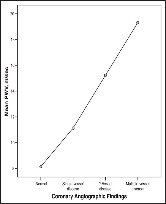
PWV was higher in patients with CAD than in those without CAD. Mean PWV in the group with normal angiographic results was 8.14±1.25 m/sec, and in patients with single‐vessel disease it was 11.13±0.91 m/sec. In those with 2‐vessel disease mean PWV was 15.22±1.11 m/sec, and in the multiple‐vessel disease group it was 19.30±2.05 m/sec (Table II). Comparison among the diseased coronary artery groups (those with single‐vessel disease, 2‐vessel disease, and multiple‐vessel disease) showed that there was a significant difference between the severity of CAD and the mean value of PWV (P<.001) (Table III).
Table II.
Measurement of Pulse Wave Velocity and Coronary Artery Disease
| Angiographic Findings | No. | Mean (SD) |
|---|---|---|
| Normal | 12 | 8.14 (1.252) |
| Single‐vessel disease | 27 | 11.13 (0.916) |
| 2‐Vessel disease | 27 | 15.22 (1.115) |
| Multiple‐vessel disease | 26 | 19.30 (2.056) |
| Total | 92 | 14.25 (4.162) |
Table III.
Comparison Between Pulse Wave Velocity and Severity of Coronary Artery Disease (N=92)
| Mean Difference (95% Confidence Interval) | P Value | ||
|---|---|---|---|
| Normal | Single‐vessel disease | −2.988 (−4.32 to 1.66) | <.001 |
| 2‐Vessel disease | −7.077 (−8.41 to 5.75) | <.001 | |
| Multiple‐vessel disease | −11.160 (−12.50 to 9.82) | <.001 | |
| Single‐vessel disease | 2‐Vessel disease | −4.089 (−5.13 to 3.05) | <.001 |
| Multiple‐vessel disease | −8.172 (−9.22 to 7.12) | <.001 | |
| 2‐Vessel disease | Multiple‐vessel disease | −4.083 (−5.14 to 3.03) | <.001 |
Values were determined by one‐way analysis of variance (ANOVA).
When the severity of CAD was expressed as single‐, 2‐, or multiple‐vessel disease, there was a linear proportional association between the severity of CAD and increased PWV (Figure).
Figure.

Comparison of adjusted means between pulse wave velocity (PWV) and severity of coronary artery disease.
PWV and Risk Factors of CAD
In a univariable analysis, but not in a multivariable analysis, PWV correlates with systolic blood pressure (P<.001) (Table IV). With blood pressure ranges between 140/90 and 160/100 mm Hg, PWV was higher (mean, 14.50 m/sec) compared with blood pressure levels <140/90 mm Hg (mean PWV, 14.22 m/sec), but this difference was not significant (P>.841).
Table IV.
Correlation Between PWV and SBP and Between PWV and DBP
| Groups | Pearson | P Value |
|---|---|---|
| PWV–SBP | 0.295 | .004 |
| PWV–DBP | 0.124 | .238 |
Abbreviations: DBP, diastolic blood pressure; PWV, pulse wave velocity; SBP, systolic blood pressure.
After controlling for age and cholesterol level, ANCOVA analysis showed that the PWV still differed significantly with different categorical severity of CAD (P<.001) (Table V).
Table V.
Adjusted Comparison Between Pulse Wave Velocity and Severity of Coronary Artery Disease (N=92)
| Mean Difference (95% Confidence Interval) | P Value | ||
|---|---|---|---|
| Normal | Single‐vessel disease | −2.807(−4.203 to 1.412) | <.001 |
| 2‐Vessel disease | −6.823(−8.272 to 5.374) | <.001 | |
| Multiple‐vessel disease | −10.834(−12.365 to 9.302) | <.001 | |
| Single‐vessel disease | 2‐Vessel disease | −4.016(−5.076 to 2.956) | <.001 |
| Multiple‐vessel disease | −8.026(−9.150 to 6.903) | <.001 | |
| 2‐Vessel disease | Multiple‐vessel disease | −4.010(−5.088 to 2.933) | <.001 |
Values were determined by analysis of covariance (ANCOVA) controlling for age and cholesterol level.
Discussion
Several noninvasive parameters have been developed that allow assessment of arterial stiffness, including PWV measurement. PWV increases as arterial stiffness rises. An increase in carotid‐femoral PWV reflects arterial stiffening as a result of structural and functional changes of the vascular tree in patients with vascular disease. Aortic stiffness is an important reflection of atherosclerotic vessel function, and PWV evaluates aortic stiffness noninvasively. Hence, clinical studies have demonstrated that PWV can be used to predict cardiovascular risk. 5 , 6 Another noninvasive parameter for measurement of arterial stiffness is the augmentation index (AI). Yasmin and Brown 7 showed that AI or PWV correlated significantly when used to measure arterial stiffness. However, in this study AI was not measured. This study showed a significant association between the severity of CAD and PWV. It is in concordance with a previous study that showed that when the severity of CAD was expressed as 1‐, 2‐, or 3‐vessel disease, PWV was significantly associated with the severity of CAD. 8 However, Ouchi and colleagues 9 showed that no significant difference was observed between PWV values in a group of patients with normal angiographic findings in comparison with patients who have CAD. In this study, the authors measured PWV from the left carotid and left femoral arteries; this was in contrast to the method used in the present study. This different technique may affect the measurement of the distance (D) that is used in the calculation of PWV.
There are several independent risk factors associated with aortic stiffness; these include sex, hypertension, diabetes mellitus, hypercholesterolemia, age, atherosclerosis, renal failure, and cerebrovascular disease. 10 , 11 Zhe and coworkers 12 reported that the metabolic syndrome was closely associated with an increased PWV, and Hansen and associates 13 noted that a higher insulin level was related to a higher PWV. In our study, we did not analyze the results with respect to the metabolic syndrome, and insulin levels were not measured. In a multivariable analysis, this study showed that there is a significant association between the severity of CAD and age and cholesterol, whereas systolic blood pressure, diastolic blood pressure, heart rate, body mass index, and random blood sugar were not significantly related to the severity of CAD. However, in a univariable analysis, PWV was correlated with systolic blood pressure. These findings concur with previous studies that reported a positive correlation between increased age, systolic blood pressure, and increased PWV. 14 Studies done by Boutouyrie and colleagues 15 and Asmar and colleagues 16 noted that an increasing low‐density lipoprotein cholesterol level was associated with increased aortic PWV; they also reported that the 2 major determinants of PWV were age and systolic blood pressure. 16 These findings are similar to our results, which showed an association between systolic blood pressure and PWV (in a univariable analysis). Although in a multivariable analysis there was no significant association between PWV and systolic blood pressure, this difference could be explained by the fact that patients recruited in the present study had a systolic blood pressure range between 90 and 160 mm Hg, whereas in the Asmar and colleagues 16 study, the systolic blood pressure was 98 to 222 mm Hg. Patients with blood pressure ranges between 140/90 and 160/100 mm Hg (n=10) showed a higher PWV when compared to patients with blood pressure levels <140/90 mm Hg, but the difference was not significant; this could be due to the small number of patients.
Our results suggest that arterial stiffness measured by PWV may be an independent marker of CAD beyond the indication provided by other well‐known cardiovascular risk factors. PWV also differed significantly with different categorical severity of CAD, even when age and total cholesterol were controlled for. Measuring aortic stiffness by means of PWV could serve as marker of end organ damage in the arterial system and help to identify patients at high risk for CAD.
Conclusion
This study showed that arterial stiffness measured through noninvasive PWV appears to be an independent and complementary cardiovascular risk factor.
Acknowledgments
Acknowledgments: The authors would like to acknowledge staff members in the interventional and noninterventional cardiology lab for their valuable assistance in making this research possible.
References
- 1. Mathur S. Epidemic of Coronary Heart Disease and its Treatment in Australia, Cardiovascular Disease Series, no. 20. Canberra: Australian Institute of Health and Welfare; 2002; pp. xi, 1, 10, 12, 20, 28–29, 51. [Google Scholar]
- 2. Australian Institute of Health and Welfare (AIHW) . Secondary Prevention and Rehabilitation after Coronary Events or stroke: A Review of Monitoring Issues. Australian Institute of Health and Welfare: Canberra, ACT; 2003; pp. 1, 12–14, 17. [Google Scholar]
- 3. Barker DJP Ischemic Heart Disease, Oxford Textbook of Medicine, 4th ed. Oxford University Press; 2003:15.4,912. [Google Scholar]
- 4. Weber T, Aue JO, Rtiurke MF, et al. Arterial stiffness, wave reflections, and the risk of coronary anery disease. Circulation. 2003;108:1–6. [DOI] [PubMed] [Google Scholar]
- 5. Boutouyrie P, Tropeano AI, Asmar R, et al. Aortic stiffness is an independent predictor of primary coronary events in hypertensive patients: A Longitudinal Study. American Heart Association. Hypertension. 2002;39:10. [DOI] [PubMed] [Google Scholar]
- 6. Rachel D, Rustam R. Microalbuminuria: how informative and reliable are individual measurement for? J Hypertension. 2003;21:1229–1233. [DOI] [PubMed] [Google Scholar]
- 7. Yasmin B, Brown MJ. Similarities and difference between augmentation index and pulse wave velocity in assessment of arterial stiffness. Q J Med. 1999;92:595–600. [DOI] [PubMed] [Google Scholar]
- 8. Lim HE, Park CG, Shin SH, et al. Aortic pulse wave velocity as an independent marker of coronary artery disease. Blood Press. 2004;13:369–375. [DOI] [PubMed] [Google Scholar]
- 9. Ouchi Y, Terashita K, Nakamura T, et al. Aortic pulse wave velocity in patients with coronary atherosclerosis – a comparison with coronary angiographic findings. Nippon Ronen Igakkai Zasshi. 1991;28(1):40–45. [DOI] [PubMed] [Google Scholar]
- 10. Nicole M. Association between arterial stiffness and atherosclerosis. The Rotterdam Study. Stroke. 2001;32:454–460. [DOI] [PubMed] [Google Scholar]
- 11. Ohmori K, Emura S, Takashima T Risk factors of atherosclerosis and aortic pulse wave velocity. Angiology. 2000: 51:53,60. [DOI] [PubMed] [Google Scholar]
- 12. Zhe XW, Zeng J, Tian XK, et al. Pulse wave velocity is associated with metabolic syndrome components in CAPD patients. Am J Nephrol. 2008;28:641–646. [DOI] [PubMed] [Google Scholar]
- 13. Hansen TW, Jeppesen J, Rasmussen S, et al. Relation between insulin and aortic stiffness. J Hum Hypertens. 2004;18:1–7. [DOI] [PubMed] [Google Scholar]
- 14. Cameron JD, Jennings GL, Dart AM. The relationship between arterial compliance. Age blood pressure and serum lipid levels. Hypertension. 1995;1.3:1718–1723. [PubMed] [Google Scholar]
- 15. Boutouyrie P, Tropeaiio AI, Asmar R, et al. Aortic stiffness is an independent predictor of primary coronary events in hypertension patients. Hypertension. 2002;39:l–1. [DOI] [PubMed] [Google Scholar]
- 16. Asmar R, Benetos A, Topouchian J, et al. Assessment of arterial distensibility by automatic pulse wave velocity measurement: validation and clinical application studies. Hypertension. 1995;26:48.5–90.5. [DOI] [PubMed] [Google Scholar]


