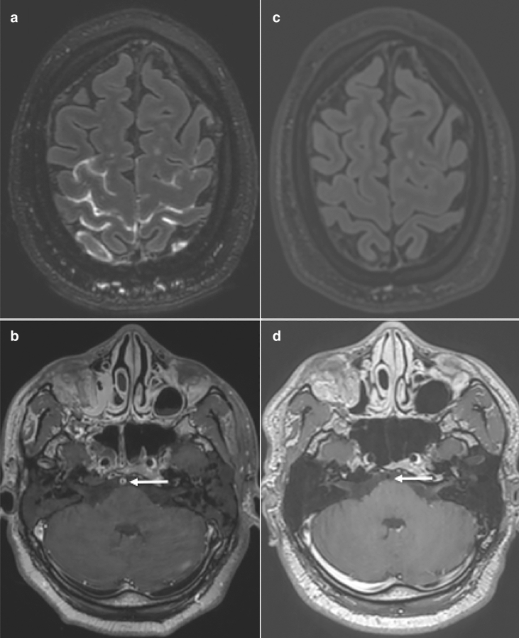
A 46-year-old man with no significant past medical history was hospitalized in the intensive care unit (ICU) due to severe coronavirus disease 2019 (COVID-19) with acute respiratory distress syndrome. He was treated with corticosteroids, antibiotics, anticoagulants, and invasive ventilation. Seven days later and shortly after weaning of both mechanical ventilation and sedation, the patient displayed various signs of delirium. No fever or other signs of meningitis were present. A brain magnetic resonance imaging (MRI) was then performed and showed a major meningeal inflammation with diffuse leptomeningeal enhancement and signs of vasculitis (Fig. 1A, B) (concentric enhancement of the vessel wall of the basilar artery). A lumbar puncture was then realized and showed no abnormalities (normal white blood cells count, no hyperproteinorachia, negative SARS-CoV-2 RT-PCR). The other virological analyses made in cerebrospinal fluid (HSV-1, HSV-2, VZV, and Enterovirus) were all negative, as well as the bacteriological cultures. No further specific therapies were started, and the patient improved rapidly. Five months later, during follow-up, another brain MRI demonstrated a spontaneous normalization of all the abnormalities previously reported (Fig. 1C, D). Aseptic meningeal inflammation and cerebral vasculitis can be found during acute COVID-19 and could spontaneously regress. They are probably para-infectious complications with immune-mediated processes trigged by SARS-CoV-2.
Fig. 1.
Axial post-contrast FLAIR (a, c), and axial contrast-enhanced T1-weighted spin-echo (b, d). Major leptomeningeal inflammation (a) with basilar artery wall enhancement (b), with spontaneous normalization during follow-up (c, d)
Author contributions
FL and SK: designed and conceptualized the study; analyzed the data; drafted the manuscript for intellectual content.
Declarations
Conflicts of interest
FL and SK report no disclosures.
Footnotes
Publisher's Note
Springer Nature remains neutral with regard to jurisdictional claims in published maps and institutional affiliations.
Contributor Information
François Lersy, Email: francois.lersy@chru-strasbourg.fr.
Stéphane Kremer, Email: stephane.kremer@chru-strasbourg.fr.


