Abstract
Objective:
The aim of this study was to investigate the changes in pulpal blood flow (PBF) and pulpal oxygen tension (PpulpO2) after injecting local anesthetics with vasoconstrictors.
Methods:
Under general anesthesia, male Japanese White rabbits were injected with 0.6 mL of 2% lidocaine with 1:80,000 epinephrine (LE) or 3% propitocaine (prilocaine) with 0.03 IU felypressin (PF) at the apical area of the lower incisor.
Results:
Relative to baseline, PBF and PpulpO2 significantly decreased 5 minutes after LE or PF injection as compared with saline. The decrease in PBF was significantly lower in the LE group than in the PF group. Although the LE group had a larger decrease in PpulpO2 relative to baseline than the PF group did, that difference was not significant. PBF and PpulpO2 recovered to baseline faster in the PF group than in the LE group.
Conclusion:
The injection of local anesthetic solutions containing vasoconstrictors (LE or PF) transiently caused significant decreases in PBF that resulted in significant decreases in PpulpO2. The recovery of PpulpO2 was faster than PBF regardless of the vasoconstrictor used.
Keywords: Pulpal blood flow; Pulpal oxygen tension; Epinephrine; Felypressin, Lidocaine, Local anesthesia
Epinephrine or felypressin are added as vasoconstrictors to local anesthetic solutions frequently used in clinical dentistry. Their vasoconstrictive action delays the local absorption into the blood vessels, thereby enhancing and prolonging the local anesthetic action.1,2 However, the vasoconstriction also causes decreased pulpal blood flow (PBF), particularly if an infiltrative technique is used.3 Because a positive correlation between tissue blood flow and oxygen tension in the oral and maxillofacial region has been observed following stellate ganglion block,4 the vasoconstrictor-induced decrease in PBF should expectedly cause a decrease in pulpal oxygen tension (PpulpO2), which, if sustained, may damage the pulpal tissues. In a study using radioisotopes to measure PBF in dogs, Kim et al3 showed that lidocaine with epinephrine reduces the PBF of maxillary canine teeth to ∼30% of control, indicating a potential risk for pulpal necrosis. However, even though injecting local anesthetic solutions containing a vasoconstrictor likely causes hypoxia in the pulp, necrosis does not normally occur. Amemiya et al5 reported that in vitro pulp cells are resistant to hypoxic conditions.
Kong et al6 used pulse oximetry to measure the oxygen dynamics of pulp tissue. Other reports have used pulse oximetry to evaluate pulp vitality in vital, traumatized, nonvital, and other teeth.7,8 However, the technique was originally intended to measure arterial oxygen saturation by passing red and infrared light through soft tissues. Questions remain about the reliability of measured values for pulpal tissues because of the differences in optical conditions, since the pulp presents as soft tissue surrounded by hard tissues such as enamel and dentin.
While studies have reported on PBF and pulpal oxygen saturation levels measured separately,3,7,8 no studies have investigated both PBF and PpulpO2 in the same subjects following injection of local anesthetic solutions containing a vasoconstrictor. As such, little is known about the relationship between the degree and duration of reduced PBF caused by vasoconstrictors and PpulpO2 dynamics. We therefore conducted this study to measure changes in PBF and PpulpO2 following administration of either 1 of 2 conventionally used local anesthetic solutions containing vasoconstrictors (lidocaine with epinephrine or propitocaine [prilocaine] with felypressin) or a saline placebo and to assess the potential relationship between PBF and PpulpO2.
METHODS AND MATERIALS
Animal Preparations
This study was approved by the animal experiment ethics committee of Tokyo Dental College (approval no. 302503). A total of 21 male Japanese White rabbits (lidocaine with epinephrine group, n = 8; propitocaine with felypressin group, n = 8; normal saline group, n = 5; weight ∼2.0 kg; Sankyo Labo, Tokyo, Japan) were used in this study and received humanitarian care in accordance with Tokyo Dental College's guidelines for the treatment of experimental animals. All experimental animals were given free access to food and water up to the morning of the day of the experiment.
All rabbits were anesthetized with 5.0% isoflurane inhaled through a mask. After each rabbit was secured in a supine position, 0.5 mL of 1% lidocaine (5 mg) was administered via infiltration, a tracheostomy was performed, and a 20-Fr pediatric tracheal tube was inserted into the trachea and secured. The right femoral artery and left posterior auricular vein were then cannulated with 22-gauge catheters as a route for arterial pressure measurement and drug administration, respectively. The rabbits were infused with Ringer's acetate solution at 8 mL/kg/h until the end of the experiment. Rocuronium was administered continuously at 14 μg/kg/min for immobilization.9 End-expiratory carbon dioxide tension (EtCO2) was measured with an anesthesia gas monitor, and mechanical ventilation was carried out to maintain an EtCO2 between 35 and 45 mm Hg. Mean arterial pressure and heart rate were continuously monitored using a pressure transducer.
Under irrigation with water, a round bur was used to make a small cavity ∼0.8-mm in diameter into the pulp chamber of the mandibular central incisor for insertion into the pulp tissues of a tissue oxygen tension probe (UOE-04T; Unique Medical, Tokyo, Japan) or a tissue blood flow probe (UHE-100; Unique Medical), which measures blood flow via the hydrogen clearance technique.10
Upon completion of the above preparations, isoflurane inhalation was stopped and sevoflurane inhalation was started. The end-expiratory concentration of sevoflurane was maintained at 1.8%. Although induction of anesthesia with sevoflurane is more rapid than isoflurane in humans, the anesthetic depth can be inadequate in rabbits, especially during the study preparations, because of sevoflurane's large minimum alveolar concentration value in rabbits.11 Thus, isoflurane was used to induce and maintain anesthesia in this study until the study preparations were completed.
Study Protocol
The tissue oxygen tension measurement probe was left for more than 1 hour after insertion into the pulp, and once the cardiovascular parameters had stabilized, observation items were recorded, consisting of heart rate, mean arterial pressure, and mandibular incisor pulpal oxygen tension (PpulpO2). Cardiovascular variables were recorded with a bio tachometer (HRM-100; Unique Medical). PpulpO2 was analyzed using a data collection and analysis system (model UCO; Unique Medical).
Supraperiosteal infiltration near the mandibular central incisor root apices was performed using 0.6 mL of 2% lidocaine with 1:80,000 epinephrine (LE) or 3% propitocaine (prilocaine) with 0.03 IU felypressin (PF). The control group was injected in the same manner and location using 0.6 mL of normal saline. Data gathering occurred before injection, 5 minutes postinjection, and at 15-minute intervals thereafter (20, 35, 50, and 65 minutes).
After measurement, the tissue oxygen tension probe was removed, and the tissue blood flow probe was inserted to the same depth in the same location. The probe was left for more than 1 hour, and once the circulation parameters had stabilized, observation items were again recorded, consisting of heart rate, mean arterial pressure, and mandibular incisor PBF. PBF was analyzed using the same data collection and analysis system. The local anesthetic administration was repeated using the same agents, volume, and injection site, and data gathering occurred at the same time intervals.
Statistical Analysis
An a priori power analysis was performed using G*Power version 3.1.9.2. The sample size for the PBF change 5 minutes after LE injection was calculated, using an α = .05, a β = .2, and an estimated effect size of 1.2 based on the PBF change compared with baseline established by data from a previous study,3 producing a minimum requirement of 8 rabbits per group.
All data are expressed as mean (standard deviation). For statistical analysis, repeated-measures analysis of variance and Dunnett test were performed for intragroup comparison of measured values compared with baseline values. Nonrepeated analysis of variance and the Student-Newman-Keuls test were performed for the intergroup comparison of measured values in the observation periods after LE, PF, or normal saline injection. The Pearson correlation coefficient was used to analyze the relationship between PBF and PpulpO2. A P value less than .05 was used to determine statistical significance. SPSS version 27 was used for the statistical analyses.
RESULTS
Injection of local anesthetic solutions containing vasoconstrictors did not affect heart rate and mean arterial pressure in either group, although it did significantly reduce PBF and PpulpO2 (Tables 1 and 2; P < .05). Comparatively, the saline placebo injection did not significantly affect heart rate, mean arterial pressure, PBF, or PpulpO2 (Table 3).
Table 1.
Hemodynamic Variables and Tissue Oxygen Tension After LE Injection (n = 8)†
|
|
Baseline
|
5 min
|
20 min
|
35 min
|
50 min
|
65 min
|
| HR, bpm | 263.3 (18.7) | 263.8 (18.2) | 263.7 (17.5) | 263.8 (19.5) | 264.4 (19.4) | 264.5 (19.5) |
| MAP, mm Hg | 70.0 (6.3) | 70.0 (5.4) | 69.0 (6.1) | 69.4 (5.8) | 70.5 (5.6) | 70.6 (5.7) |
| PpulpO2, mm Hg | 38.2 (3.0) | 32.7 (4.3)* | 32.8 (4.1)* | 37.2 (4.0) | 39.7 (2.7) | 40.0 (2.7) |
| PBF, mL/100 g/min | 31.1 (3.5) | 21.2 (3.3)* | 24.8 (4.4)* | 26.1 (3.6)* | 30.2 (3.7) | 31.4 (4.0) |
Data are presented as mean (standard deviation). HR indicates heart rate; MAP, mean arterial pressure; PBF, pulpal blood flow; PpulpO2, pulpal oxygen tension.
P < .05 as compared with baseline.
Table 2.
Hemodynamic Variables and Tissue Oxygen Tension After PF Injection (n = 8)†
|
|
Baseline
|
5 min
|
20 min
|
35 min
|
50 min
|
65 min
|
| HR, bpm | 260.1 (16.5) | 258.4 (16.1) | 258.9 (15.9) | 260.1 (15.5) | 260.6 (16.0) | 259.8 (16.8) |
| MAP, mm Hg | 67.3 (8.5) | 67.1 (8.0) | 66.8 (8.2) | 67.0 (8.7) | 67.4 (8.5) | 67.1 (7.9) |
| PpulpO2, mm Hg | 36.0 (6.0) | 32.4 (5.3)* | 35.5 (6.1) | 35.9 (6.2) | 35.8 (6.1) | 36.2 (6.1) |
| PBF, mL/100 g/min | 30.0 (2.8) | 23.4 (2.3)* | 27.3 (3.0)* | 30.2 (2.9) | 30.1 (2.9) | 30.0 (2.6) |
Data are presented as mean (standard deviation). HR indicates heart rate; MAP, mean arterial pressure; PBF, pulpal blood flow; PpulpO2, pulpal oxygen tension.
P < .05 as compared with baseline.
Table 3.
Hemodynamic Variables and Tissue Oxygen Tension After Normal Saline Injection (n = 5)†
|
|
Baseline
|
5 min
|
20 min
|
35 min
|
50 min
|
65 min
|
| HR, bpm | 267.0 (26.8) | 268.7 (24.7) | 267.4 (26.7) | 266.2 (27.6) | 264.9 (24.4) | 265.0 (22.1) |
| MAP, mm Hg) | 68.3 (5.1) | 68.0 (4.0) | 67.6 (4.1) | 68.1 (6.2) | 68.0 (6.7) | 67.7 (5.1) |
| PpulpO2, mm Hg | 37.9 (5.3) | 38.3 (5.3) | 37.3 (6.7) | 37.2 (6.5) | 37.3 (5.9) | 38.0 (5.5) |
| PBF, mL/100 g/min | 29.4 (3.4) | 28.8 (3.9) | 29.7 (4.3) | 29.2 (4.5) | 29.9 (3.7) | 29.8 (3.8) |
Data are presented as mean (standard deviation). HR indicates heart rate; MAP, mean arterial pressure; PBF, pulpal blood flow; PpulpO2, pulpal oxygen tension.
P < .05 as compared with baseline.
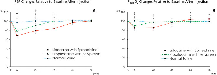
Although significant decreases in PBF were noted following injections with either test local anesthetic compared with baseline and with the saline placebo group, the LE group had significantly larger PBF decreases than the PF group did for most time intervals (Figure 1A). The reduction in PBF relative to baseline 5 minutes after injection was ∼10% larger in the LE group than in the PF group (∼70% vs ∼80%, respectively; P < .05).
Figure 1.
(A) Pulpal blood flow (PBF) changes relative to baseline after injection. PBF was significantly lower for 2% lidocaine with 1:80,000 epinephrine (LE) as compared with propitocaine (prilocaine) with 0.03 IU felypressin (PF) and saline placebo for 35 minutes. PBF was significantly lower for PF compared with saline placebo for 20 minutes. PBF did not change after the saline placebo injection. (B) Pulpal oxygen tension (PpulpO2) changes relative to baseline after injection. PpulpO2 was significantly lower for LE and PF compared with saline placebo at 5 minutes. PpulpO2 was significantly lower for LE compared with PF and saline at 20 minutes. PpulpO2 did not change after the saline placebo injection. *LE versus PF. †LE versus normal saline. ‡PF versus normal saline. P < .05.
Similarly, significant decreases in PpulpO2 were observed after injections in both the LE and PF groups compared with baseline and with the saline group. The reduction in PpulpO2 relative to baseline 5 minutes after injection was ∼5% larger in the LE group than in the PF group (∼85% vs ∼90%, respectively), although that difference was insignificant. Of note, a significant increase in PpulpO2 was observed in the LE group relative to the PF and saline groups at 50 minutes; however, that increase was insignificant compared with baseline (Figure 1B).
Comparing observation periods after the injections, PBF remained significantly lower for a longer period in the LE group than in the PF group. Relative to baseline, PBF recovered in 50 minutes after LE injection and 35 minutes after PF injection (Figure 1A). Similarly, PpulpO2 remained lower for a longer period after injection of LE than PF. Relative to baseline, PpulpO2 recovered 20 minutes after PF injection and 35 minutes after LE injection (Figure 1B). Regardless of the vasoconstrictor used, PpulpO2 recovered faster than PBF did.
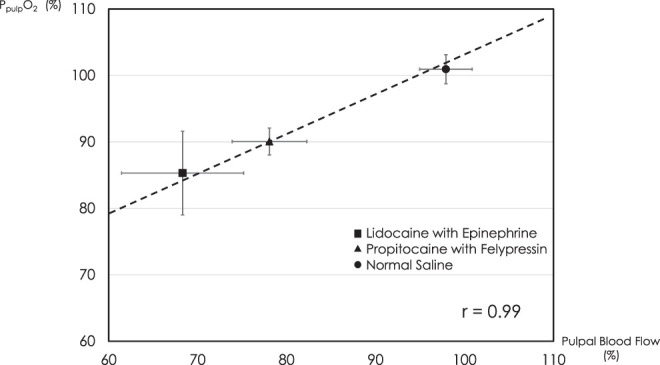
A linear relationship was observed between PBF and PpulpO2 values 5 minutes after the injection (r = 0.99; Figure 2). That same linear relationship was not found at the other time points.
Figure 2.

Relationship between Pulpal blood flow (PBF) and pulpal oxygen tension (PpulpO2) 5 minutes after injection. A linear relationship was observed between PBF and PpulpO2 values 5 minutes after the injection (r = 0.99).
DISCUSSION
In this study, injections of LE or PF transiently produced significant reductions in PBF and PpulpO2. PBF significantly decreased to a lower level from baseline 5–35 minutes after LE injection as compared with after PF injection. PpulpO2 was significantly decreased 5 minutes after both injections with vasoconstrictors and recovered faster after PF as compared with LE (20 minutes vs 35 minutes). Accordingly, our findings regarding the duration of PF being shorter than that of LE are similar to those already reported.12,13
The blood vessels of dental pulp are a microcirculatory system made up of arterioles and venules, and the main determinant of PBF is vascular resistance.14 The local anesthetic solutions used in this study contained the vasoconstrictors epinephrine or felypressin. Felypressin is a synthetic hormone with a structure similar to arginine vasopressin and is a direct-acting agonist that stimulates vascular smooth muscle. It acts on the vasopressin V1 receptor subtype, which activates phospholipase C and initiates the cascade of intracellular events that lead to vascular contraction.15 Felypressin is widely used among dental practitioners in Japan, Europe, and elsewhere. Comparing the 2 vasoconstrictors, epinephrine acts mainly on pulpal arterioles, while felypressin acts preferentially on pulpal venules.15,16 Epinephrine is likely to have a stronger vasoconstricting action and longer duration of action than felypressin. Thus, PBF and PpulpO2 may be reduced to a lesser extent after injection with felypressin as compared with epinephrine.
Terakawa et al4 reported that after stellate ganglion block, tissue blood flow and tissue oxygen tension in the oral and maxillofacial region increased on the injection side and decreased on the contralateral side. This indicates a positive correlation between tissue blood flow and tissue oxygen tension. Kaneko et al17 administered LE or PF to the central incisors of adult volunteers and examined changes in PBF and oxidized and reduced hemoglobin in the pulp using a near-infrared analyzer, finding that PBF decreased and caused a concomitant decrease in pulpal oxidized hemoglobin. The results of that study support our results, namely, that PpulpO2 decreased as a result of decreased PBF due to vasoconstrictor administration.
Although the decrease in PpulpO2 occurred along with the reduction in PBF, it did return to baseline faster. Studies on dental pulp and retinal tissues have shown that although tissue blood flow decreases in proportion to increases in inhaled oxygen concentration, tissue oxygen tension does not change, suggesting the presence of an oxygen-dependent mechanism that regulates blood flow locally.18,19 In addition, the fact that oxygen is more easily transported to sites where blood flow has decreased as a result of injury20 further supports the existence of such a mechanism. In this study, the fact that PpulpO2 recovered ahead of PBF despite the transient decrease in PpulpO2 accompanying the vasoconstrictor-induced decrease in PBF supports the concept of a mechanism in dental pulp involved with automatically regulating PpulpO2.
In a previous study, injection of epinephrine at doses greater than 1.8 μg into the mental foramen of dogs caused a decrease in blood flow in cuspid pulps.21 Another study noted that the metabolic activity of dental pulp cells did not decrease while incubated under hypoxic conditions for 1–4 days.5 It has also been reported that a vasoconstrictor-induced decrease in PBF is unlikely to cause strong pulpal ischemia, considering that the decrease in oxidized hemoglobin in the pulp after LE administration in rabbits was about a half of the decrease noted when PBF was completely blocked by bilateral carotid artery ligation.17 In this study, the recovery of PBF 50 minutes after LE administration and a decrease in PpulpO2 no greater than ∼15% suggests that the pulpal tissues were not subjected to substantial ischemic conditions. The previous reports and the data from this study suggest that dental pulp is unlikely to suffer irreversible injury during the short period of hypoxia caused by administration of a local anesthetic with a vasoconstrictor.
In this study, PBF decreased to ∼70% of baseline after LE administration. In contrast, Kim et al3 reported that PBF decreased to ∼30% of the control value when 1 mL of 2% lidocaine with 1:100,000 epinephrine (a 10 μg epinephrine dose) was administered to the maxillary canine teeth of dogs weighing approximately 10 kg. Because rabbit teeth grow perpetually, the anatomical form of their root apices resembles that of human immature permanent teeth.22 The anatomical form of the root apices of dog teeth, on the other hand, resembles that of human permanent teeth in being close together.22 Since the dose of epinephrine locally administered to rabbits in this study was 7.5 μg, it is possible that the vasoconstrictor-induced decrease in PBF was affected not only by the vasoconstrictor dose but also by the anatomical form of the root apices.
A strong relationship between PBF and PpulpO2 5 minutes after epinephrine or felypressin injection was observed in this study. However, this relationship was not observed after the injection 20 minutes and later. This is thought to be attributed to the faster recovery of PpulpO2 as compared with that of PBF. These results suggest the possible presence of compensatory mechanisms that act to reduce the metabolic rate to better balance oxygen consumption and oxygen supply that become apparent when the PBF is reduced. If such mechanisms exist, they might explain the increase in PpulpO2 above baseline noted 50 minutes after LE injection. Although epinephrine might affect the metabolic processes in pulpal tissues and act to decrease tissue oxygen consumption, that is beyond the scope of this study and remains unknown.
Limitations
One of the potential weaknesses of this study was the order in which measurements were performed. Although the monitored parameters were allowed to return to baseline before the PBF was calculated, it is possible that the first experimental step (PpulpO2 probe insertion/measurement and injection) altered the sensitivity of the vasoconstrictor receptors for the second experimental step (PBF probe insertion/measurement and injection). Thus, future investigations could confirm the results of this study by assessing the potential impact of the order in which PBF and PpulpO2 measurements are obtained.
CONCLUSION
Supraperiosteal injection of local anesthetic solutions containing the vasoconstrictors epinephrine or felypressin caused significant decreases in PBF and PpulpO2. A linear relationship was observed between PBF and PpulpO2 values 5 minutes after the injections, which supports the positive correlation between blood flow and oxygen tension. However, PpulpO2 recovered faster than PBF, regardless of whether epinephrine or felypressin was administered.
Disclosure
The authors have declared no conflicts of interest.
REFERENCES
- 1.Ohkado S, Ichinohe T, Kaneko Y. Comparative study on anesthetic potency depending on concentrations of lidocaine and epinephrine: assessment of dental local anesthetics using the jaw-opening reflex. Anesth Prog . 2001;48:16–20. [PMC free article] [PubMed] [Google Scholar]
- 2.Sisk AL. Vasoconstrictors in local anesthesia for dentistry. Anesth Prog . 1992;39:187–193. [PMC free article] [PubMed] [Google Scholar]
- 3.Kim S, Edwall L, Trowbridge H, Chien S. Effects of local anesthetics on pulpal blood flow in dogs. J Dent Res . 1984;63:650–652. doi: 10.1177/00220345840630050801. [DOI] [PubMed] [Google Scholar]
- 4.Terakawa Y, Ichinohe T, Kaneko Y. Relationship between oral tissue blood flow and oxygen tension in rabbit. Bull Tokyo Dent Coll . 2009;50:83–90. doi: 10.2209/tdcpublication.50.83. [DOI] [PubMed] [Google Scholar]
- 5.Amemiya K, Kaneko Y, Muramatsu T, Shimono M, Inoue T. Pulp cell responses during hypoxia and reoxygenation in vitro. Eur J Oral Sci . 2003;111:332–338. doi: 10.1034/j.1600-0722.2003.00047.x. [DOI] [PubMed] [Google Scholar]
- 6.Kong HJ, Shin TJ, Hyun HK, Kim YJ, Kim JW, Shon WJ. Oxygen saturation and perfusion index from pulse oximetry in adult volunteers with viable incisors. Acta Odontol Scand . 2016;74:411–415. doi: 10.3109/00016357.2016.1171898. [DOI] [PubMed] [Google Scholar]
- 7.Caldeira CL, Barletta FB, Ilha MC, Abrao CV, Gavini G. Pulse oximetry: a useful test for evaluating pulp vitality in traumatized teeth. Dent Traumatol . 2016;32:385–389. doi: 10.1111/edt.12279. [DOI] [PubMed] [Google Scholar]
- 8.Riehl J, Hetzel SJ, Snyder CJ, Soukup JW. Detection of pulpal blood flow in vivo with pulse oximetry in dogs. Front Vet Sci . 2016. 20:Article36, 1–9. [DOI] [PMC free article] [PubMed]
- 9.Terakawa Y, Ichinohe T, Kaneko Y. Rocuronium and vecuronium do not affect mandibular bone marrow and masseter muscular blood flow in rabbits. J Oral Maxillofac Surg . 2010;68:15–20. doi: 10.1016/j.joms.2009.04.040. [DOI] [PubMed] [Google Scholar]
- 10.Aukland K, Bower BF, Berliner RW. Measurement of local blood flow with hydrogen gas. Circulation Res . 1964;14:164. doi: 10.1161/01.res.14.2.164. [DOI] [PubMed] [Google Scholar]
- 11.Scheller MS, Saidman LJ, Partridge BL. MAC of sevoflurane in humans and the New Zealand White rabbit. Can J Anaesth . 1988;35:153–156. doi: 10.1007/BF03010656. [DOI] [PubMed] [Google Scholar]
- 12.Miyoshi T, Aida H, Kaneko Y. Comparative study on anesthetic potency of dental local anesthetics assessed by the jaw-opening reflex in rabbits. Anesth Prog . 2000;47:35–41. [PMC free article] [PubMed] [Google Scholar]
- 13.Shinzaki H, Sunada K. Advantages of anterior inferior alveolar nerve block with felypressin-propitocaine over conventional epinephrine-lidocaine: an efficacy and safety study. J Dent Anesth Pain Med . 2015;15:63–68. doi: 10.17245/jdapm.2015.15.2.63. [DOI] [PMC free article] [PubMed] [Google Scholar]
- 14.Ikeda H, Suda H. Seltzer and Bender's Dental Pulp 2nd ed. Chicago: Quintessence; 2012. [Google Scholar]
- 15.Jastak JT, Yagiela JA, Donaldson D. Pharmacology of vasoconstrictors. Local Anesthesia of the Oral Cavity . 1995. pp. 61–85. Philadelphia: W.B. Saunders.
- 16.Ichinohe T. Vasoconstrictor [in Japanes] In: Fukushima K, editor. Dental Anesthesiology 8th ed. Tokyo: Ishiyaku; 2019. pp. 126–140. [Google Scholar]
- 17.Kaneko Y, Ichinohe T, Ishikawa T, et al. Effects of hypoxia caused by vasoconstrictor contained in dental local anesthetic solution on pulp [in Japanese] Projects for Promoting Private Universities HigherLevel Academic Research 19962000. Tokyo: Tokyo Dental College; 2001. pp. 274–278. [Google Scholar]
- 18.Yu CY, Boyd NM, Cringle SJ, Alder VA, Yu DY. Tissue oxygen tension and blood-flow changes in rat incisor pulp with graded systemic hyperoxia. Arch Oral Biol . 2002;47:239–246. doi: 10.1016/s0003-9969(01)00108-x. [DOI] [PubMed] [Google Scholar]
- 19.Strenn K, Menapace R, Rainer G, Findl O, Wolzt M, Schmetterer L. Reproducibility and sensitivity of scanning laser Doppler flowmetry during graded changes in PO2. Br J Ophthalmol . 1997;81:360–364. doi: 10.1136/bjo.81.5.360. [DOI] [PMC free article] [PubMed] [Google Scholar]
- 20.Budinger GRS, Mutlu GM. Balancing the risks and benefits of oxygen therapy in critically ill adults. Chest . 2013;143:1151–1162. doi: 10.1378/chest.12-1215. [DOI] [PMC free article] [PubMed] [Google Scholar]
- 21.Savoie SS, Lemay H, Taleb L. The effect of epinephrine on pulpal microcirculation. J Dent Res . 1979;58:2074–2079. doi: 10.1177/00220345790580110601. [DOI] [PubMed] [Google Scholar]
- 22.Okuda A. An Atlas of Veterinary Dental Radiology [in Japanese] Tokyo: Gakusosha I; 2003. [Google Scholar]


