Abstract
TP53 is one of the most ubiquitously altered genes in human cancer. The biological impact of rare variants, particularly those located within noncoding regions, remains poorly understood. From interrogation of clinical massively parallel sequencing data from over 55,000 tumors, which included 23,330 tumors with known TP53 mutations, TP53 intron 4 nucleotide substitutions at position c.375+5G were identified in 45 tumors (0.2% of TP53‐mutated cancers), comprising cancers of different organ sites. Loss‐of‐heterozygosity or a second‐hit somatic TP53 mutation was observed in 34 of 40 (85%) informative cases. RT‐PCR analysis showed the c.375+5G>T variant to be associated with aberrantly spliced TP53 mRNA transcripts with concomitant loss of the normal transcript. Immunohistochemical staining for p53 was performed on a representative subset of tumors with TP53 c.375+5G variants (n = 14), all of which showed loss of protein expression (100%; n = 13 complete loss, n = 1 subclonal loss). Our data are consistent with classification of TP53 c.375+5G variants as deleterious intronic mutations that interfere with proper mRNA splicing, ultimately resulting in loss of expression of functional p53 protein. The clinical scenario of a tumor with loss of p53 immunohistochemical staining, yet lacking a detectable TP53 exonic mutation, should therefore prompt consideration of splice‐altering intronic variants.
Keywords: p53, TP53, intronic mutation, pathogenic variant, cancer
Introduction
The TP53 tumor suppressor gene is the most frequently somatically altered gene in human cancer [1]. The majority of mutations occur in the DNA‐binding domain, resulting in loss of transcription factor function [1, 2]. Most of these are missense mutations, clustered at a few hot‐spots, although nonsense and frameshift mutations, causing truncation of the protein, constitute approximately 20–25% of mutations.
Immunohistochemical staining for p53 protein expression serves as a useful surrogate marker for mutation status [3, 4]. The pattern of immunohistochemical staining correlates with the type of TP53 mutation. Missense mutations render the protein resistant to proteolytic degradation, resulting in nuclear accumulation of protein, manifested as diffuse immunohistochemical staining throughout the tumor. In contrast, truncating mutations result in loss of protein expression and complete absence of staining.
The advent of massively parallel sequencing has led to increased recognition of variants in noncoding regions. Mutations at canonical splice sites (i.e. conserved donor and acceptor sites) at the exon–intron boundaries result in aberrant splicing and are estimated to represent 2% of TP53 mutations [2]. However, the effects of nucleotide variants at other positions on splicing and protein expression remain poorly understood, and tumor sequencing assays generally only report coding and canonical splice site mutations, omitting these deeper intronic variants from clinical reports.
The prototype of a TP53 mutation‐driven cancer is high‐grade serous ovarian carcinoma, which has the highest prevalence of somatic TP53 genetic alterations across human cancer types [1]. With mutational prevalence reported to be 95–100% across various studies [5, 6], TP53 mutation is considered by many to be an essential diagnostic feature of this tumor subtype [7]. We recently conducted a retrospective analysis of our institutional cohort of high‐grade serous ovarian cancer patients subjected to tumor mutational profiling using the Memorial Sloan Kettering ‐ Integrated Mutation Profiling of Actionable Cancer Targets (MSK‐IMPACT) platform to identify rare cases that lack TP53 genetic alterations [6]. Through this effort, we observed a recurrent intronic variant, c. 375+5G>T, in several tumors which did not harbor any other TP53 sequence alteration. As the biological significance of this variant was unknown, the original tumor sequencing report had documented these cases as negative for TP53 somatic genetic alterations. To explore the possibility that intronic nucleotide substitutions at position c.375+5G represent an alternative mechanism for disruption of the TP53 gene, in this study we performed a comprehensive interrogation of these variants across a pan‐cancer cohort, complemented by mRNA and immunohistochemical analyses.
Materials and methods
Case selection and massively parallel sequencing data analysis
We performed a retrospective search of tumor samples, across all cancer types, from 55,772 patients subjected to clinical targeted massively parallel sequencing using the MSK‐IMPACT platform, to determine the prevalence of somatic TP53 c. 375+5G variants. Specific details of panel design, capture protocol, sequencing, quality control, read alignment, and variant calling for the MSK‐IMPACT assay have been described [8]. Sequencing data from tumors harboring these variant alleles were analyzed for coexisting TP53 exonic mutations. Fraction and Allele‐Specific Copy Number Estimates from Tumor Sequencing (FACETS) was used to infer copy number alterations and loss‐of‐heterozygosity (LOH) [9].
TP53 mRNA transcript analysis
RNA extraction and reverse transcription were performed on formalin‐fixed paraffin‐embedded (FFPE) tumor and adjacent normal tissue obtained from the surgical resection specimen of a high‐grade serous ovarian carcinoma with a TP53 c. 375+5G>T variant. RT‐PCR was performed on cDNA from matched patient tumor and normal samples, and from peripheral blood mononuclear cells pooled from healthy individuals, followed by capillary electrophoresis and Sanger sequencing. To assess for aberrant splicing of intron 4, the following M13‐tagged primers were used: exon 3‐F: GTAAAACGACGGCCAGTCAGACCTATGGAAACTACTTCCTG; exon 4‐F: GTAAAACGACGGCCAGTCAATGGATGATTTGATGCTG; and exon 5‐R: CAGGAAACAGCT ATGACGGCAAAACATCTTGTTGAGG.
Immunohistochemistry
Immunohistochemical staining for p53 was performed on FFPE tissue sections with the monoclonal antibody clone DO‐7 (Ventana Medical Systems, Tucson, AZ, USA; retrieval: CC1 32 min; incubation: 16 min; ready to use dilution) with external tissue controls (including tumors with known TP53 mutations) for each run. Assessment of p53 immunostaining was performed by a pathologist (MHC) and classified as wild‐type (heterogeneous staining) or aberrant (diffuse or complete absence of staining) expression pattern.
Results and discussion
Interrogation of our institutional database of 55,772 tumors from patients subjected to clinical somatic mutational profiling revealed 45 cases (0.08%) with nucleotide substitutions in intron 4 of TP53 at position c. 375+5G (G>T, n = 23; G>A, n = 15; and G>C, n = 7). These were distributed over cancer types across organ sites, including those with a high frequency of TP53 genetic alterations, such as ovarian high‐grade serous, colorectal, pancreatic, gastroesophageal, and lung carcinomas. For each tumor type, the prevalence was <1% (Table 1). The prevalence of known TP53 mutations across all cancer types in our institutional cohort was 42% (n = 23,330), and thus c.375+5G somatic variants represent approximately 0.2% of all TP53 mutations. Germline testing performed on 23,111 cancer patients at our institution revealed 53 patients with pathogenic TP53 germline variants, and none with a c.375+5G germline variant.
Table 1.
Distribution of TP53 c.375+5G intronic variants across tumor types.
| TP53 c.375+5G substitution (n) | Frequency (%) | |||
|---|---|---|---|---|
| G>T | G>A | G>C | ||
| Breast carcinoma | 1 | 1 | 3 | 5/5,887 (0.08%) |
| Colorectal adenocarcinoma | 3 | 2 | 2 | 7/4,859 (0.14%) |
| Esophageal/gastroesophageal junction adenocarcinoma | 1 | 1 | 1 | 3/943 (0.32%) |
| Pancreatic adenocarcinoma | 3 | 2 | 0 | 5/2,520 (0.20%) |
| Anal squamous cell carcinoma | 1 | 0 | 0 | 1/126 (0.79%) |
| Gliomas * | 2 | 4 | 0 | 6/2,037 (0.29%) |
| Lung carcinomas † | 5 | 2 | 0 | 7/7,488 (0.09%) |
| Ovarian high‐grade serous carcinoma | 5 | 0 | 1 | 6/1,441 (0.42%) |
| Uterine leiomyosarcoma | 1 | 0 | 0 | 1/191 (0.52%) |
| Urothelial carcinoma | 0 | 2 | 0 | 2/1,910 (0.10%) |
| Merkel cell carcinoma | 0 | 1 | 0 | 1/138 (0.72%) |
| Cancer of unknown primary | 1 | 0 | 0 | 1/790 (0.13%) |
| Other tumor types | 0 | 0 | 0 | 0/39,131 (0%) |
| Total | 23 | 15 | 7 | 45/55,772 (0.08%) |
Gliomas comprising anaplastic astrocytoma (n = 1), low‐grade glioma (n = 1), and glioblastoma (n = 4).
Lung carcinomas comprising adenocarcinoma (n = 4), small cell carcinoma (n = 2), and large cell neuroendocrine carcinoma (n = 1).
LOH of the TP53 locus was observed in 30 of 40 (75%) evaluable cases (five cases were inconclusive due to insufficient heterozygous SNP markers in this genomic region). In 4 of the 10 tumors without LOH, a second TP53 mutation was detected. Given that TP53 is a prototypic tumor suppressor that conforms to the two‐hit model, the data support genetic inactivation of the remaining allele in a total of 34 of 40 (85%) cases, and generally consistent with c.375+5G variants being driver alterations. For the remaining 15%, the functional status of the remaining allele was unknown.
To assess the impact of nucleotide substitution at c.375+5G on mRNA splicing, we analyzed TP53 mRNA transcripts in a high‐grade serous ovarian carcinoma harboring the c.375+5G>T variant. Using exon 3 (forward) and exon 5 (reverse) primers flanking the intronic variant site, RT‐PCR was performed on mRNA extracted from tumor and normal controls. Capillary electrophoresis of RT‐PCR products revealed loss of the 382‐bp band corresponding to the wild‐type transcript, and the presence of two additional bands (103 and 182 bp), in tumor tissue (Figure 1A). Direct sequencing revealed corresponding transcripts with partial (r.176_375del) and complete (r.97_375del) exon 4 deletions, consistent with aberrant splicing of intron 4 (Figure 1B,C). The r.176_375del transcript introduces a premature stop codon, which, if translated, results in a truncated protein (p.Gly59Valfs*23), while exon 4 skipping is predicted to lead to an abnormal p53 protein product lacking part of the DNA‐binding domain (p.Ser33_Thr125del). Of note, the r.176_375del transcript was also detected when RT‐PCR was performed using an alternate exon 4 forward primer, and this transcript was previously reported in lung cancer, in association with a c.375+5G>A variant [10]. It is important to note that TP53 splicing is extremely complex and our targeted approach may miss other aberrantly spliced transcripts not targeted by our analysis. Nevertheless, our results are consistent with an intact c.375+5G site being essential for generating the full‐length mRNA transcript, with nucleotide substitution being associated with truncated transcripts that may undergo nonsense‐mediated decay.
Figure 1.

mRNA splicing analysis of the TP53 c.375+5G>T variant. (A) RT‐PCR was performed using exon 3 and exon 5 primers (top), followed by capillary electrophoresis (bottom). (B and C) Direct sequencing of PCR products characterizing the aberrant transcripts.
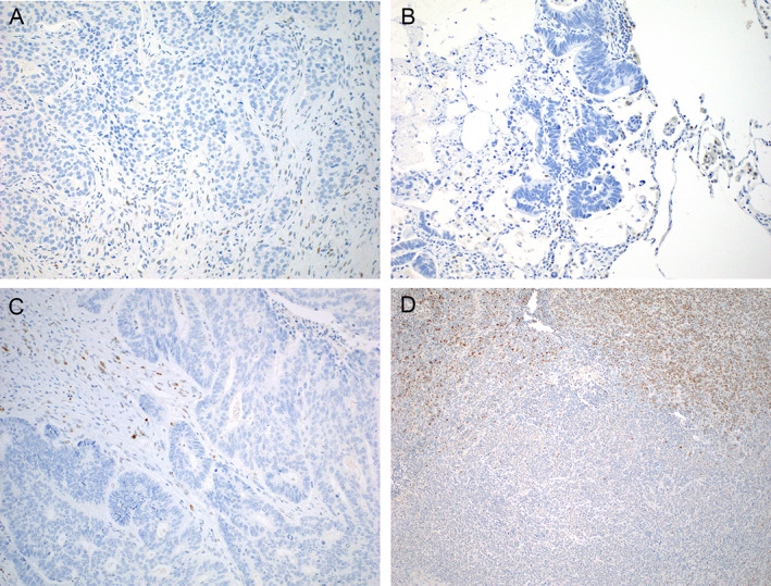
Immunohistochemical staining for p53 was performed on tumor tissues from 14 cases (c.375+5G>T, n = 7; G>A, n = 4; G>C, n = 3). Complete loss of p53 protein expression throughout the tumor section was observed in 13 of these cases (93%; Figure 2A–C). The remaining case was a Merkel cell carcinoma demonstrating a heterogeneous staining pattern, with areas showing a wild‐type p53 expression pattern and distinct areas with complete absence of staining, suggestive of subclonal loss (Figure 2D). The variant allelic fraction for c.375+5G>A in this tumor was only 20% (compared to 81% for a coexisting BCOR mutation), and TP53 LOH was present, which further support this interpretation. Overall, the immunohistochemistry results strongly support the notion that TP53 c.375+5G somatic variants result in loss of p53 expression.
Figure 2.

Immunohistochemical staining of p53 in representative tumors harboring TP53 c.375+5G variants. While scattered normal stromal cells and lymphocytes in the stroma show weak nuclear staining, serving as an internal control, complete absence of p53 expression is seen in (A) invasive ductal carcinoma of breast (G>C), (B) metastatic colorectal adenocarcinoma (G>A), and (C) high‐grade serous ovarian carcinoma (G>T). In a case of (D) Merkel cell carcinoma with a subclonal G>A variant, there are distinct areas with loss of p53 expression adjacent to areas with intact expression.
This study provides compelling evidence to support c.375+5G variants as deleterious in nature and observed across a range of tumor types. In silico analysis of all human splice sites across the genome has demonstrated guanine to be conserved at the +5 position in 77% of splice donor sites [11]. Our data show that, for the TP53 gene, nucleotide substitutions at c.375+5G cause aberrant mRNA splicing, ultimately resulting in loss‐of‐function. Previous studies have demonstrated examples of intronic variants located some distance away from splice junctions to elicit aberrant mRNA splicing, likely through disruption of existing regulatory elements (e.g. splice enhancers or silencers) or creation of cryptic splice sites [10, 12]. For instance, a notable clinically relevant example is provided by MET exon 14 skipping in lung adenocarcinoma which can be caused by somatic mutations well into introns 13 and 14 of MET [13]. Furthermore, loss of p53 expression by immunohistochemistry should prompt consideration of an intronic splice‐altering mutation when a nonsense or frameshift mutation in the coding sequence is not identified. Diagnostically, identification of a TP53 c.375+5G somatic mutation allows molecular confirmation of the histopathologic diagnosis when an exonic mutation is not present in a cancer type known to have a high prevalence of TP53 genetic alterations, such as high‐grade serous ovarian carcinoma. Failure to identify TP53 alterations may also have prognostic and therapeutic implications, as TP53 inactivation is prognostically unfavorable across a wide spectrum of cancer types [1] and correctly establishing TP53 status may soon become critical in clinical trials of MDM2 inhibitors. Finally, our findings highlight the opportunity provided by large‐scale, systematic clinical genomic profiling to identify novel or underappreciated driver alterations even in TP53, the most heavily sequenced cancer gene.
Author contributions statement
MHC, ML and DM were involved in study design, data analysis, data interpretation, and writing of the manuscript. CY and VR carried out the experiments. NM, AZ and AM‐B analyzed the data. All authors had final approval of the submitted and published versions.
Acknowledgement
This work was supported by a NIH/NCI Cancer Center Support Grant P30 CA008748.
No conflicts of interest were declared.
Contributor Information
M Herman Chui, Email: chuim1@mskcc.org.
Diana Mandelker, Email: mandelkd@mskcc.org.
References
- 1. Donehower LA, Soussi T, Korkut A, et al. Integrated analysis of TP53 gene and pathway alterations in the Cancer Genome Atlas. Cell Rep 2019; 28: 1370–1384.e5. [DOI] [PMC free article] [PubMed] [Google Scholar]
- 2. Bouaoun L, Sonkin D, Ardin M, et al. TP53 variations in human cancers: new lessons from the IARC TP53 database and genomics data. Hum Mutat 2016; 37: 865–876. [DOI] [PubMed] [Google Scholar]
- 3. Köbel M, Piskorz AM, Lee S, et al. Optimized p53 immunohistochemistry is an accurate predictor of TP53 mutation in ovarian carcinoma. J Pathol Clin Res 2016; 2: 247–258. [DOI] [PMC free article] [PubMed] [Google Scholar]
- 4. Singh N, Piskorz AM, Bosse T, et al. p53 Immunohistochemistry is an accurate surrogate for TP53 mutational analysis in endometrial carcinoma biopsies. J Pathol 2020; 250: 336–345. [DOI] [PubMed] [Google Scholar]
- 5. Cancer Genome Atlas Research Network . Integrated genomic analyses of ovarian carcinoma. Nature 2011; 474: 609–615. [DOI] [PMC free article] [PubMed] [Google Scholar]
- 6. Chui MH, Momeni Boroujeni A, Mandelker D, et al. Characterization of TP53‐wildtype tubo‐ovarian high‐grade serous carcinomas: rare exceptions to the binary classification of ovarian serous carcinoma. Mod Pathol 2021; 34: 490–501. [DOI] [PMC free article] [PubMed] [Google Scholar]
- 7. Vang R, Levine DA, Soslow RA, et al. Molecular alterations of TP53 are a defining feature of ovarian high‐grade serous carcinoma: a rereview of cases lacking TP53 mutations in the Cancer Genome Atlas Ovarian Study. Int J Gynecol Pathol 2016; 35: 48–55. [DOI] [PMC free article] [PubMed] [Google Scholar]
- 8. Cheng DT, Mitchell TN, Zehir A, et al. Memorial Sloan Kettering‐Integrated Mutation Profiling of Actionable Cancer Targets (MSK‐IMPACT): a hybridization capture‐based next‐generation sequencing clinical assay for solid tumor molecular oncology. J Mol Diagn 2015; 17: 251–264. [DOI] [PMC free article] [PubMed] [Google Scholar]
- 9. Shen R, Seshan VE. FACETS: allele‐specific copy number and clonal heterogeneity analysis tool for high‐throughput DNA sequencing. Nucleic Acids Res 2016; 44: e131. [DOI] [PMC free article] [PubMed] [Google Scholar]
- 10. Smardova J, Liskova K, Ravcukova B, et al. Complex analysis of the p53 tumor suppressor in lung carcinoma. Oncol Rep 2016; 35: 1859–1867. [DOI] [PubMed] [Google Scholar]
- 11. Ma SL, Vega‐Warner V, Gillies C, et al. Whole exome sequencing reveals novel PHEX splice site mutations in patients with hypophosphatemic rickets. PLoS One 2015; 10: e0130729. [DOI] [PMC free article] [PubMed] [Google Scholar]
- 12. Anna A, Monika G. Splicing mutations in human genetic disorders: examples, detection, and confirmation. J Appl Genet 2018; 59: 253–268. [DOI] [PMC free article] [PubMed] [Google Scholar]
- 13. Paik PK, Drilon A, Fan PD, et al. Response to MET inhibitors in patients with stage IV lung adenocarcinomas harboring MET mutations causing exon 14 skipping. Cancer Discov 2015; 5: 842–849. [DOI] [PMC free article] [PubMed] [Google Scholar]


