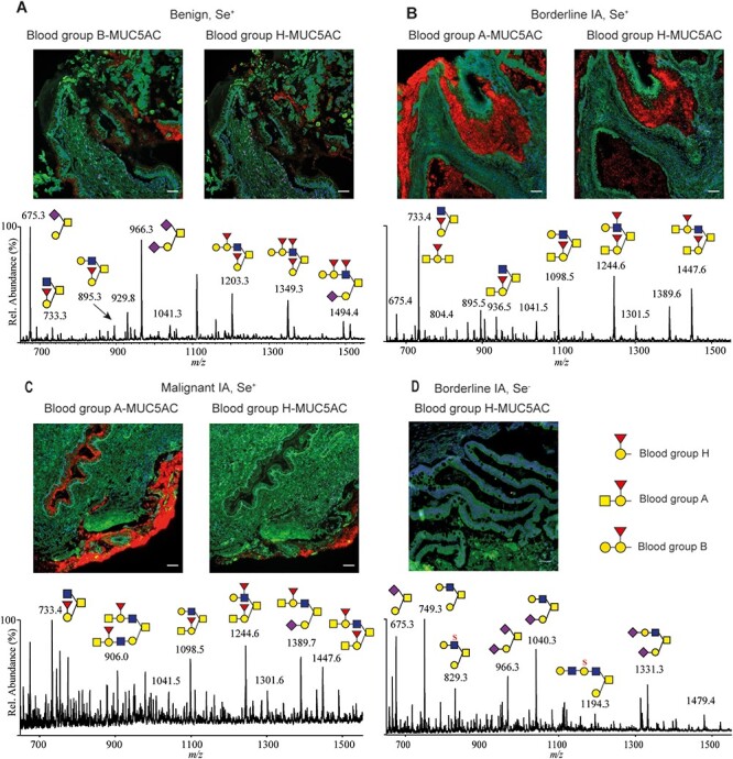
Fig. 2.

Ovarian cyst fluid ABH O-linked oligosaccharides glycans matched with in situ PLA-positive ABH/MUC5AC-staining. Positive staining from secretor individuals (Se+) from benign (A), borderline (B) and malignant (C) mucinous tumor tissue using PLA matching the patient blood group and MUC5AC as well as blood group H/MUC5AC PLA. The mass spectrometric profiles below images show the presence of oligosaccharides identified containing A, B or H determinants from the patients’ cysts fluid (data from (Vitiazeva et al. 2015). (D) A nonsecretor (Se−) mucinous OC patients’ tissue negative for blood group H/MUC5AC PLA without blood group ABH determinants in the patient’s cysts fluids O-linked oligosaccharides is displayed. Inserted white scale bars represent 50 μm.
