Abstract
We present a case in which a 55-year-old male with a past medical history of systemic lupus erythematosus (SLE) and rheumatoid arthritis (RA) presented with sharp, worsening right-sided abdominal pain radiating across the entire abdomen after eating peanuts. Computed tomography (CT) imaging showed evidence of acute sigmoid diverticulitis complicated by a walled-off perforation. The patient’s past medical history suggested previous recurrent episodes of diverticulitis. Our patient underwent exploratory laparotomy, sigmoid colon resection with low anterior anastomosis and proctocolectomy, and loop ileostomy. During treatment, the sigmoid colon was found to be very indurated and abnormally going all the way down to the peritoneal reflection. Appropriate identification of the patient’s condition and timely intervention resulted in a successful outcome.
Keywords: complicated diverticulitis, exploratory laparotomy, sigmoid resection, sigmoid diverticulitis, diverticular disease
Introduction
Diverticulitis is inflammation of one or more diverticula, which are outpouchings of the colon. Together with diverticulosis (the formation of diverticula), diverticulitis is described under the umbrella categorization of diverticular disease, but is more technically identified as symptomatic diverticulosis [1]; approximately 15%-20% of diverticulosis are symptomatic, i.e. become inflamed and transform into diverticulitis [2]. Risk factors for diverticular disease include advanced age (in the United States, almost 80% of individuals over the age of 85 years are estimated to be affected by diverticular disease [3]), immunodeficiency (for instance due to medical management of autoimmune conditions), and lifestyle factors such as a diet low in fibers and high in red meat [2,4-7].
Obstruction of diverticula and inflammatory processes are thought to be a part of the pathophysiology of diverticulitis [8], and research has also shown that diverticula form at areas of weakness in the gut wall which are susceptible to herniation but are also thinner due to muscle layer atrophy [8,9], which predisposes these areas to perforation not just by inflammatory processes but luminal trauma by a variety of items including foreign bodies and ingested items [10,11]. Traditionally, patients at risk for or diagnosed with diverticular disease were recommended to avoid hard foods such as nuts (including peanuts), seeds, corn, and popcorn [12,13] for concerns of entrapment and perforation by mechanisms such as luminal trauma.
Diverticulitis is a clinical diagnosis; work-up typically includes CT imaging, a determination as to whether the diverticulitis is complicated or uncomplicated, and if uncomplicated, a determination of whether or not the patient meets systemic inflammatory response syndrome (SIRS) criteria [5]. Uncomplicated diverticulitis is localized diverticular inflammation, whereas complicated diverticulitis is diverticular inflammation with an abscess, phlegmon, fistula, obstruction, bleeding, or perforation [5,7]. The SIRS criteria requires meeting at least two of the following four conditions: temperature greater than 100.4°F or less than 96.8°F; heart rate greater than 90 beats per minute; respiratory rate greater than 20 breaths per minute or PaCO2 less than 32 mm Hg; white blood cell count of greater than 12,000 per mm2, less than 4,000 per mm2, or more than 10% band forms [5]. Cases of diverticulitis that are complicated are managed by surgery, or percutaneous drainage if the complication is an abscess [5-7], regardless of SIRS criteria. Patients with uncomplicated diverticulitis and who do not meet SIRS criteria are typically not admitted but usually sent home with instructions for a course of antibiotics for 7-10 days and a clear liquid diet for 2-3 days; patients with uncomplicated diverticulitis who meet SIRS criteria are admitted, placed on IV antibiotics, and made nil per os (NPO, meaning no oral intake). Admitted patients with uncomplicated diverticulitis that fail to improve with IV antibiotics are managed by urgent colectomy with end colostomy [5].
Here we discuss the case of a 55-year-old male with a past medical history of systemic lupus erythematosus (SLE), rheumatoid arthritis (RA), and suggestive of recurrent diverticulitis presenting with sharp right-sided abdominal pain radiating across the entire abdomen after eating peanuts; he was found to have evidence of acute sigmoid diverticulitis on CT with evidence of a walled-off microperforation, making his condition a complicated diverticulitis.
Case presentation
Chief complaint
Our patient is a 55-year-old male reporting sharp right-sided abdominal pain with onset one day prior to presentation.
History of present illness
The patient had seen his primary care physician for constipation 11 days prior to admission, which was treated with polyethylene glycol 3350 (MiraLAX, Braintree Laboratories Inc., Braintree, MA, USA) at the time. The patient reports that his symptoms started suddenly on the day prior to the presentation which was preceded by a large, soft bowel movement. The patient denied any associated chest pain, shortness of breath, fevers/chills, cough, or any other signs of infection. The patient presented to the emergency department because he was unable to get an appointment with his primary care physician. The patient also reported eating peanuts (which he consumes frequently) on the day prior to presentation.
Past medical history
The patient’s past medical history includes a deep venous thrombosis (DVT) in his left leg over 20 years ago, coagulopathy controlled by warfarin, SLE, and RA; both autoimmune conditions (SLE and RA) are controlled by hydroxychlorquine. Of note, the patient also reported that he had similar sharp, stabbing pains in the past, most recently at his last appointment with his primary care physician; the most recent of these pains spontaneously resolved with daily MiraLAX intake. The patient has never smoked tobacco.
Examination
On examination, the patient was found to have mild lower abdominal tenderness without rebound, but the examination was otherwise unremarkable.
Investigations
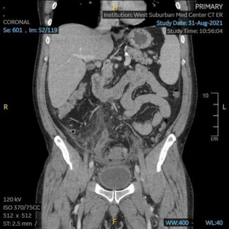
While in the emergency department, the patient underwent a CT scan, which showed acute sigmoid diverticulitis with a walled-off perforation, making the patient’s diverticulitis a complicated diverticulitis (Figure 1). The perforation was suspected to be a microperforation, but as the patient did not appear acutely toxic, consultations were made with other specialists to discuss the possibility of bowel resection after the resolution of the patient’s acute symptoms. During his entire inpatient stay, the patient’s white blood cell count remained low and stable.
Figure 1. Abdominal CT (coronal) showing bowel wall thickening and small foci of gas next to the sigmoid colon suggestive of perforation.
Preoperative diagnosis
Based on the patient’s history, associated investigations, and imaging, the preoperative diagnosis was complicated diverticulitis.
Treatment
The patient underwent an exploratory laparotomy, sigmoid colon resection with low anterior anastomosis and proctocolectomy, placement of two Jackson-Pratt (JP) drains, and loop ileostomy. Notable pathology during the operation consisted of a very indurated abnormal sigmoid colon going all the way down to the peritoneal reflection, which was dissected out. The descending colon was transected above the level of inflammation with a gastrointestinal anastomosis (GIA) stapler (US Surgical Corp., Nonvalk, CT, USA) and then the affected colon was dissected out along its mesenteric attachments with Sonicision (Covidien, Mansfield, MA, USA) below the level of the chronic inflammation. After this, it was transected with a thoracic-abdominal (TA) stapling device (US Surgical Corp., Nonvalk, CT, USA) and the removed portion of the colon was sent to pathology for further analysis for possible neoplasia. Due to the very low nature of the anastomosis, it was decided to perform a diverting loop ileostomy.
Post-operative diagnosis
The postoperative diagnosis was complicated diverticulitis.
Outcome/progress
After recovering from anesthesia, the patient was followed for four days in an inpatient setting, the course of which was uneventful. Samples sent to pathology from the operation were negative for tumors but revealed multiple diverticula and a serosal abscess cavity measuring 3.2 x 2.6 x 1.1 cm which was filled with green-yellow pus. The patient was discharged home on the fourth postoperative day and followed in an outpatient setting. In the outpatient clinic follow-up eight weeks after his procedure, the patient reported complete resolution of his symptoms.
Discussion
Diverticular disease is traditionally taught and understood as a condition of the elderly (i.e. over 65 years of age), and known risk factors include advanced age, obesity, immunodeficiency, and diets that are low in fiber and high in fat and/or red meat [2,4-7]. Of these, age is typically considered the most prognostic factor [2,4,14]; acute diverticulitis is uncommon in patients under the age of 40 and typically occurs in patients over the age of 60 [15]. However, recent research has observed an increasing trend of hospital admissions in the United States for diverticulitis and diverticular disease for patients under the age of 60 [16-18]. This trend has not been limited to the United States; at least one study of an Italian population has found a similar trend of more individuals younger than the traditional age range for diverticular disease being admitted for diverticulitis, a majority of these individuals being male [19]. Further, some research has suggested a seasonal or sinusoidal pattern for diverticulitis admissions in the United States, with a peak during the summer months [20]. A number of studies suggest that the traditional understanding of diverticulitis and its management need updating to accommodate more recent research suggesting an increased rate of diverticulitis among individuals under the age of 65, and that diverticulitis management should be based on disease severity rather than the age of the patient [4,7,8,17-19,21].
Among the traditional recommendations for patients at risk for or diagnosed with diverticular disease was avoidance of hard foods such as nuts (including peanuts), seeds, corn, and popcorn [12,13]. However, recent research such as the study by Strate et al. (2008) suggests that nut, corn, and popcorn consumption is not associated with an increased risk of diverticular disease or complications thereof [13]. As a result, these and other sources advise against avoiding foods such as nuts and seeds, asserting that there is little or no evidence in the literature to support the traditional recommendation [12,13,22,23]. However, these sources are unable to address certain aspects of the physical mechanics that underlie diverticula formation, notably that diverticula occur at areas of weakness in the gut wall [8,9], predisposing these areas to perforation not just by inflammatory processes but also luminal trauma by a variety of items including both foreign bodies and ingested items [10,11]. Therefore, these sources, as Strate et al. themselves observe, are unable to rule out luminal trauma from hard foods such as nuts as a cause of diverticular perforation [23].
Of the traditional risk factors for diverticular disease, our patient has one (immunodeficiency), but other circumstances of his case are consistent with some recent research related to diverticulitis presentations in that he is younger than the age ranges typically associated with diverticulitis, is male, and was admitted in a summer month (August). In contrast with other recent research, however, the mechanisms by which our patient developed diverticulitis at an age earlier than typical presentations may also be related to his high peanut consumption. Based on the findings in our case, particularly the multiple diverticula present in the sigmoid colon resected from our patient, it is likely that our patient’s sigmoid colon wall was weakened and compromised well before, potentially years before, the onset of episodic pain. It is possible that the onset of symptoms in our patient was precipitated by luminal trauma to an already-compromised colon wall by frequent peanut consumption. The walled-off nature of our patient’s perforation may have contributed to relief of symptoms in the past with daily MiraLAX intake. In addition, the recurrent episodes of flare-ups of pain may have also been due to disturbance of the walled-off perforation by the passage of hard foods such as peanuts.
The management of our patient was based on imaging findings, physical examination, and patient history, and in spite of his younger age and autoimmune conditions, he was managed and treated in an appropriate and timely manner, resulting in a successful outcome.
Conclusions
Diverticulitis, i.e. symptomatic diverticulosis, was traditionally considered a condition of the geriatric population over the age of 65 years but is also associated with other risk factors, including a high-fat/red meat diet and immunodeficiency. However, some recent research has shown a marked increase in hospital admissions for diverticulitis in individuals under the age of 60 years in the United States, many of whom lack the other traditionally associated risk factors as well. Other recent research has failed to support the traditional recommendation that individuals with or at risk of diverticular disease avoid certain hard foods such as nuts (including peanuts), seeds, corn, and popcorn; these sources, however, do not account for the physical mechanisms underlying the pathophysiology of diverticular perforation and are therefore unable to rule out luminal trauma due to passage of hard foods and other substances as a mechanism of diverticular perforation. Our case demonstrates the need to appropriately diagnose and manage diverticulitis in any population, regardless of age, and also demonstrates a possible mechanism by which the traditional recommendations for diverticular disease may remain valid for patients with risk factors for diverticular disease such as autoimmune conditions treated by medications with immunosuppressant effects.
The content published in Cureus is the result of clinical experience and/or research by independent individuals or organizations. Cureus is not responsible for the scientific accuracy or reliability of data or conclusions published herein. All content published within Cureus is intended only for educational, research and reference purposes. Additionally, articles published within Cureus should not be deemed a suitable substitute for the advice of a qualified health care professional. Do not disregard or avoid professional medical advice due to content published within Cureus.
The authors have declared that no competing interests exist.
Human Ethics
Consent was obtained or waived by all participants in this study
References
- 1.Taylor BES, Arnell TD. Surgery: A Case-Based Clinical Review. New York, NY: Springer; 2015. Bright red blood per rectum; pp. 203–212. [Google Scholar]
- 2.Diverticular disease in the elderly. Comparato G, Pilotto A, Franzè A, Franceschi M, Di Mario F. Dig Dis. 2007;25:151–159. doi: 10.1159/000099480. [DOI] [PubMed] [Google Scholar]
- 3.Acute diverticulitis. Ferzoco LB, Raptopoulos V, Silen W. N Engl J Med. 1998;338:1521–1526. doi: 10.1056/NEJM199805213382107. [DOI] [PubMed] [Google Scholar]
- 4.Acute diverticulitis in younger patients: any rationale for a different approach? Faria GR, Almeida AB, Moreira H, Pinto-de-Sousa J, Correia-da-Silva P, Pimenta AP. World J Gastroenterol. 2011;17:207–212. doi: 10.3748/wjg.v17.i2.207. [DOI] [PMC free article] [PubMed] [Google Scholar]
- 5.Frank PN, de Virgilio C, Grigorian A. Surgery: A Case-Based Clinical Review. New York, NY: Springer; 2015. Left lower quadrant pain and fever; pp. 245–252. [Google Scholar]
- 6.Ad hoc Practice Parameters Committee of the American College of Gastroenterology: diagnosis and management of diverticular disease of the colon in adults. Stollman NH, Raskin JB. Am J Gastroenterol. 1999;94:3110–3121. doi: 10.1111/j.1572-0241.1999.01501.x. [DOI] [PubMed] [Google Scholar]
- 7.Diagnosis and management of acute diverticulitis. Wilkins T, Embry K, George R. https://www.aafp.org/afp/2013/0501/p612.html. Am Fam Physician. 2013;87:612–620. [PubMed] [Google Scholar]
- 8.Epidemiology and pathophysiology of diverticular disease. Matrana MR, Margolin DA. Clin Colon Rectal Surg. 2009;22:141–146. doi: 10.1055/s-0029-1236157. [DOI] [PMC free article] [PubMed] [Google Scholar]
- 9.Postmortem survey of diverticular disease of the colon. I. Diverticulosis and diverticulitis. Hughes LE. Gut. 1969;10:336–344. doi: 10.1136/gut.10.5.336. [DOI] [PMC free article] [PubMed] [Google Scholar]
- 10.Diverticular perforation secondary to a chicken bone: food for thought. Eastment JG, Butler N, Slater K. Cureus. 2019;11:0. doi: 10.7759/cureus.4273. [DOI] [PMC free article] [PubMed] [Google Scholar]
- 11.Foreign bodies in sigmoid colon diverticulosis. Ross E, McKenna P, Anderson JH. Clin J Gastroenterol. 2017;10:491–497. doi: 10.1007/s12328-017-0786-4. [DOI] [PubMed] [Google Scholar]
- 12.Krok K. Complicated Cases in GI. West Deptford: SLACK Incorporated; 2009. Colonic obstruction in a patient with multiple medical problems; pp. 113–116. [Google Scholar]
- 13.Nut, corn, and popcorn consumption and the incidence of diverticular disease. Strate LL, Liu YL, Syngal S, Aldoori WH, Giovannucci EL. JAMA. 2008;300:907–914. doi: 10.1001/jama.300.8.907. [DOI] [PMC free article] [PubMed] [Google Scholar]
- 14.Complicated diverticulitis: is it time to rethink the rules? Chapman J, Davies M, Wolff B, Dozois E, Tessier D, Harrington J, Larson D. Ann Surg. 2005;242:576–581. doi: 10.1097/01.sla.0000184843.89836.35. [DOI] [PMC free article] [PubMed] [Google Scholar]
- 15.Acute diverticulitis in patients 40 years of age and younger. Marinella MA, Mustafa M. Am J Emerg Med. 2000;18:140–142. doi: 10.1016/s0735-6757(00)90004-4. [DOI] [PubMed] [Google Scholar]
- 16.Diverticulitis in the United States: 1998-2005: changing patterns of disease and treatment. Etzioni DA, Mack TM, Beart RW Jr, Kaiser AM. Ann Surg. 2009;249:210–217. doi: 10.1097/SLA.0b013e3181952888. [DOI] [PubMed] [Google Scholar]
- 17.Trends in diverticulitis management in the United States from 2002 to 2007. Masoomi H, Buchberg BS, Magno C, Mills SD, Stamos MJ. Arch Surg. 2011;146:400–406. doi: 10.1001/archsurg.2010.276. [DOI] [PubMed] [Google Scholar]
- 18.Epidemiological trends and geographic variation in hospital admissions for diverticulitis in the United States. Nguyen GC, Sam J, Anand N. World J Gastroenterol. 2011;17:1600–1605. doi: 10.3748/wjg.v17.i12.1600. [DOI] [PMC free article] [PubMed] [Google Scholar]
- 19.Trends in hospital admission for acute diverticulitis in Italy from 2008 to 2015. Binda GA, Mataloni F, Bruzzone M, et al. Tech Coloproctol. 2018;22:597–604. doi: 10.1007/s10151-018-1840-z. [DOI] [PubMed] [Google Scholar]
- 20.Cyclical increase in diverticulitis during the summer months. Ricciardi R, Roberts PL, Read TE, Marcello PW, Hall JF, Schoetz DJ, Foley EF. Arch Surg. 2011;146:319–323. doi: 10.1001/archsurg.2011.27. [DOI] [PubMed] [Google Scholar]
- 21.Diverticulitis: the effect of age and location on the course of disease. Reisman Y, Ziv Y, Kravrovitc D, Negri M, Wolloch Y, Halevy A. Int J Colorect Dis. 1999;14:250–254. doi: 10.1007/s003840050219. [DOI] [PubMed] [Google Scholar]
- 22.Are there trigger foods I should avoid to prevent diverticulitis attacks? [ Nov; 2021 ];Zeratsky Zeratsky, K K. https://www.mayoclinic.org/healthy-lifestyle/nutrition-and-healthy-eating/expert-answers/diverticulitis-diet/faq-20058293 2021
- 23.Avoiding nuts and seeds for better gut health? You shouldn’t. [ Nov; 2021 ];Godman H. https://www.health.harvard.edu/blog/avoiding-nuts-and-seeds-for-better-gut-health-you-shouldnt-2019090317593 Harvard Health Letter, September. 2019 September


